



CA224

Based on 1 Customer Validation
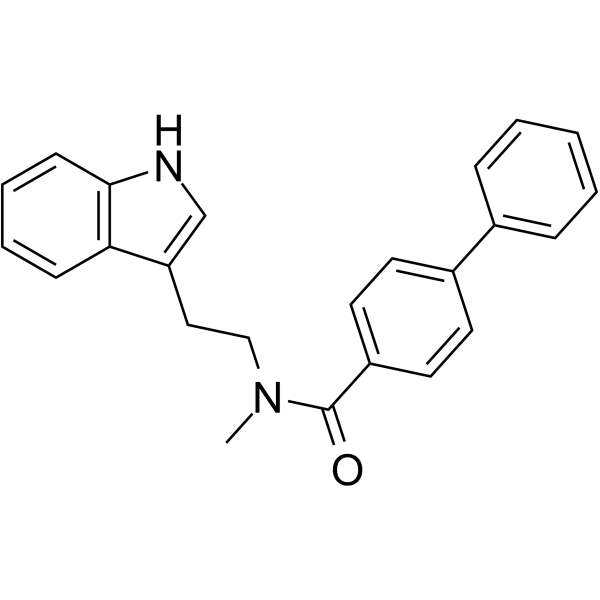
CA224 (Compound 1) is a selective and orally active Cdk4–cyclin D1 inhibitor with an IC50 of 6.2 µM. CA224 induces cell apoptosis and shows antitumor activity.
For research use only. We do not sell to patients.
- Purity : 99.94%
- CAS No.: 883561-04-4
- Formula: C24H22N2O
- Molecular Weight:354.44
-
Storage:Powder -20°C, 3 years , 4°C, 2 years ; In solvent -80°C, 6 months , -20°C, 1 month

To place orders, for customer services and technical support, please contact: MedChemExpress USA
Tel: 609-228-6898 E-mail: [email protected] [email protected]


-
 Biological Activity
-
Chemical Information
-
Solvent & Solubility
- Protocols
- Purity & Documentation
- References
-
 Help & FAQs




Biological Activity
Description
IC50 & Target
|
cdk4-cyclin D1 6.2 μM (IC50) |
cdk2-cyclin A 521 μM (IC50) |
Cellular Effect
|
Cell Line

|
Type | Value | Description | References |
|---|---|---|---|---|
| A549 | IC50 |
3.5 μM
Compound: 1, CA224
|
Growth inhibition of p53+, pRb+ human A549 cells after 48 hrs by MTT assay
Growth inhibition of p53+, pRb+ human A549 cells after 48 hrs by MTT assay
|
[PMID: 25368960] |
| A549 | IC50 |
3.5 μM
Compound: CA224
|
Antiproliferative activity against A549 cells
Antiproliferative activity against A549 cells
|
[PMID: 16750360] |
| Calu-1 | IC50 |
11.5 μM
Compound: 1, CA224
|
Growth inhibition of p53 null, pRb+ human Calu1 cells after 48 hrs by MTT assay
Growth inhibition of p53 null, pRb+ human Calu1 cells after 48 hrs by MTT assay
|
[PMID: 25368960] |
| Calu-1 | IC50 |
11.5 μM
Compound: CA224
|
Antiproliferative activity against Calu1 cells
Antiproliferative activity against Calu1 cells
|
[PMID: 16750360] |
| LS174T | IC50 |
3.5 μM
Compound: 1, CA224
|
Growth inhibition of p53+, pRb+ human LS 174T cells after 48 hrs by MTT assay
Growth inhibition of p53+, pRb+ human LS 174T cells after 48 hrs by MTT assay
|
[PMID: 25368960] |
| LS174T | IC50 |
3.5 μM
Compound: CA224
|
Antiproliferative activity against LS174T cells
Antiproliferative activity against LS174T cells
|
[PMID: 16750360] |
| NCI-H1299 | IC50 |
2.5 μM
Compound: 1, CA224
|
Growth inhibition of p53 null, pRb+ human NCI-H1299 cells after 48 hrs by MTT assay
Growth inhibition of p53 null, pRb+ human NCI-H1299 cells after 48 hrs by MTT assay
|
[PMID: 25368960] |
| NCI-H358 | IC50 |
2.2 μM
Compound: 1, CA224
|
Growth inhibition of p53 null, pRb null human NCI-H358 cells after 48 hrs by MTT assay
Growth inhibition of p53 null, pRb null human NCI-H358 cells after 48 hrs by MTT assay
|
[PMID: 25368960] |
| NCI-H460 | IC50 |
2 μM
Compound: 1, CA224
|
Growth inhibition of p53+, pRb+ human NCI-H460 cells after 48 hrs by MTT assay
Growth inhibition of p53+, pRb+ human NCI-H460 cells after 48 hrs by MTT assay
|
[PMID: 25368960] |
| PC-3 | IC50 |
6.2 μM
Compound: 1, CA224
|
Growth inhibition of p53 null,pRb+ human PC3 cells after 48 hrs by MTT assay
Growth inhibition of p53 null,pRb+ human PC3 cells after 48 hrs by MTT assay
|
[PMID: 25368960] |
| PC-3 | IC50 |
6.2 μM
Compound: CA224
|
Antiproliferative activity against PC3 cells
Antiproliferative activity against PC3 cells
|
[PMID: 16750360] |
| Sf9 | IC50 |
521 μM
Compound: 2
|
Inhibition of human CDK2/GST-cyclin A expressed in Sf9 cells assessed as histone phosphorylation
Inhibition of human CDK2/GST-cyclin A expressed in Sf9 cells assessed as histone phosphorylation
|
[PMID: 18650093] |
| Sf9 | IC50 |
6 μM
Compound: 2
|
Inhibition of human CDK4/GST-cyclin D1 expressed in Sf9 cells assessed as retinoblastoma GST-RB152 phosphorylation
Inhibition of human CDK4/GST-cyclin D1 expressed in Sf9 cells assessed as retinoblastoma GST-RB152 phosphorylation
|
[PMID: 18650093] |
In Vitro
CA224 (Compound 1) (48 h) shows antiproliferation activity against human cancer cell lines[1].
CA224 (18-48 h) blocks the growth of cancer cells at G0/G1 and G2/M phase of the cell cycle, and selectively kills SV40 large T-antigen transformed normal mouse embryonic liver cells (BNL SV A.8)[1][2].
CA224 (0-4 µM, 30 min) inhibits tubulin polymerization and enhances the depolymerization of stabilized tubulin protein[1].
CA224 (0-72 h) induces cell apoptosis in cancer cells[1].
CA224 (10 μM) shows 50%, 14%, 51% and 19% inhibition of CYP3A4, CYP2D6, CYP2C9, and CYP2C19, respectively[1].
MedChemExpress (MCE) has not independently confirmed the accuracy of these methods. They are for reference only.
-
Cell Line:LS174T, PC-3, MiaPaCa, A549, Calu-1, NCI-H460, NCI-H1299, NCI-H358, BNL CL2 and BNL SV A.8
-
Concentration:
-
Incubation Time:48 h
-
Result:Showed antiproliferation activity with IC50 values of 3.5, 6.2, 4.0, 3.5, 11.5, 2.0, 2.5, 2.2, 2.6 and 3.8 uM against LS174T, PC-3, MiaPaCa, A549, Calu-1, NCI-H460, NCI-H1299, NCI-H358, BNL CL2 and BNL SV A.8, respectively.
-
Cell Line:A549, NCI-H1299, NCI-H358, BNL CL2, BNL SV A.8 and Calu-1
-
Concentration:IC50 concentration (IC70 for Calu-1)
-
Incubation Time:24 h for A549, NCI-H1299 and Calu-1, 18 h for NCI-H358, 48 h for BNL CL2 and BNL SV A.8
-
Result:Induced a profound block at G2/M in A549 and NCI-H1299 cells. Maintained nocodazole- and paclitaxel-induced G2/M block in NCI-H358 cells. Exhibited prominent G2/M arrest in BNL CL2 cells. 31% of cells were detected in sub-G1 phase (control: 0%) in BNL SV A.8 cells. Retained the G0/G1 block in serum-starved p53-null Calu-1 cells.
-
Cell Line:A549 and LS174T
-
Concentration:IC50 concentration; 1, 2, 3 and 4 µM for tubulin polymerization
-
Incubation Time:24 h; 30 min for tubulin polymerization in A549 cells
-
Result:Induced p53, p21, and p27. Downregulated cyclin B1 and Cdk1. Inhibited tubulin polymerization in a dose-dependent manner and resulted in accumulation of unassembled tubulin in the supernatant.
-
Cell Line:A549, NCI-H460, NCI-H358, and NCI-H1299
-
Concentration:IC50 and IC70 concentration
-
Incubation Time:24, 48 and 72 h
-
Result:Induces apoptotic cell death in a dose- and time-dependent manner.
In Vivo
MedChemExpress (MCE) has not independently confirmed the accuracy of these methods. They are for reference only.
-
Animal Model:The severe combined immunodeficient (SCID) mouse, lacking both T and B immune cells. Male mice weighing 18−25 g, 6−8 weeks of age for subcutaneous injection of HCT-116, female mice weighing 15−24 g, 6−8 weeks of age for subcutaneous injection of NCI-H460[1]
-
Dosage:100 mg/kg
-
Administration:Intraperitoneal injection, once a day for 9 consecutive days
-
Result:Showed significant tumor growth inhibition in both HCT-116 and NCI-H460 tumor models without significant bodyweight loss.
-
Animal Model:BALB/c mice[1]
-
Dosage:10 mg/kg (oral administration) or 1.0 mg/kg (intravenous injection)
-
Administration:Oral or intravenous injection (Pharmacokinetics Analysis)
-
Result:Pharmacokinetics parameters determined for CA224 after IV and PO administration[1].
Parameter IV (1 mg/kg) Oral (10 mg/kg) t1/2,β (h) 0.33 1.16 AUC0-t (ng·h/mL) 187 172 AUC0-∞ (ng·h/mL) 189 182 Cmax (ng/mL) 371 190 Vd (L/Kg) 2.52 nd Vdss (L/Kg) 1.76 nd CL (mL/min/kg) 88.3 nd Bioavailability - 9.6% Time points considered
for t1/2,β calculation0.5-2 h 1-4 h


Chemical Information
-
CAS No. 883561-04-4
-
Appearance Solid
-
Molecular Weight 354.44
-
Formula C24H22N2O
-
Color White to off-white
-
SMILES
O=C(C1=CC=C(C2=CC=CC=C2)C=C1)N(CCC3=CNC4=C3C=CC=C4)C
-
Shipping
Room temperature in continental US; may vary elsewhere.
-
Storage
Powder -20°C 3 years 4°C 2 years In solvent -80°C 6 months -20°C 1 month


Solvent & Solubility
In Vitro:
DMSO : 100 mg/mL (282.14 mM; Need ultrasonic; Hygroscopic DMSO has a significant impact on the solubility of product, please use newly opened DMSO)
Please refer to the solubility information to select the appropriate solvent. Once prepared, please aliquot and store the solution to prevent product inactivation from repeated freeze-thaw cycles.
Storage method and period of stock solution: -80°C, 6 months; -20°C, 1 month. When stored at -80°C, please use it within 6 months. When stored at -20°C, please use it within 1 month.
Please refer to the solubility information to select the appropriate solvent. Once prepared, please aliquot and store the solution to prevent product inactivation from repeated freeze-thaw cycles.
Storage method and period of stock solution: -80°C, 6 months; -20°C, 1 month. When stored at -80°C, please use it within 6 months. When stored at -20°C, please use it within 1 month.
Concentration (start) × Volume (start) = Concentration (final) × Volume (final)
In Vivo:
Select the appropriate dissolution method based on your experimental animal and administration route.
- For the following dissolution methods, please ensure to first prepare a clear stock solution using an In Vitro approach and then sequentially add co-solvents:
- To ensure reliable experimental results, the clarified stock solution can be appropriately stored based on storage conditions. As for the working solution for In Vivo experiments, it is recommended to prepare freshly and use it on the same day.
- The percentages shown for the solvents indicate their volumetric ratio in the final prepared solution. If precipitation or phase separation occurs during preparation, heat and/or sonication can be used to aid dissolution.
In Vivo Dissolution Calculator
Please enter the basic information of animal experiments:
-
-
-
-
Recommended: Prepare an additional quantity of animals to account for potential losses during experiments.
Please enter your animal formula composition:
-
%DMSO +
Recommended: Keep the proportion of DMSO in working solution below 2% if your animal is weak.
-
%+
-
+%Tween-80 + +
-
%Saline +
The co-solvents required include: DMSO, . All of co-solvents are available by MedChemExpress (MCE). , Tween 80. All of co-solvents are available by MedChemExpress (MCE).
Working solution concentration: 0.22 mg/mL
Method for preparing stock solution: mg drug dissolved in μL DMSO. Stock solution concentration: mg/mL.
1. Take μL DMSO stock solution;
2. Add μL .
μL , mix evenly;
3. Then add μL Tween 80, mix evenly;
4. Then add μL
Please ensure that the stock solution in the first step is dissolved to a clear state, and add co-solvents in sequence. You can use ultrasonic heating (ultrasonic cleaner, recommended frequency 20-40 kHz), vortexing, etc. to assist dissolution.


Purity & Documentation
-
Data Sheet (286 KB)
-
SDS (251 KB)
- English - EN (251 KB)
- Français - FR (251 KB)
- Deutsch - DE (251 KB)
- Norwegian - NO (251 KB)
- Español - ES (251 KB)
- Swedish - SV (251 KB)
- Italian - IT (251 KB)
- Korean - KR (251 KB)
- Portuguese - PT (251 KB)
-
Handling Instructions (2659 KB)
References


Complete Stock Solution Preparation Table
Please refer to the solubility information to select the appropriate solvent. Once prepared, please aliquot and store the solution to prevent product inactivation from repeated freeze-thaw cycles.
Storage method and period of stock solution: -80°C, 6 months; -20°C, 1 month. When stored at -80°C, please use it within 6 months. When stored at -20°C, please use it within 1 month.
| Optional Solvent | Concentration Solvent Mass | 1 mg | 5 mg | 10 mg | 25 mg |
|---|---|---|---|---|---|
| DMSO | 1 mM | 2.8214 mL | 14.1068 mL | 28.2135 mL | 70.5338 mL |
| 5 mM | 0.5643 mL | 2.8214 mL | 5.6427 mL | 14.1068 mL | |
| 10 mM | 0.2821 mL | 1.4107 mL | 2.8214 mL | 7.0534 mL | |
| 15 mM | 0.1881 mL | 0.9405 mL | 1.8809 mL | 4.7023 mL | |
| 20 mM | 0.1411 mL | 0.7053 mL | 1.4107 mL | 3.5267 mL | |
| 25 mM | 0.1129 mL | 0.5643 mL | 1.1285 mL | 2.8214 mL | |
| 30 mM | 0.0940 mL | 0.4702 mL | 0.9405 mL | 2.3511 mL | |
| 40 mM | 0.0705 mL | 0.3527 mL | 0.7053 mL | 1.7633 mL | |
| 50 mM | 0.0564 mL | 0.2821 mL | 0.5643 mL | 1.4107 mL | |
| 60 mM | 0.0470 mL | 0.2351 mL | 0.4702 mL | 1.1756 mL | |
| 80 mM | 0.0353 mL | 0.1763 mL | 0.3527 mL | 0.8817 mL | |
| 100 mM | 0.0282 mL | 0.1411 mL | 0.2821 mL | 0.7053 mL |


CA224 Related Classifications
HY-111207 Related Classifications
 Powered by Bioz