



H1k
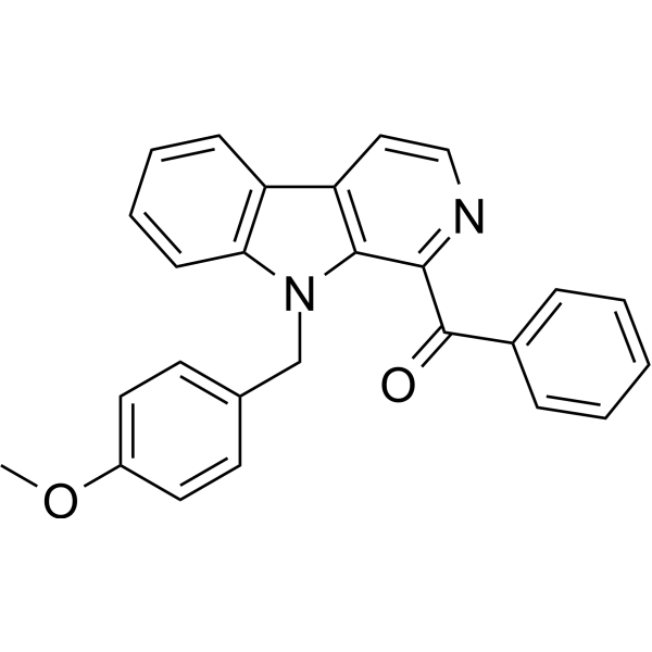
H1k, a Eudistomin Y derivative, is a lysosome-targeted antiproliferation agent. H1k increases the autophagy signal and downregulate the expression of cyclin-dependent kinase (CDK1) and cyclin B1. H1k can be used in research of cancer.
For research use only. We do not sell to patients.
- CAS No.: 3039928-21-4
- Formula: C26H20N2O2
- Molecular Weight:392.45
-
Storage:
Please store the product under the recommended conditions in the Certificate of Analysis.

To place orders, for customer services and technical support, please contact: MedChemExpress USA
Tel: 609-228-6898 E-mail: [email protected] [email protected]


-
 Biological Activity
-
Chemical Information
- Protocols
- Purity & Documentation
- References
-
 Help & FAQs




Biological Activity
Description
Cellular Effect
|
Cell Line

|
Type | Value | Description | References |
|---|---|---|---|---|
| A549 | IC50 |
14.8 μM
Compound: H1k
|
Cytotoxicity against human A549 cells assessed as cell inhibition measured after 72 hrs by MTT assay
Cytotoxicity against human A549 cells assessed as cell inhibition measured after 72 hrs by MTT assay
|
[PMID: 36774698] |
| HepG2 | IC50 |
9.6 μM
Compound: H1k
|
Cytotoxicity against human HepG2 cells assessed as cell inhibition measured after 72 hrs by MTT assay
Cytotoxicity against human HepG2 cells assessed as cell inhibition measured after 72 hrs by MTT assay
|
[PMID: 36774698] |
| LS180 | IC50 |
2.9 μM
Compound: H1k
|
Cytotoxicity against human LS180 cells assessed as cell inhibition measured after 72 hrs by MTT assay
Cytotoxicity against human LS180 cells assessed as cell inhibition measured after 72 hrs by MTT assay
|
[PMID: 36774698] |
| MDA-MB-231 | IC50 |
20.5 μM
Compound: H1k
|
Cytotoxicity against human MDA-MB-231 cells assessed as cell inhibition measured after 72 hrs by MTT assay
Cytotoxicity against human MDA-MB-231 cells assessed as cell inhibition measured after 72 hrs by MTT assay
|
[PMID: 36774698] |
| SGC-7901 | IC50 |
12.1 μM
Compound: H1k
|
Cytotoxicity against human SGC-7901 cells assessed as cell inhibition measured after 72 hrs by MTT assay
Cytotoxicity against human SGC-7901 cells assessed as cell inhibition measured after 72 hrs by MTT assay
|
[PMID: 36774698] |
In Vitro
H1k (72 h) has antiproliferation activity against LS-180, HepG-2, SGC-7901, A549 and MDA-MB-231 cells with IC50 values of 2.9, 9.6, 12.1, 14.8, and 20.5 μM, respectively[1].
H1k (0-50 μM; 24 h) triggers a distinct G2-M arrest in the MDA-MB-231 and SGC-7901 cells in a dose-dependent manner[1].
H1k (0-20 μM; 6 h; MDA-MB-231 cells) induces autophagy to exert its antiproliferative activity, and lysosomes are its functional targets[1].
MedChemExpress (MCE) has not independently confirmed the accuracy of these methods. They are for reference only.
-
Cell Line:MDA-MB-231 and SGC-7901 cells
-
Concentration:0, 5, 10, 20, 50 μM
-
Incubation Time:24 hours
-
Result:Arrested cell cycle at G2-M period in a dose-dependent manner.
-
Cell Line:MDA-MB-231 and SGC-7901 cells
-
Concentration:0, 5, 10, and 20 μM
-
Incubation Time:6 hours
-
Result:Increased the LC3B-I and LC3B-II levels at MDA-MB-231 cells in a dose-dependent manner.
-
Cell Line:MDA-MB-231 and SGC-7901 cells
-
Concentration:0, 5, 10, 20, 50 μM
-
Incubation Time:6 hours
-
Result:Inhibited antiproliferation of cancer cells and the downregulation of CDK1 and cyclin B1.


Chemical Information
-
CAS No. 3039928-21-4
-
Molecular Weight 392.45
-
Formula C26H20N2O2
-
SMILES
COC1=CC=C(C=C1)CN2C3=C(C4=C2C(C(C5=CC=CC=C5)=O)=NC=C4)C=CC=C3
-
Shipping
Room temperature in continental US; may vary elsewhere.
-
Storage
Please store the product under the recommended conditions in the Certificate of Analysis.


Purity & Documentation
References

Calculators
Concentration (start) × Volume (start) = Concentration (final) × Volume (final)


H1k Related Classifications
HY-149261 Related Classifications
 Powered by Bioz