



CDK1-IN-3
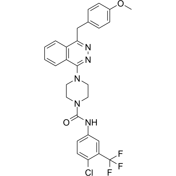
CDK1-IN-3 (8g) is a selective CDK1 inhibitor with IC50s of 36.8, 305.17 and 369.37 nM for CDK1, CDK2 and CDK5, respectively. CDK1-IN-3 inhibits the growth of cancer cells by affecting cell cycle. CDK1-IN-3 can be used for the research of cancer.
商品は「研究用試薬」です。人や動物の医療用・臨床診断用・食品用の製品ではありません。
研究用途以外に使用した場合、当社は一切の責任を負いかねます。
- CAS 番号: 3032997-12-6
- 分子式: C28H25ClF3N5O2
- 分子量:555.98
-
保管条件:
Please store the product under the recommended conditions in the Certificate of Analysis.

To place orders, for customer services and technical support, please contact: MedChemExpress USA
Tel: 609-228-6898 E-mail: [email protected] [email protected]


-
 生物活性
生物活性
-
化学情報
- プロトコル
- 純度とドキュメンテーション
- 参考文献
-
 Help & FAQs




生物活性
製品説明
IC50 & Target
|
CDK1 36.8 nM (IC50) |
CDK2 305.17 nM (IC50) |
CDK5 369.37 nM (IC50) |
体外実験
CDK1-IN-3 (0-10 μM; 24 h) inhibits the growth of PDAC, melanoma, leukemia, colon and breast cancer cells[1]. CDK1-IN-3 (0-1 μM) inhibits CDK1, CDK2 and CDK5 with IC50s of 36.8, 305.17 and 369.37 nM, respectively[1]. CDK1-IN-3 (0-10 μM) inhibits AXL, PTK2B, FGFR, JAK1, IGF1R and BRAF kinases with IC50s of 5655, 3632, 4626, 5265, 5514 and 2829 nM, respectively[1]. CDK1-IN-3 (0.51 μM; 24 h) decreases CDK1 protein level in virto and affects cell cycle[1].
MedChemExpress (MCE) has not independently confirmed the accuracy of these methods. They are for reference only.
-
Cell Line:PDAC, melanoma, leukemia, colon and breast cancer cell lines
-
Concentration:0-10 μM
-
Incubation Time:24 hours
-
Result:Inhibited cell growth of PDAC, melanoma, leukemia, colon, and breast cancer cells over 61%, and inhibited MDA-PATC53 and PL45 cells with IC50s of 0.51 and 0.74 μM, respectively.
-
Cell Line:MDA-PATC53 cell line
-
Concentration:0.51 μM
-
Incubation Time:24 hours
-
Result:Downregulated CDK1 protein level compared to untreated cells.
-
Cell Line:MDA-PATC53 cell line
-
Concentration:0.51 μM
-
Incubation Time:24 hours
-
Result:Significantly arrested in G2/M phase of the cell cycle compared with the untreated cells.


化学情報
-
CAS 番号 3032997-12-6
-
分子量 555.98
-
分子式 C28H25ClF3N5O2
-
SMILES
O=C(N1CCN(C2=NN=C(CC3=CC=C(OC)C=C3)C4=C2C=CC=C4)CC1)NC5=CC=C(Cl)C(C(F)(F)F)=C5
-
輸送条件
Room temperature in continental US; may vary elsewhere.
-
保管条件
Please store the product under the recommended conditions in the Certificate of Analysis.


純度とドキュメンテーション
参考文献

Calculators
濃度 (開始) × 体積 (開始) = 濃度 (終了) × 体積 (終了)


CDK1-IN-3 関連分類
HY-151407 関連分類
 Powered by Bioz