



Coelonin

Based on 1 Customer Validation
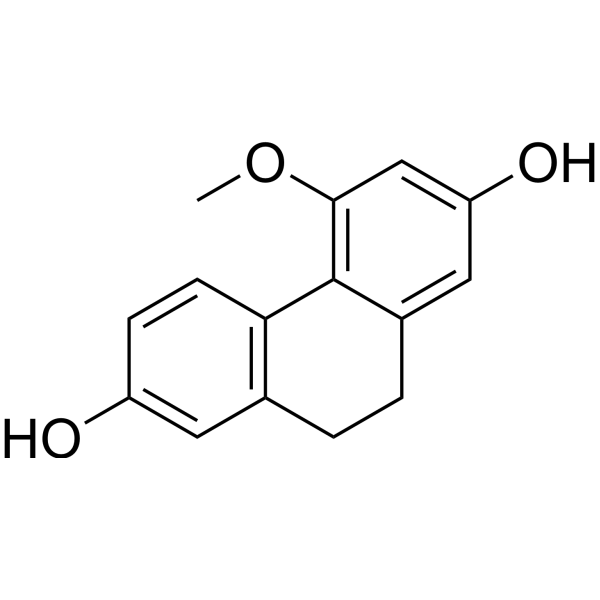
Coelonin is a dihydrophenanthrene with anti-inflammation activity. Coelonin inhibits LPS-induced PTEN phosphorylation. Coelonin inhibits NF-κB activation and p27Kip1 degradation by regulating the PI3K/AKT pathway negatively. Coelonin can inhibit IκBα phosphorylation and degradation and increases the expression of IκBα protein.
商品は「研究用試薬」です。人や動物の医療用・臨床診断用・食品用の製品ではありません。
研究用途以外に使用した場合、当社は一切の責任を負いかねます。
- 純度 : 98.0%
- CAS 番号: 82344-82-9
- 分子式: C15H14O3
- 分子量:242.27
-
保管条件:
-20°C, sealed storage, away from moisture and light
* In solvent : -80°C, 6 months; -20°C, 1 month (sealed storage, away from moisture and light)

To place orders, for customer services and technical support, please contact: MedChemExpress USA
Tel: 609-228-6898 E-mail: [email protected] [email protected]


-
 生物活性
生物活性
-
化学情報
- プロトコル
- 純度とドキュメンテーション
- 参考文献
-
 Help & FAQs




生物活性
製品説明
IC50 & Target
|
Akt |
NF-κB |
IL-1β |
IL-6 |
Cellular Effect
|
Cell Line

|
Type | Value | Description | References |
|---|---|---|---|---|
| A549 | EC50 |
5.05 μg/mL
Compound: 10
|
Cytotoxicity against human A549 cells after 3 days by sulforhodamine B assay
Cytotoxicity against human A549 cells after 3 days by sulforhodamine B assay
|
[PMID: 19193043] |
| BV-2 | IC50 |
15.2 μM
Compound: 16
|
Antineuroinflammatory activity against mouse BV2 cells assessed as inhibition of LPS-induced NO production incubated for 24 hrs by Griess assay
Antineuroinflammatory activity against mouse BV2 cells assessed as inhibition of LPS-induced NO production incubated for 24 hrs by Griess assay
|
[PMID: 31415170] |
| DU-145 | EC50 |
4.79 μg/mL
Compound: 10
|
Cytotoxicity against human DU145 cells after 3 days by sulforhodamine B assay
Cytotoxicity against human DU145 cells after 3 days by sulforhodamine B assay
|
[PMID: 19193043] |
| HCT-8 | EC50 |
3.84 μg/mL
Compound: 10
|
Cytotoxicity against human HCT8 cells after 3 days by sulforhodamine B assay
Cytotoxicity against human HCT8 cells after 3 days by sulforhodamine B assay
|
[PMID: 19193043] |
| KB | EC50 |
3.4 μg/mL
Compound: 10
|
Cytotoxicity against human KB cells after 3 days by sulforhodamine B assay
Cytotoxicity against human KB cells after 3 days by sulforhodamine B assay
|
[PMID: 19193043] |
| MCF7 | EC50 |
1.8 μg/mL
Compound: 10
|
Cytotoxicity against human MCF7 cells after 3 days by sulforhodamine B assay
Cytotoxicity against human MCF7 cells after 3 days by sulforhodamine B assay
|
[PMID: 19193043] |
| Neutrophil | IC50 |
>10 μM
Compound: 15
|
Antiinflammatory activity in human neutrophils assessed as inhibition of fMLP/CB-induced elastase release using MeO-Suc-Ala-Ala-Pro-Val-p-nitroanilide as elastase substrate preincubated for 5 mins followed by fMLP/CB-induction
Antiinflammatory activity in human neutrophils assessed as inhibition of fMLP/CB-induced elastase release using MeO-Suc-Ala-Ala-Pro-Val-p-nitroanilide as elastase substrate preincubated for 5 mins followed by fMLP/CB-induction
|
[PMID: 27525452] |
| Neutrophil | IC50 |
0.4 μM
Compound: 15
|
Antiinflammatory activity in human neutrophils assessed as inhibition of FMLP/cytochalasin B-induced superoxide anion generation by measuring superoxide dismutase-inhibitable reduction of ferricytochrome c preincubated for 5 mins followed by FMLP/cytochal
Antiinflammatory activity in human neutrophils assessed as inhibition of FMLP/cytochalasin B-induced superoxide anion generation by measuring superoxide dismutase-inhibitable reduction of ferricytochrome c preincubated for 5 mins followed by FMLP/cytochal
|
[PMID: 27525452] |
| PC-3 | EC50 |
2.82 μg/mL
Compound: 10
|
Cytotoxicity against human PC3 cells after 3 days by sulforhodamine B assay
Cytotoxicity against human PC3 cells after 3 days by sulforhodamine B assay
|
[PMID: 19193043] |
体外実験
Coelonin (2.5 μg/mL) significantly reduces both NF-κB p65 and p105/50 phosphorylation levels[1].
Coelonin (0-5 μg/mL, 1.5 h) dose dependently reduces the increase of the phosphorylation of PTEN, AKT and IκBa induced by LPS[1].
Coelonin (10 and 20 μg/ml) mitigates particulate matter 2.5 (PM2.5)-induced inflammation by reducing the generation of inflammatory factors, including interleukin-6 (IL-6) and tumor necrosis factor-α (TNF-α)[2].
The inhibition of IL-1β, IL-6 and TNF-α expression by Coelonin is independent of PTEN, whereas the inhibition of p27Kip1 degradation results in cell-cycle arrest in the G1 phase, which is dependent on PTEN[1].
MedChemExpress (MCE) has not independently confirmed the accuracy of these methods. They are for reference only. Further protocols information, click here.
-
Cell Line:RAW264.7 cells
-
Concentration:0, 1, 2.5, and 5 μg/mL
-
Incubation Time:1.5 h
-
Result:Dose dependently reduced the increase of p65 accumulation in the nucleus induced by LPS. Dose dependently reversed LPS-induced iNOS and COX2 expression. LPS (200 ng/mL) significantly increased the phosphorylation of PTEN, AKT and inhibitor of NF-κB (IκBa), which was dose-dependently reduced by coelonin pre-treatment.


化学情報
-
CAS 番号 82344-82-9
-
性状 Solid
-
分子量 242.27
-
分子式 C15H14O3
-
Color Light yellow to light brown
-
SMILES
OC(C=C1CC2)=CC(OC)=C1C3=C2C=C(O)C=C3
-
Structure Classification
-
Initial Source
-
輸送条件
Room temperature in continental US; may vary elsewhere.
-
保管条件
-20°C, sealed storage, away from moisture and light
* In solvent : -80°C, 6 months; -20°C, 1 month (sealed storage, away from moisture and light)

プロトコル
-
Kinase activity and phosphorylation assays
Kinase activity assays measure the ability of kinases to transfer phosphate groups from ATP to specific substrates, while phosphorylation assays detect the presence and levels of phosphorylated proteins. Common methods include radiolabeled ATP incorporation (e. g. ,), ADP release detection via bioluminescence (e. g. ,[3]), enzyme-linked immunosorbent assays (ELISA) for phospho-specific epitopes (e. g. ,[6]), and microtiter-based formats for high-throughput screening (e. g. ,[8]). The ADP-Glo assay quantifies kinase activity by measuring ADP produced during phosphorylation using a luciferase-based system. Radiometric assays involve autoradiography or scintillation counting after incorporation of 32P-labeled ATP into substrate proteins. ELISA-based approaches rely on phospho-specific antibodies to detect activated kinases in cell lysates or purified samples.
-
Western Blot
Western blotting (WB) is a commonly used experimental method in molecular biology, biochemistry, and immunogenetics for identifying and quantifying target proteins. It combines gel electrophoresis with immunoassay, enabling researchers to analyze protein expression, post-translational modifications, and molecular weight.
-
RNA extraction experimental
By lysing cells, releasing RNA, and removing impurities such as proteins and DNA, high-purity RNA products are finally obtained. The commonly used traditional method is the guanidine isothiocyanate/phenol/chloroform method (Trizol), which is suitable for a variety of animal materials including animal tissues, microorganisms, cultured cells, etc., and most plant materials.
-
Research Protocol for Inflammation-related Diseases
The NLRP3 inflammasome is a cytosolic innate immune signaling platform that integrates priming signals and danger-signal activation to promote caspase-1 activation, maturation of IL-1β and IL-18, and gasdermin D-mediated pyroptotic cell death. The core experimental logic is to determine whether inflammatory disease phenotypes are driven by increased NLRP3 expression, ASC-containing inflammasome assembly, caspase-1 cleavage, GSDMD cleavage, and extracellular release of IL-1β/IL-18 rather than by nonspecific cell injury alone. The pathway is strongly linked to inflammation-related disease phenotypes because monosodium urate crystals activate NALP3/NLRP3 inflammasome signaling in gout-like crystal inflammation, cholesterol crystals activate NLRP3 inflammasomes in atherogenesis models, and DSS-induced intestinal inflammation has been reported to involve NLRP3 inflammasome activity. However, experimental colitis studies also show context-dependent protective effects of NLRP3 inflammasome co
-
Protocol for Kinase activity and phosphorylation assays
Kinase activity assays measure transfer of phosphate from ATP to a protein or peptide substrate, generating phosphorylated substrate, ADP, or incorporated radiolabeled phosphate as the readout; phosphorylation assays measure site-specific phosphorylation in cells or tissues as a proxy for kinase-pathway activation, inhibition, or substrate regulation. Phosphorylation can be detected by phospho-specific Western blot, immunoprecipitation kinase assay, phospho-immunofluorescence, phospho-flow cytometry, luminescent ADP detection, radiolabeled ATP incorporation, or reporter-based pathway assays, and these readouts can be applied to cancer cells, primary neurons, mouse tumors, organoids, inflammatory macrophages, ferroptosis studies, and mitophagy studies when the kinase target is biologically relevant.


純度とドキュメンテーション
-
データシート (273 KB)
-
SDS (536 KB)
- English - EN (536 KB)
- Français - FR (536 KB)
- Deutsch - DE (536 KB)
- Norwegian - NO (536 KB)
- Español - ES (536 KB)
- Swedish - SV (536 KB)
- Italian - IT (536 KB)
- Korean - KR (536 KB)
- Portuguese - PT (536 KB)
-
取扱説明書 (2659 KB)
参考文献
[1]. Jiang F, et al. Coelonin, an Anti-Inflammation Active Component of Bletilla striata and Its Potential Mechanism. Int J Mol Sci. 2019 Sep 8;20(18):4422. [Content Brief]
[2]. Cheng W, et al. Inhibition of inflammation-induced injury and cell migration by coelonin and militarine in PM2.5-exposed human lung alveolar epithelial A549 cells. Eur J Pharmacol. 2021 Apr 5;896:173931. [Content Brief]

Calculators
濃度 (開始) × 体積 (開始) = 濃度 (終了) × 体積 (終了)


Coelonin 関連分類
HY-N8884 関連分類
 Powered by Bioz