



Mocravimod hydrochloride
Based on 2 publication(s) in Google Scholar
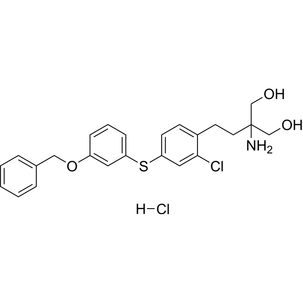
Mocravimod (hydrochloride) is an orally active sphingosine-1-phosphate receptor (S1PR) modulator that blocks the signal required by T cells to egress from lymph nodes and other lymphoid organs. Mocravimod (hydrochloride) preferentially binds to S1PR1 over S1PR2 and S1PR3 in cardiomyocytes. Mocravimod (hydrochloride) significantly lowered the concentration of reactive oxygen species (ROS), prevented mitochondrial permeability transition pore opening, boosted mitochondrial membrane potential (MMP), and increased phosphorylation of AKT, EKR, GSK-3β, JAK2, and STAT3. Mocravimod (hydrochloride) retains T cell effector function. Mocravimod (hydrochloride) can be used for the study of acute myelogenous leukemia, diabetes and Myocardial Ischemia-Reperfusion Injury (MIRI).
For research use only. We do not sell to patients.
- Purity : 99.99%
- CAS No.: 509088-69-1
- Formula: C24H27Cl2NO3S
- Molecular Weight:480.45
-
Storage:
4°C, sealed storage, away from moisture
* In solvent : -80°C, 6 months; -20°C, 1 month (sealed storage, away from moisture)

To place orders, for customer services and technical support, please contact: MedChemExpress USA
Tel: 609-228-6898 E-mail: [email protected] [email protected]


-
 Biological Activity
-
Chemical Information
-
Solvent & Solubility
- Protocols
- Purity & Documentation
- References
-
 Help & FAQs


-
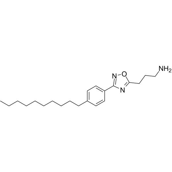
HY-149004
SLF1081851Spns2 Inhibitor -
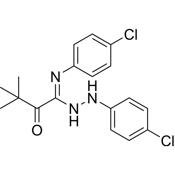
HY-19736
TY-52156LPL Receptor Antagonist -
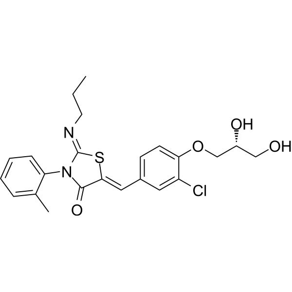
HY-10569
PonesimodS1P1 Agonist -
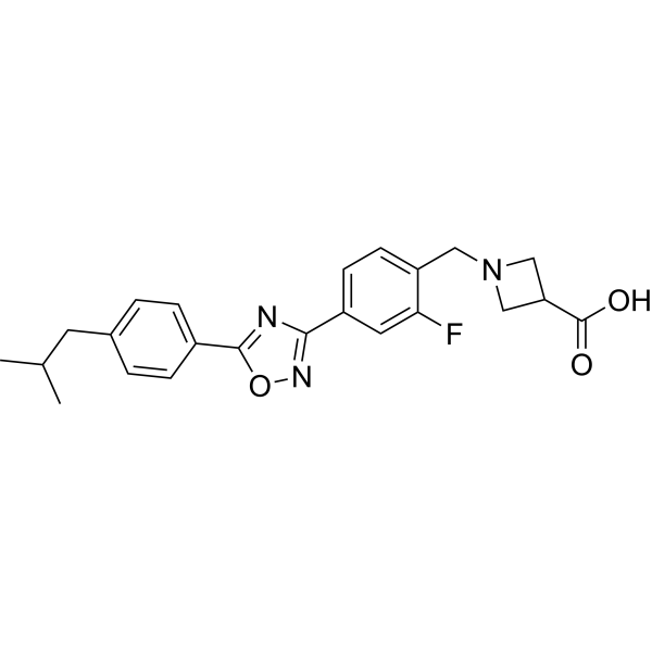
HY-101265
IcanbelimodS1P Receptor Agonist -
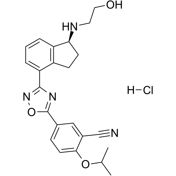
HY-12288A
Ozanimod hydrochlorideS1P Receptor Modulator -
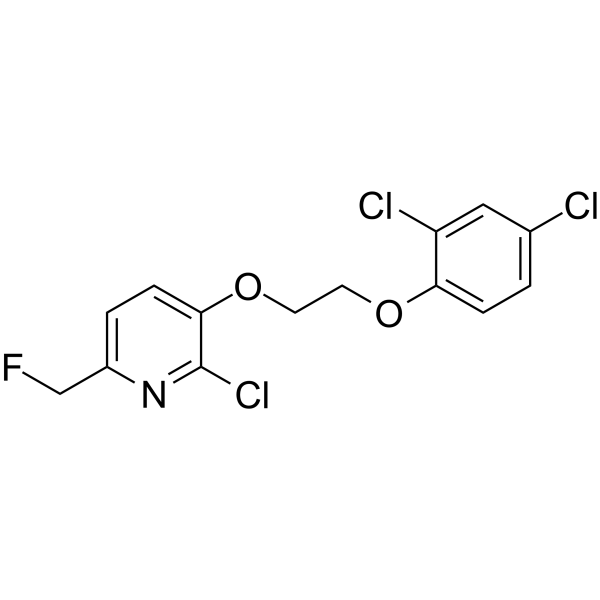
HY-108494
CYM50260S1P4 Receptor Agonist -
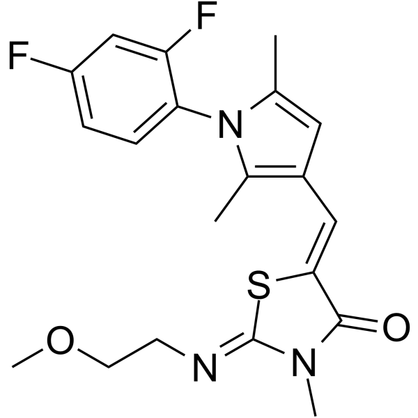
HY-108495
CYM50308S1P4 Receptor Agonist -
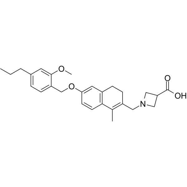
HY-12685
CeralifimodLPL Receptor Agonist -
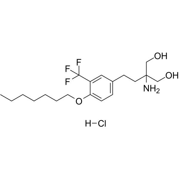
HY-16734A
Amiselimod hydrochlorideS1PR1 Modulator -
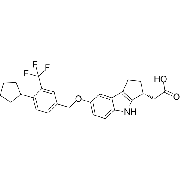
HY-12789
EtrasimodS1P1 Receptor Antagonist -
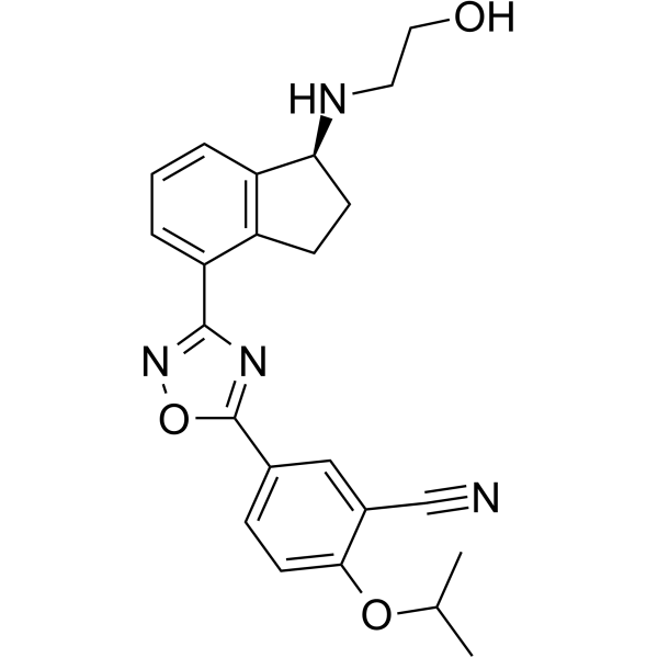
HY-12288
OzanimodS1P Receptor Modulator -
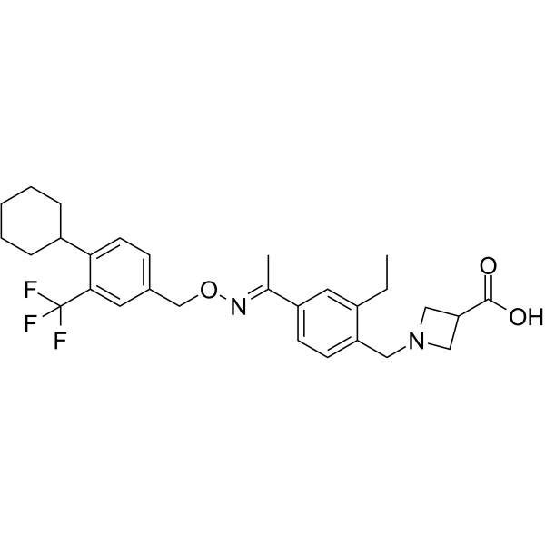
HY-12355
SiponimodS1P1/S1P5 Agonist
Publications Citing Use of MedChemExpress (MCE) Mocravimod hydrochloride
More

Biological Activity
Description
IC50 & Target
S1PR1[1]
In Vitro
Mocravimod (KRP203) (0.2-5 μM, 24 h) (hydrochloride) significantly improves the survival rate of the hypoxia-reoxygenation (H/R) H9c2 cells, reduces lactate dehydrogenase (LDH) release, and decreases apoptosis[4].
Mocravimod (0.2-1 μM, 24 h) (hydrochloride) significantly reduces reactive oxygen species (ROS) levels, improves mitochondrial membrane potential, and inhibits the opening of the mitochondrial permeability transition pore (mPTP) in hypoxia-reoxygenation (H/R) H9c2 cells[4].
Mocravimod (0.2-1 μM, 24 h) (hydrochloride) inhibits the expression of mitochondrial-mediated apoptosis pathway proteins such as cytochrome C, caspase-9, and caspase-3[4].
Mocravimod (0.2-1 μM, 24 h) (hydrochloride) significantly increases the phosphorylation levels of key signaling pathway proteins in hypoxia-reoxygenation (H/R) H9c2 cells, including AKT (Ser473), ERK (Thr202/Tyr204), GSK-3β (Ser9), JAK2 (Tyr1007/1008), and STAT3 (Ser727)[4].
MedChemExpress (MCE) has not independently confirmed the accuracy of these methods. They are for reference only. Further protocols information, click here.
-
Cell Line:Hypoxia-Reoxygenation (H/R) H9c2 Cells
-
Concentration:0.2 μM, 1 μM, 5 μM
-
Incubation Time:24 h
-
Result:Induced apoptosis.
-
Cell Line:Hypoxia-Reoxygenation (H/R) H9c2 Cells
-
Concentration:0.2 μM, 1 μM
-
Incubation Time:24 h
-
Result:Inhibited the expression of mitochondrial-mediated apoptosis pathway proteins such as cytochrome C, caspase-9, and caspase-3.
In Vivo
Mocravimod (3.0 mg/kg, i.p., thrice weekly) (hydrochloride) significantly inhibits the development of atherosclerotic lesions in mice on a high-cholesterol diet, and this effect was independent of changes in blood lipids, but rather by regulating the infiltration of inflammatory cells within plaques[3].
MedChemExpress (MCE) has not independently confirmed the accuracy of these methods. They are for reference only.
-
Animal Model:Low-density lipoprotein receptor-deficient mice (LDL-R-/-) on cholesterol-rich diet[3].
-
Dosage:3 mg/kg
-
Administration:I.p., thrice weekly for 6-16 weeks
-
Result:Significantly reduced the lesion area of the aortic root and the entire aorta.
Decreased levels of macrophages (MOMA-2 positive) and T cells (CD3 positive) in plaques.
No effect on plasma lipid levels (total cholesterol, HDL cholesterol, triglycerides), body weight, or liver and kidney function parameters (such as ALT, BUN, creatinine).
Decreased CD4+ and CD8+ T cells in the spleen and lymph nodes, with decreased expression of the activation marker CD69.
Decreased MHC-II expression (activation marker) in F4/80+ macrophages and CD11c+ dendritic cells; reduced cytokine (TNF-α, MCP-1, IL-6) production after in vitro stimulation.
Clinical Trial
| NCT Number | Sponsor | Condition | Start Date |
Phase

|
|---|---|---|---|---|
| NCT01329991 | Plexxikon| | 2011-05 | PHASE1 |


Chemical Information
-
CAS No. 509088-69-1
-
Appearance Solid
-
Molecular Weight 480.45
-
Formula C24H27Cl2NO3S
-
Color White to off-white
-
SMILES
OCC(CCC1=CC=C(SC2=CC=CC(OCC3=CC=CC=C3)=C2)C=C1Cl)(N)CO.[H]Cl
-
Synonyms
KRP-203
-
Shipping
Room temperature in continental US; may vary elsewhere.
-
Storage
4°C, sealed storage, away from moisture
* In solvent : -80°C, 6 months; -20°C, 1 month (sealed storage, away from moisture)


Publications (2)
-
Journal Impact Factor
-
Most Recent
-
Cell Biol Toxicol
The novel sphingosine-1-phosphate receptor modulator KRP-203 prevents myocardial ischemia-reperfusion injury by preserving mitochondrial function through activation of the RISK and SAFE signaling pathways. [Abstract]2025 Nov 27;41(1):159. PMID: 41307722 -
bioRxiv
Sphingosine-1-phosphate receptor modulators resensitize FLT3-ITD acute myeloid leukemia cells with NRAS mutations to FLT3 inhibitors. [Abstract]2025 Nov 24:2025.11.21.689510. PMID: 41341164


Solvent & Solubility
In Vitro:
DMSO : 200 mg/mL (416.28 mM; Need ultrasonic; Hygroscopic DMSO has a significant impact on the solubility of product, please use newly opened DMSO)
Please refer to the solubility information to select the appropriate solvent. Once prepared, please aliquot and store the solution to prevent product inactivation from repeated freeze-thaw cycles.
Storage method and period of stock solution: -80°C, 6 months; -20°C, 1 month (sealed storage, away from moisture). When stored at -80°C, please use it within 6 months. When stored at -20°C, please use it within 1 month.
Please refer to the solubility information to select the appropriate solvent. Once prepared, please aliquot and store the solution to prevent product inactivation from repeated freeze-thaw cycles.
Storage method and period of stock solution: -80°C, 6 months; -20°C, 1 month (sealed storage, away from moisture). When stored at -80°C, please use it within 6 months. When stored at -20°C, please use it within 1 month.
Concentration (start) × Volume (start) = Concentration (final) × Volume (final)
In Vivo:
Select the appropriate dissolution method based on your experimental animal and administration route.
- For the following dissolution methods, please ensure to first prepare a clear stock solution using an In Vitro approach and then sequentially add co-solvents:
- To ensure reliable experimental results, the clarified stock solution can be appropriately stored based on storage conditions. As for the working solution for In Vivo experiments, it is recommended to prepare freshly and use it on the same day.
- The percentages shown for the solvents indicate their volumetric ratio in the final prepared solution. If precipitation or phase separation occurs during preparation, heat and/or sonication can be used to aid dissolution.
Add each solvent one by one: 10% DMSO 40% PEG300 5% Tween-80 45% Saline
Solubility: ≥ 2.5 mg/mL (5.20 mM); Clear solution
This protocol yields a clear solution of ≥ 2.5 mg/mL (saturation unknown).
Taking 1 mL working solution as an example, add 100 μL DMSO stock solution (25.0 mg/mL) to 400 μL PEG300, and mix evenly; then add 50 μL Tween-80 and mix evenly; then add 450 μL Saline to adjust the volume to 1 mL.
Preparation of Saline: Dissolve 0.9 g sodium chloride in ddH₂O and dilute to 100 mL to obtain a clear Saline solution.
Add each solvent one by one: 10% DMSO 90% (20% SBE-β-CD in Saline)
Solubility: 2.5 mg/mL (5.20 mM); Suspended solution; Need ultrasonic
This protocol yields a suspended solution of 2.5 mg/mL. Suspended solution can be used for oral and intraperitoneal injection.
Taking 1 mL working solution as an example, add 100 μL DMSO stock solution (25.0 mg/mL) to 900 μL 20% SBE-β-CD in Saline, and mix evenly.
Preparation of 20% SBE-β-CD in Saline (4°C, storage for one week): 2 g SBE-β-CD powder is dissolved in 10 mL Saline, completely dissolve until clear.
In Vivo Dissolution Calculator
Please enter the basic information of animal experiments:
-
-
-
-
Recommended: Prepare an additional quantity of animals to account for potential losses during experiments.
Please enter your animal formula composition:
-
%DMSO +
Recommended: Keep the proportion of DMSO in working solution below 2% if your animal is weak.
-
%+
-
+%Tween-80 + +
-
%Saline +
The co-solvents required include: DMSO, . All of co-solvents are available by MedChemExpress (MCE). , Tween 80. All of co-solvents are available by MedChemExpress (MCE).
Working solution concentration: 0.22 mg/mL
Method for preparing stock solution: mg drug dissolved in μL DMSO. Stock solution concentration: mg/mL. * In solvent : -80°C, 6 months; -20°C, 1 month (sealed storage, away from moisture)
1. Take μL DMSO stock solution;
2. Add μL .
μL , mix evenly;
3. Then add μL Tween 80, mix evenly;
4. Then add μL
Please ensure that the stock solution in the first step is dissolved to a clear state, and add co-solvents in sequence. You can use ultrasonic heating (ultrasonic cleaner, recommended frequency 20-40 kHz), vortexing, etc. to assist dissolution.

Protocols
-
Kinase activity and phosphorylation assays
Kinase activity assays measure the ability of kinases to transfer phosphate groups from ATP to specific substrates, while phosphorylation assays detect the presence and levels of phosphorylated proteins. Common methods include radiolabeled ATP incorporation (e. g. ,), ADP release detection via bioluminescence (e. g. ,[3]), enzyme-linked immunosorbent assays (ELISA) for phospho-specific epitopes (e. g. ,[6]), and microtiter-based formats for high-throughput screening (e. g. ,[8]). The ADP-Glo assay quantifies kinase activity by measuring ADP produced during phosphorylation using a luciferase-based system. Radiometric assays involve autoradiography or scintillation counting after incorporation of 32P-labeled ATP into substrate proteins. ELISA-based approaches rely on phospho-specific antibodies to detect activated kinases in cell lysates or purified samples.
-
Western Blot
Western blotting (WB) is a commonly used experimental method in molecular biology, biochemistry, and immunogenetics for identifying and quantifying target proteins. It combines gel electrophoresis with immunoassay, enabling researchers to analyze protein expression, post-translational modifications, and molecular weight.
-
Mitochondrial membrane-potential fluorescent assay
Mitochondrial membrane potential fluorescent assays estimate ΔΨm in living cells using lipophilic cationic dyes such as TMRM, TMRE, rhodamine 123, and JC-1, which accumulate in mitochondria according to membrane polarization; loss of signal after FCCP or CCCP treatment is interpreted as mitochondrial depolarization. TMRM/TMRE and rhodamine 123 are commonly used for semi-quantitative live-cell microscopy or flow cytometry, while JC-1 can report a shift from red aggregate fluorescence to green monomer fluorescence during depolarization; interpretation requires controls because dye concentration, quenching mode, cell type, dye efflux, and mitochondrial mass can affect fluorescence independently of ΔΨm.
-
ROS/oxidative-stress fluorescent staining
ROS/oxidative-stress fluorescent staining uses cell-permeant fluorogenic probes that become fluorescent after oxidation inside cells or tissues; commonly used examples include DCFH-DA/DCFDA for broad cellular oxidant detection, DHE for superoxide-related signal detection, MitoSOX for mitochondrial superoxide-related signal detection, and CellROX probes for oxidative-stress-associated fluorescence readouts. The assay detects probe oxidation rather than a single ROS species unless the probe and analysis method have been chemically validated for that species. DCFH-DA enters cells, is deacetylated by intracellular esterases to DCFH, and produces fluorescent DCF after oxidation, so the readout is used as an operational measure of total cellular oxidative stress rather than a species-specific ROS measurement. DHE and MitoSOX can report superoxide-related oxidation, but red fluorescence alone can include non-specific ethidium-like oxidation products; HPLC or optimized spectral approaches are
-
Fluorescent plasma-membrane potential dye assay
Fluorescent plasma-membrane potential dye assays measure changes in cell membrane potential using voltage-sensitive dyes whose fluorescence changes when cells depolarize or hyperpolarize. Anionic bis-oxonol dyes such as DiBAC4(3) enter depolarized cells more readily and show increased fluorescence after intracellular binding, while hyperpolarization reduces dye accumulation and fluorescence. FMP/FLIPR membrane-potential dyes are used for faster, homogeneous microplate assays of ion-channel or receptor-mediated membrane-potential changes.
-
Mitochondrial membrane-potential and mitochondrial mass staining
Mitochondrial membrane potential staining measures the electrochemical polarization across the mitochondrial inner membrane in live cells using lipophilic cationic fluorescent probes; early rhodamine-based work showed that selective mitochondrial dye accumulation is lost when the mitochondrial transmembrane potential is dissipated. JC-1 reports mitochondrial polarization by shifting from green monomer fluorescence to red J-aggregate fluorescence as dye concentration increases within energized mitochondria; therefore, the red/green fluorescence ratio is used as a relative readout of mitochondrial membrane potential. TMRE or TMRM staining provides a single-channel relative readout because these cationic rhodamine esters accumulate in polarized mitochondria, and lower fluorescence indicates reduced mitochondrial polarization when acquisition and dye-loading conditions are controlled. Mitochondrial mass staining is commonly performed with MitoTracker Green FM or related MitoTracker dyes as
-
Transepithelial/transendothelial electrical resistance assay
TEER measures electrical resistance across epithelial or endothelial monolayers cultured on permeable supports, and the readout reflects ionic conductance through the cell barrier, especially the paracellular pathway regulated by junctional integrity. TEER can be measured without destroying the monolayer and is commonly used before or during transport, permeability, barrier-disruption, and barrier-maturation experiments. TEER values are influenced by biological maturation and technical conditions; reported factors include temperature, medium formulation, passage number, electrode geometry, membrane properties, and junctional length during early monolayer maturation. Therefore, TEER should be interpreted with blank-insert subtraction, area normalization, repeated readings, and, when possible, orthogonal barrier readouts such as FITC-dextran flux or tight-junction staining.
-
Protocol for Kinase activity and phosphorylation assays
Kinase activity assays measure transfer of phosphate from ATP to a protein or peptide substrate, generating phosphorylated substrate, ADP, or incorporated radiolabeled phosphate as the readout; phosphorylation assays measure site-specific phosphorylation in cells or tissues as a proxy for kinase-pathway activation, inhibition, or substrate regulation. Phosphorylation can be detected by phospho-specific Western blot, immunoprecipitation kinase assay, phospho-immunofluorescence, phospho-flow cytometry, luminescent ADP detection, radiolabeled ATP incorporation, or reporter-based pathway assays, and these readouts can be applied to cancer cells, primary neurons, mouse tumors, organoids, inflammatory macrophages, ferroptosis studies, and mitophagy studies when the kinase target is biologically relevant.
-
Research Protocol for Metabolic Diseases
AMP-activated protein kinase, AMPK, is a conserved cellular energy sensor that responds to reduced cellular energy status and coordinates metabolism by increasing ATP-generating catabolic pathways while suppressing ATP-consuming anabolic processes. In metabolic disease research, the AMPK pathway is experimentally relevant because it regulates hepatic lipid synthesis, fatty acid oxidation, glucose production, skeletal-muscle glucose disposal, mTORC1-linked biosynthesis, autophagy, mitochondrial homeostasis, and whole-body energy balance. The central pathway logic is that energy stress, metformin, exercise-like stimulation, or direct AMPK activators increase AMPKα Thr172 phosphorylation and downstream substrate phosphorylation, including ACC and RAPTOR. Phosphorylation of ACC suppresses lipogenesis and supports fatty acid oxidation, whereas phosphorylation of RAPTOR suppresses mTORC1 signaling and links cellular energy status to growth and protein synthesis control. The pathway is linked
-
Research Protocol for Cancer Immunology
Cancer immunology studies how the immune system recognizes, suppresses, edits, or fails to eliminate malignant cells through tumor antigen release, antigen presentation, T-cell priming, immune trafficking, tumor-cell killing, and feedback inhibition in the tumor microenvironment. The cancer-immunity cycle links tumor antigenicity, dendritic-cell priming, CD8+ T-cell infiltration, cytotoxic function, and immune-checkpoint regulation to tumor rejection or immune escape. Immune-checkpoint pathways such as PD-1/PD-L1 and CTLA-4 suppress antitumor T-cell activity and can be therapeutically blocked, but many tumors remain resistant because of poor antigen presentation, weak T-cell infiltration, suppressive myeloid cells, regulatory T cells, and tumor-intrinsic immune-exclusion programs. Unresolved questions include which immune-cell states predict response, how tumor-intrinsic pathways exclude immune cells, how myeloid suppression limits checkpoint blockade, and which combination strategies


Purity & Documentation
-
Data Sheet (279 KB)
-
SDS (393 KB)
- English - EN (393 KB)
- Français - FR (393 KB)
- Deutsch - DE (393 KB)
- Norwegian - NO (393 KB)
- Español - ES (393 KB)
- Swedish - SV (393 KB)
- Italian - IT (393 KB)
- Korean - KR (393 KB)
- Portuguese - PT (393 KB)
-
Handling Instructions (2659 KB)
References
[1]. Dertschnig S, et al. Mocravimod, a Selective Sphingosine-1-Phosphate Receptor Modulator, in Allogeneic Hematopoietic Stem Cell Transplantation for Malignancy. Transplant Cell Ther. 2023 Jan;29(1):41.e1-41.e9. [Content Brief]
[3]. Potì F, et al. Mocravimod, sphingosine 1-phosphate receptor type 1 agonist, ameliorates atherosclerosis in LDL-R-/- mice. Arterioscler Thromb Vasc Biol. 2013 Jul;33(7):1505-12. [Content Brief]
[4]. Wu N, et al. The novel sphingosine-1-phosphate receptor modulator Mocravimod prevents myocardial ischemia-reperfusion injury by preserving mitochondrial function through activation of the RISK and SAFE signaling pathways. Cell Biol Toxicol. 2025 Nov 27;41(1):159. [Content Brief]


Complete Stock Solution Preparation Table
Please refer to the solubility information to select the appropriate solvent. Once prepared, please aliquot and store the solution to prevent product inactivation from repeated freeze-thaw cycles.
Storage method and period of stock solution: -80°C, 6 months; -20°C, 1 month (sealed storage, away from moisture). When stored at -80°C, please use it within 6 months. When stored at -20°C, please use it within 1 month.
| Optional Solvent | Concentration Solvent Mass | 1 mg | 5 mg | 10 mg | 25 mg |
|---|---|---|---|---|---|
| DMSO | 1 mM | 2.0814 mL | 10.4069 mL | 20.8138 mL | 52.0345 mL |
| 5 mM | 0.4163 mL | 2.0814 mL | 4.1628 mL | 10.4069 mL | |
| 10 mM | 0.2081 mL | 1.0407 mL | 2.0814 mL | 5.2035 mL | |
| 15 mM | 0.1388 mL | 0.6938 mL | 1.3876 mL | 3.4690 mL | |
| 20 mM | 0.1041 mL | 0.5203 mL | 1.0407 mL | 2.6017 mL | |
| 25 mM | 0.0833 mL | 0.4163 mL | 0.8326 mL | 2.0814 mL | |
| 30 mM | 0.0694 mL | 0.3469 mL | 0.6938 mL | 1.7345 mL | |
| 40 mM | 0.0520 mL | 0.2602 mL | 0.5203 mL | 1.3009 mL | |
| 50 mM | 0.0416 mL | 0.2081 mL | 0.4163 mL | 1.0407 mL | |
| 60 mM | 0.0347 mL | 0.1734 mL | 0.3469 mL | 0.8672 mL | |
| 80 mM | 0.0260 mL | 0.1301 mL | 0.2602 mL | 0.6504 mL | |
| 100 mM | 0.0208 mL | 0.1041 mL | 0.2081 mL | 0.5203 mL |


Mocravimod hydrochloride Related Classifications
HY-13660 Related Classifications
 Powered by Bioz
Keywords
- Mocravimod
- 509088-69-1
- KRP-203
- KRP203
- KRP 203
- LPL Receptor
- Reactive Oxygen Species (ROS)
- Akt
- GSK-3
- JAK
- STAT
- S1PR
- Sphingosine-1-phosphate receptor agonist
- Myocardial ischemia–reperfusion injury
- Mitochondrial function
- Reperfusion injury salvage kinase signaling pathway
- Survivor activating factor enhancement signaling pathway
- Inhibitor
- inhibitor
- inhibit