



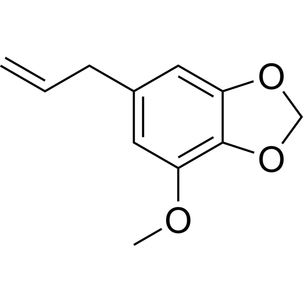
Myristicin

Based on 1 Customer Validation
Myristicine is an orally bioavailable serotonin receptor antagonist and weak monoamine oxidase (MAO) inhibitor. Myristicine also exerts anti-cancer effects on gastric cancer cells by inhibiting the EGFR/ERK signaling pathway. Myristicine is the main component of nutmeg essential oil and has anti-cancer, anti-proliferative, antibacterial, anti-inflammatory and apoptosis-inducing effects. Myristicine abuse can produce hallucinogenic effects, organ damage, etc.
For research use only. We do not sell to patients.
- Purity : 99.48%
- CAS No.: 607-91-0
- Formula: C11H12O3
- Molecular Weight:192.21
-
Storage:
4°C, stored under nitrogen
* In solvent : -80°C, 6 months; -20°C, 1 month (stored under nitrogen)

To place orders, for customer services and technical support, please contact: MedChemExpress USA
Tel: 609-228-6898 E-mail: [email protected] [email protected]


-
 Biological Activity
-
Chemical Information
-
Solvent & Solubility
- Protocols
- Purity & Documentation
- References
-
 Help & FAQs





Biological Activity
Description
IC50 & Target
[4]|
EGFR |
ERK |
Cellular Effect
|
Cell Line

|
Type | Value | Description | References |
|---|---|---|---|---|
| KB | IC50 |
75 μg/mL
Compound: 27
|
Antitumor activity against human KB cells assessed as cell growth inhibition
Antitumor activity against human KB cells assessed as cell growth inhibition
|
[PMID: 36858050] |
| RAW264.7 | IC50 |
48.6 μM
Compound: 21
|
Inhibition of LPS-induced NO production in mouse RAW264.7 cells incubated for 24 hrs in presence of LPS by Griess reagent based assay
Inhibition of LPS-induced NO production in mouse RAW264.7 cells incubated for 24 hrs in presence of LPS by Griess reagent based assay
|
[PMID: 31642320] |
In Vitro
Myristicin (7.8-31.25 μM; 48 h) can delay the proliferation of gastric cancer cells and induce endoplasmic reticulum (ER) stress and apoptosis; at the same time, Myristicin reduces the expression of cell cycle proteins, increases the expression of Bax, and activates caspase enzyme, and enhances the release of cytochrome C and mitochondrial ROS levels [3].
MedChemExpress (MCE) has not independently confirmed the accuracy of these methods. They are for reference only. Further protocols information, click here.
-
Cell Line:Human gastric cancer cells
-
Concentration:7.8125, 15.625, and 31.25 μM
-
Incubation Time:48 h
-
Result:Significantly decreased expression of cyclins, increased Bax expression.
In Vivo
Myristicin (200 mg/kg; po; single dose) treated 6 hours in advance, on acetic acid (AA)-induced small Mouse experimental colitis model has preventive and protective effect[4].
MedChemExpress (MCE) has not independently confirmed the accuracy of these methods. They are for reference only.
-
Animal Model:Experimental colitis model in mice[4]
-
Dosage:200 mg/kg
-
Administration:po; single dose, 6 h before Rectal AA (colitis inducer) instillation
-
Result:Reduced cellular oxidative stress induced by AA, and relieved the histopathological colonic damage, alleviated inflammation in the AA-induced colitis.


Chemical Information
-
CAS No. 607-91-0
-
Appearance Liquid (Density: 1.1437 g/cm3)
-
Molecular Weight 192.21
-
Formula C11H12O3
-
Color Colorless to light yellow
-
SMILES
C=CCC1=CC(OC)=C2OCOC2=C1
-
Synonyms
Myristicine
-
Structure Classification
-
Initial Source
-
Shipping
Room temperature in continental US; may vary elsewhere.
-
Storage
4°C, stored under nitrogen
* In solvent : -80°C, 6 months; -20°C, 1 month (stored under nitrogen)


Solvent & Solubility
In Vitro:
DMSO : 100 mg/mL (520.26 mM; Need ultrasonic; Hygroscopic DMSO has a significant impact on the solubility of product, please use newly opened DMSO)
Please refer to the solubility information to select the appropriate solvent. Once prepared, please aliquot and store the solution to prevent product inactivation from repeated freeze-thaw cycles.
Storage method and period of stock solution: -80°C, 6 months; -20°C, 1 month (stored under nitrogen). When stored at -80°C, please use it within 6 months. When stored at -20°C, please use it within 1 month.
Please refer to the solubility information to select the appropriate solvent. Once prepared, please aliquot and store the solution to prevent product inactivation from repeated freeze-thaw cycles.
Storage method and period of stock solution: -80°C, 6 months; -20°C, 1 month (stored under nitrogen). When stored at -80°C, please use it within 6 months. When stored at -20°C, please use it within 1 month.
Concentration (start) × Volume (start) = Concentration (final) × Volume (final)
In Vivo:
Select the appropriate dissolution method based on your experimental animal and administration route.
- For the following dissolution methods, please ensure to first prepare a clear stock solution using an In Vitro approach and then sequentially add co-solvents:
- To ensure reliable experimental results, the clarified stock solution can be appropriately stored based on storage conditions. As for the working solution for In Vivo experiments, it is recommended to prepare freshly and use it on the same day.
- The percentages shown for the solvents indicate their volumetric ratio in the final prepared solution. If precipitation or phase separation occurs during preparation, heat and/or sonication can be used to aid dissolution.
Add each solvent one by one: 10% DMSO 40% PEG300 5% Tween-80 45% Saline
Solubility: ≥ 2.5 mg/mL (13.01 mM); Clear solution
This protocol yields a clear solution of ≥ 2.5 mg/mL (saturation unknown).
Taking 1 mL working solution as an example, add 100 μL DMSO stock solution (25.0 mg/mL) to 400 μL PEG300, and mix evenly; then add 50 μL Tween-80 and mix evenly; then add 450 μL Saline to adjust the volume to 1 mL.
Preparation of Saline: Dissolve 0.9 g sodium chloride in ddH₂O and dilute to 100 mL to obtain a clear Saline solution.
Add each solvent one by one: 10% DMSO 90% (20% SBE-β-CD in Saline)
Solubility: ≥ 2.5 mg/mL (13.01 mM); Clear solution
This protocol yields a clear solution of ≥ 2.5 mg/mL (saturation unknown).
Taking 1 mL working solution as an example, add 100 μL DMSO stock solution (25.0 mg/mL) to 900 μL 20% SBE-β-CD in Saline, and mix evenly.
Preparation of 20% SBE-β-CD in Saline (4°C, storage for one week): 2 g SBE-β-CD powder is dissolved in 10 mL Saline, completely dissolve until clear.
In Vivo Dissolution Calculator
Please enter the basic information of animal experiments:
-
-
-
-
Recommended: Prepare an additional quantity of animals to account for potential losses during experiments.
Please enter your animal formula composition:
-
%DMSO +
Recommended: Keep the proportion of DMSO in working solution below 2% if your animal is weak.
-
%+
-
+%Tween-80 + +
-
%Saline +
The co-solvents required include: DMSO, . All of co-solvents are available by MedChemExpress (MCE). , Tween 80. All of co-solvents are available by MedChemExpress (MCE).
Working solution concentration: 0.22 mg/mL
Method for preparing stock solution: mg drug dissolved in μL DMSO. Stock solution concentration: mg/mL. * In solvent : -80°C, 6 months; -20°C, 1 month (stored under nitrogen)
1. Take μL DMSO stock solution;
2. Add μL .
μL , mix evenly;
3. Then add μL Tween 80, mix evenly;
4. Then add μL
Please ensure that the stock solution in the first step is dissolved to a clear state, and add co-solvents in sequence. You can use ultrasonic heating (ultrasonic cleaner, recommended frequency 20-40 kHz), vortexing, etc. to assist dissolution.

Protocols
-
Research Protocol for Infectious Diseases
Infectious-disease experiments test how pathogens interact with host barriers, innate immune receptors, inflammatory signaling, pathogen replication, and tissue injury; pattern-recognition receptors such as TLRs, RIG-I-like receptors, NOD-like receptors, and inflammasomes detect microbial molecules and activate NF-κB, interferon, and cytokine responses. The central hypothesis is that infection severity reflects the balance between pathogen burden and host response: protective inflammation restricts pathogen growth, whereas excessive or mislocalized inflammation contributes to tissue damage and disease phenotype. Unresolved questions include which host pathways are protective versus pathogenic, why some infection models fail to translate to human disease, and which combined readouts best predict clinically relevant infection outcomes.
-
Apoptosis
Apoptosis, also called programmed cell death, is generally characterized by distinct morphological characteristics.
-
TUNEL staining for apoptotic DNA fragmentation
TUNEL staining detects DNA strand breaks by using terminal deoxynucleotidyl transferase to add labeled nucleotides to exposed 3′-OH DNA termini, generating either microscopic staining in fixed cells or tissue sections, or fluorescence/cytometric signal in cell suspensions. TUNEL positivity reflects DNA fragmentation but should not be interpreted alone as definitive apoptosis, because TUNEL can also label necrotic, autolytic, mechanically damaged, or DNA-repair-associated DNA breaks.
-
Annexin V plus membrane-impermeant dye apoptosis staining
Annexin V-based apoptosis assays rely on the detection of phosphatidylserine (PS) externalization from the inner leaflet of the plasma membrane to the outer leaflet, an early biochemical hallmark of apoptosis. Fluorescently labeled Annexin V binds PS in a calcium-dependent manner, enabling identification of early apoptotic cells by flow cytometry or fluorescence microscopy. When combined with a membrane-impermeant DNA-binding dye (e. g. , propidium iodide), this approach allows discrimination between viable (Annexin V−/dye−), early apoptotic (Annexin V+/dye−), and late apoptotic or necrotic (Annexin V+/dye+) cell populations by assessing membrane integrity and PS exposure.
-
Apoptosis Solutions
Apoptosis is a regulated, generally non-lytic cell-death pathway that removes unwanted, damaged, infected, or abnormal cells through coordinated morphological changes, caspase activation, DNA fragmentation, and membrane remodeling. The intrinsic apoptosis pathway is controlled mainly by mitochondrial outer membrane permeabilization, BCL-2 family proteins, cytochrome c release, apoptosome formation, caspase-9 activation, and downstream executioner caspase-3/7 activation. The extrinsic apoptosis pathway is initiated by death receptors such as Fas, TNFR, and TRAIL receptors, which recruit adaptor proteins and activate caspase-8 before engaging executioner caspases or mitochondrial amplification through BID cleavage. Apoptosis is linked to many phenotypes, including cancer cell killing, tissue homeostasis, immune regulation, neurodegeneration, infection response, and treatment-induced cytotoxicity; unresolved questions include how apoptosis interacts with necroptosis, pyroptosis, ferroptos
-
Research Protocol for Inflammation-related Diseases
The NLRP3 inflammasome is a cytosolic innate immune signaling platform that integrates priming signals and danger-signal activation to promote caspase-1 activation, maturation of IL-1β and IL-18, and gasdermin D-mediated pyroptotic cell death. The core experimental logic is to determine whether inflammatory disease phenotypes are driven by increased NLRP3 expression, ASC-containing inflammasome assembly, caspase-1 cleavage, GSDMD cleavage, and extracellular release of IL-1β/IL-18 rather than by nonspecific cell injury alone. The pathway is strongly linked to inflammation-related disease phenotypes because monosodium urate crystals activate NALP3/NLRP3 inflammasome signaling in gout-like crystal inflammation, cholesterol crystals activate NLRP3 inflammasomes in atherogenesis models, and DSS-induced intestinal inflammation has been reported to involve NLRP3 inflammasome activity. However, experimental colitis studies also show context-dependent protective effects of NLRP3 inflammasome co
-
Cell Viability Determination by MTT Colorimetric Assay
The following protocol uses the MTT colorimetric assay as a classic literature-established method for assessing cell viability/metabolic activity in cultured mammalian cells. MTT[3-(4,5-dimethylthiazol-2-yl)-2,5-diphenyltetrazolium bromide] is reduced by metabolically active cells to a colored formazan product; the amount of formazan is quantified spectrophotometrically and provides an indirect measure of metabolically active viable cells. Importantly, MTT reduction reflects cellular oxidoreductase/metabolic activity rather than an absolute direct count of living cells, so changes in cellular metabolism can alter the signal independently of cell number.


Purity & Documentation
-
Data Sheet (276 KB)
-
SDS (254 KB)
- English - EN (254 KB)
- Français - FR (254 KB)
- Deutsch - DE (254 KB)
- Norwegian - NO (254 KB)
- Español - ES (254 KB)
- Swedish - SV (254 KB)
- Italian - IT (254 KB)
- Korean - KR (254 KB)
- Portuguese - PT (254 KB)
-
Handling Instructions (2659 KB)
References
[1]. Kalbhen DA, et al. Nutmeg as a narcotic. A contribution to the chemistry and pharmacology of nutmeg (Myristica fragrans).Angew Chem Int Ed Engl. 1971 Jun;10(6):370-4. [Content Brief]
[2]. Dawidowicz AL, et al. Simple and rapid determination of myristicin in human serum.Forensic Toxicol. 2013 Jan;31(1):119-123. Epub 2012 Aug 15. [Content Brief]
[4]. Song J, et al. Myristicin Suppresses Gastric Cancer Growth via Targeting the EGFR/ ERK Signaling Pathway. Curr Mol Pharmacol. 2023;16(7):712-724. [Content Brief]


Complete Stock Solution Preparation Table
Please refer to the solubility information to select the appropriate solvent. Once prepared, please aliquot and store the solution to prevent product inactivation from repeated freeze-thaw cycles.
Storage method and period of stock solution: -80°C, 6 months; -20°C, 1 month (stored under nitrogen). When stored at -80°C, please use it within 6 months. When stored at -20°C, please use it within 1 month.
| Optional Solvent | Concentration Solvent Mass | 1 mg | 5 mg | 10 mg | 25 mg |
|---|---|---|---|---|---|
| DMSO | 1 mM | 5.2026 mL | 26.0132 mL | 52.0264 mL | 130.0661 mL |
| 5 mM | 1.0405 mL | 5.2026 mL | 10.4053 mL | 26.0132 mL | |
| 10 mM | 0.5203 mL | 2.6013 mL | 5.2026 mL | 13.0066 mL | |
| 15 mM | 0.3468 mL | 1.7342 mL | 3.4684 mL | 8.6711 mL | |
| 20 mM | 0.2601 mL | 1.3007 mL | 2.6013 mL | 6.5033 mL | |
| 25 mM | 0.2081 mL | 1.0405 mL | 2.0811 mL | 5.2026 mL | |
| 30 mM | 0.1734 mL | 0.8671 mL | 1.7342 mL | 4.3355 mL | |
| 40 mM | 0.1301 mL | 0.6503 mL | 1.3007 mL | 3.2517 mL | |
| 50 mM | 0.1041 mL | 0.5203 mL | 1.0405 mL | 2.6013 mL | |
| 60 mM | 0.0867 mL | 0.4336 mL | 0.8671 mL | 2.1678 mL | |
| 80 mM | 0.0650 mL | 0.3252 mL | 0.6503 mL | 1.6258 mL | |
| 100 mM | 0.0520 mL | 0.2601 mL | 0.5203 mL | 1.3007 mL |


Myristicin Related Classifications
HY-N2510 Related Classifications
 Powered by Bioz