



NSC194598

Based on 1 Customer Validation
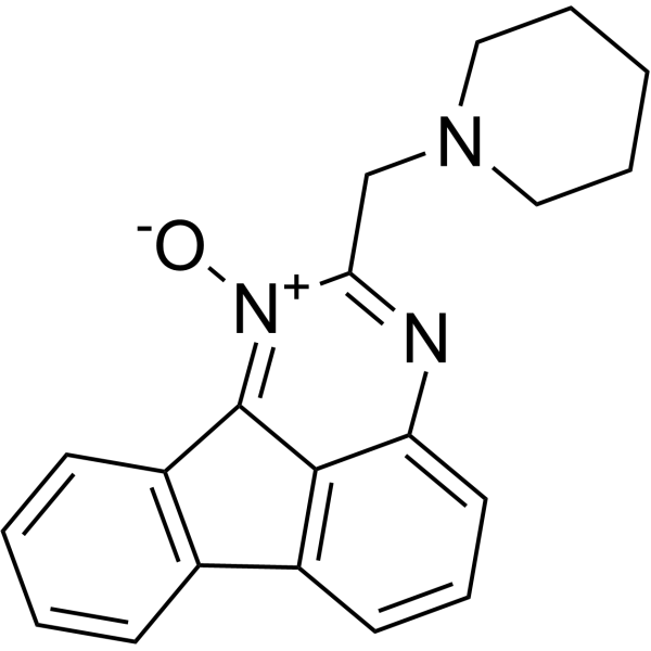
NSC194598 is a p53 DNA-binding inhibitor with an IC50 value of 180 nM. NSC194598 inhibits p53 DNA binding and induction of target genesn when p53 is stabilized and activated by irradiation or chemotherapy. NSC194598 can interfere with transcriptional activation of mutated rearranged during transfection (RET) gene, induce apoptosis and G0/G1 phase arrest. NSC194598 can be used for the researches of acute radiation toxicity and medullary thyroid carcinoma.
For research use only. We do not sell to patients.
- Purity : 98.9%
- CAS No.: 5358-76-9
- Formula: C20H19N3O
- Molecular Weight:317.38
-
Storage:Powder -20°C, 3 years , 4°C, 2 years ; In solvent -80°C, 6 months , -20°C, 1 month

To place orders, for customer services and technical support, please contact: MedChemExpress USA
Tel: 609-228-6898 E-mail: [email protected] [email protected]


-
 Biological Activity
-
Chemical Information
-
Solvent & Solubility
- Protocols
- Purity & Documentation
- References
-
 Help & FAQs





Biological Activity
Description
IC50 & Target
[2]|
Caspase 3 |
In Vitro
NSC194598 (1-2 μM; 35 min) inhibits p53, p63γ, and p73β DNA binding[1].
NSC194598 inhibits the DNA-binding activity of p53-Cluc in a cell-free luciferase fragment competition assay with an IC50 of 0.18 μM[1].
NSC194598 inhibits a p53 quadruple mutant (p53-
M133L/V203A/N239Y/N268D) with an IC50 of 0.06 μM[1].
NSC194598 (2-40 μM; 4 h) inhibits IR-induced p53 DNA binding and target gene expression in U2OS cells[1].
NSC194598 (2-40 μM; 8 h) inhibits p21 induction by activated p53A138V in H1299-p53A138V cells without altering p53 levels[1].
NSC194598 (5-20 μM; 32 h) causes weak p53 accumulation and p21 induction in SJSA cells, and inhibits Nutlin-induced p21 expression[1].
NSC194598 (0.5-20 μM) stabilizes the wild-type RET promoter G-quadruplex structure without altering its parallel folding pattern[2].
NSC194598 (0.6-2.5 μg/mL; 24 h) inhibits wild-type RET promoter-driven luciferase activity in HEK293 WT cells by over 60% after 24 h treatment at 2.5 μg/mL, with concentration-dependent effects[2].
NSC194598 (1.2-2.5 μg/mL; 24-48 h) reduces RET mRNA expression in TT cells[2].
NSC194598 (2.5 μM; 24 h) inhibits recruitment of RNA polymerase II, Sp1, and nucleolin to the RET promoter at 2.5 μM for 24 h in TT cells[2].
NSC194598 (0.3-2.5 μg/mL; 24 h) reduces TT cell growth and increases caspase 3 activity[2].
NSC194598 (0.6-2.5 μg/mL; 24 h) induces dose-dependent increases in late apoptotic/necrotic TT cells, and induces G0/G1 phase arrest[2].
MedChemExpress (MCE) has not independently confirmed the accuracy of these methods. They are for reference only. Further protocols information, click here.
-
Cell Line:H1299-p53-A138V human lung cancer cells
-
Concentration:2, 5, 10, 20, 40 μM
-
Incubation Time:8 h
-
Result:Inhibited p21 protein expression without changing p53 protein levels.
-
Cell Line:Human medullary thyroid carcinoma TT cells
-
Concentration:1.2, 2.5 μg/mL
-
Incubation Time:24 h, 48 h
-
Result:Reduced RET mRNA expression by 90% at concentrations below 2.5 μg/mL after 24 and 48 h.
-
Cell Line:Human medullary thyroid carcinoma TT cells
-
Concentration:1.2, 2.5 μg/mL
-
Incubation Time:24 h, 48 h
-
Result:Induced corresponding reductions in RET protein levels alongside reduced RET mRNA expression.
-
Cell Line:Human medullary thyroid carcinoma TT cells
-
Concentration:0.3, 0.6, 1.2, 2.5 μg/mL
-
Incubation Time:24 h
-
Result:Reduced cell number to 70% of control at 2.5 μg/mL.
-
Cell Line:Human medullary thyroid carcinoma TT cells
-
Concentration:0.6, 1.2, 2.5 μg/mL
-
Incubation Time:24 h
-
Result:Induced dose-dependent increases in propidium iodide-stained unfixed cells.
-
Cell Line:Human medullary thyroid carcinoma TT cells
-
Concentration:0.6, 1.2, 2.5 μg/mL
-
Incubation Time:24 h
-
Result:Induced a dose-dependent increase in sub-G0/G1 apoptotic cells.
In Vivo
MedChemExpress (MCE) has not independently confirmed the accuracy of these methods. They are for reference only.
-
Animal Model:C57BL/6 (male and female, ~2 months old)[1]
-
Dosage:80 μg
-
Administration:i.p.; single dose, before 8 Gy gamma radiation
-
Result:Produced a statistically significant improvement in survival over 40 days after irradiation.
Did not prevent initial body weight loss but increased the probability of weight recovery after the initial decrease.


Chemical Information
-
CAS No. 5358-76-9
-
Appearance Solid
-
Molecular Weight 317.38
-
Formula C20H19N3O
-
Color Light yellow to yellow
-
SMILES
[O-][N+](C(CN1CCCCC1)=N2)=C3C4=C(C=CC=C4)C5=C3C2=CC=C5
-
Shipping
Room temperature in continental US; may vary elsewhere.
-
Storage
Powder -20°C 3 years 4°C 2 years In solvent -80°C 6 months -20°C 1 month


Solvent & Solubility
In Vitro:
DMSO : 8.3 mg/mL (26.15 mM; Need ultrasonic and warming; Hygroscopic DMSO has a significant impact on the solubility of product, please use newly opened DMSO)
Please refer to the solubility information to select the appropriate solvent. Once prepared, please aliquot and store the solution to prevent product inactivation from repeated freeze-thaw cycles.
Storage method and period of stock solution: -80°C, 6 months; -20°C, 1 month. When stored at -80°C, please use it within 6 months. When stored at -20°C, please use it within 1 month.
Please refer to the solubility information to select the appropriate solvent. Once prepared, please aliquot and store the solution to prevent product inactivation from repeated freeze-thaw cycles.
Storage method and period of stock solution: -80°C, 6 months; -20°C, 1 month. When stored at -80°C, please use it within 6 months. When stored at -20°C, please use it within 1 month.
Concentration (start) × Volume (start) = Concentration (final) × Volume (final)

Protocols
-
Protocol for Electrophoretic Mobility Shift Assay (EMSA)
Electrophoretic mobility shift assay detects protein-nucleic acid binding by incubating a labeled DNA or RNA probe with purified protein or cell extract, then separating free probe from slower-migrating protein-probe complexes on a native gel. For cancer cells, primary neurons, mouse tumor samples, intestinal organoids, inflammatory macrophages, or drug-treated samples, EMSA can measure transcription-factor DNA binding or RNA-binding protein activity in extracts, but it does not directly measure transcription, protein expression, or chromatin occupancy in intact cells. Specificity is judged by competition with unlabeled wild-type probe, failure of mutated or unrelated competitors to compete, and antibody supershift or disruption when the binding protein identity must be confirmed.
-
Apoptosis
Apoptosis, also called programmed cell death, is generally characterized by distinct morphological characteristics.
-
TUNEL staining for apoptotic DNA fragmentation
TUNEL staining detects DNA strand breaks by using terminal deoxynucleotidyl transferase to add labeled nucleotides to exposed 3′-OH DNA termini, generating either microscopic staining in fixed cells or tissue sections, or fluorescence/cytometric signal in cell suspensions. TUNEL positivity reflects DNA fragmentation but should not be interpreted alone as definitive apoptosis, because TUNEL can also label necrotic, autolytic, mechanically damaged, or DNA-repair-associated DNA breaks.
-
Cell Cytotoxicity Assay
Cytotoxicity assays are usually based on the assessment of cell membrane damage, which can also be indirectly detected by measuring cell viability. Detection methods include MTT assay, CKK-8 assay, LDH assay and ATP assay, etc.
-
Flow cytometric DNA-content cell-cycle staining
Flow cytometric DNA-content cell-cycle staining measures the fluorescence intensity of DNA-bound fluorochromes in single cells or nuclei to estimate DNA content distributions, allowing assignment of populations to G0/G1, S, and G2/M phases by DNA histogram deconvolution. Propidium iodide (PI) intercalates into DNA, and PI fluorescence is proportional to cellular DNA content when staining is performed under conditions that make DNA accessible and minimize non-DNA signal. Cells with G2/M DNA content are expected to show approximately twice the fluorescence intensity of G0/G1 cells, while S-phase cells occupy intermediate fluorescence values. PI-based DNA-content analysis can also detect cells with fractional DNA content, often reported as sub-G1, when DNA fragmentation and extraction during staining reduce retained DNA signal in apoptotic cells. DAPI is an alternative DNA fluorochrome for univariate DNA-content analysis, while bivariate approaches combining DNA content with proliferation
-
Annexin V plus membrane-impermeant dye apoptosis staining
Annexin V-based apoptosis assays rely on the detection of phosphatidylserine (PS) externalization from the inner leaflet of the plasma membrane to the outer leaflet, an early biochemical hallmark of apoptosis. Fluorescently labeled Annexin V binds PS in a calcium-dependent manner, enabling identification of early apoptotic cells by flow cytometry or fluorescence microscopy. When combined with a membrane-impermeant DNA-binding dye (e. g. , propidium iodide), this approach allows discrimination between viable (Annexin V−/dye−), early apoptotic (Annexin V+/dye−), and late apoptotic or necrotic (Annexin V+/dye+) cell populations by assessing membrane integrity and PS exposure.
-
Research Protocol for Endocrine Diseases
Endocrine diseases often arise from disrupted hormone production, hormone signaling, or target-tissue responsiveness; for diabetes-focused endocrine disease models, insulin signaling regulates glucose uptake, hepatic glucose output, lipid metabolism, and β-cell compensation. Type 2 diabetes develops through interacting defects in insulin resistance, β-cell dysfunction, adipose inflammation, hepatic glucose overproduction, altered incretin signaling, and ectopic lipid metabolism. A major unresolved question is whether endocrine dysfunction is driven primarily by target-tissue insulin resistance, intrinsic β-cell failure, immune/inflammatory stress, or combined multi-organ failure that differs by disease stage.
-
Apoptosis Solutions
Apoptosis is a regulated, generally non-lytic cell-death pathway that removes unwanted, damaged, infected, or abnormal cells through coordinated morphological changes, caspase activation, DNA fragmentation, and membrane remodeling. The intrinsic apoptosis pathway is controlled mainly by mitochondrial outer membrane permeabilization, BCL-2 family proteins, cytochrome c release, apoptosome formation, caspase-9 activation, and downstream executioner caspase-3/7 activation. The extrinsic apoptosis pathway is initiated by death receptors such as Fas, TNFR, and TRAIL receptors, which recruit adaptor proteins and activate caspase-8 before engaging executioner caspases or mitochondrial amplification through BID cleavage. Apoptosis is linked to many phenotypes, including cancer cell killing, tissue homeostasis, immune regulation, neurodegeneration, infection response, and treatment-induced cytotoxicity; unresolved questions include how apoptosis interacts with necroptosis, pyroptosis, ferroptos
-
Protocol for Cell Cycle
Cell-cycle analysis by flow cytometry measures DNA content in single cells to estimate the fraction of cells in G0/G1, S, and G2/M phases. Propidium iodide intercalates into DNA, and after RNA removal with RNase, fluorescence intensity reflects cellular DNA content: 2N cells are assigned to G0/G1, cells between 2N and 4N to S phase, and 4N cells to G2/M. DNA-content analysis alone cannot reliably separate G0 from G1 or G2 from M. Ki-67 can distinguish quiescent G0 cells from cycling cells, EdU or BrdU incorporation marks active DNA synthesis in S phase, and phospho-histone H3 staining identifies mitotic cells within the 4N population.


Purity & Documentation
-
Data Sheet (289 KB)
-
SDS (251 KB)
- English - EN (251 KB)
- Français - FR (251 KB)
- Deutsch - DE (251 KB)
- Norwegian - NO (251 KB)
- Español - ES (251 KB)
- Swedish - SV (251 KB)
- Italian - IT (251 KB)
- Korean - KR (251 KB)
- Portuguese - PT (251 KB)
-
Handling Instructions (2659 KB)
References
[1]. Li Q, et al. Inhibition of p53 DNA binding by a small molecule protects mice from radiation toxicity. Oncogene. 2020;39(29):5187-5200. [Content Brief]
[2]. Shin YJ, et al. Involvement of G-quadruplex structures in regulation of human RET gene expression by small molecules in human medullary thyroid carcinoma TT cells. Oncogene. 2015;34(10):1292-1299. [Content Brief]


Complete Stock Solution Preparation Table
Please refer to the solubility information to select the appropriate solvent. Once prepared, please aliquot and store the solution to prevent product inactivation from repeated freeze-thaw cycles.
Storage method and period of stock solution: -80°C, 6 months; -20°C, 1 month. When stored at -80°C, please use it within 6 months. When stored at -20°C, please use it within 1 month.
| Optional Solvent | Concentration Solvent Mass | 1 mg | 5 mg | 10 mg | 25 mg |
|---|---|---|---|---|---|
| DMSO | 1 mM | 3.1508 mL | 15.7540 mL | 31.5080 mL | 78.7699 mL |
| 5 mM | 0.6302 mL | 3.1508 mL | 6.3016 mL | 15.7540 mL | |
| 10 mM | 0.3151 mL | 1.5754 mL | 3.1508 mL | 7.8770 mL | |
| 15 mM | 0.2101 mL | 1.0503 mL | 2.1005 mL | 5.2513 mL | |
| 20 mM | 0.1575 mL | 0.7877 mL | 1.5754 mL | 3.9385 mL | |
| 25 mM | 0.1260 mL | 0.6302 mL | 1.2603 mL | 3.1508 mL |


NSC194598 Related Classifications
HY-156135 Related Classifications
 Powered by Bioz