



Periplogenin
Based on 1 publication(s) in Google Scholar
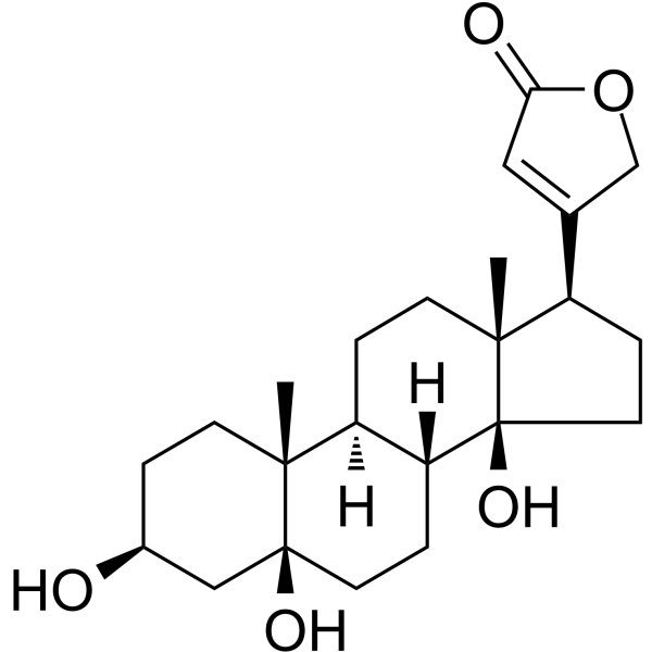
Periplogenin is an orally active cardiac glycoside found in Cortex periplocae. Periplogenin can induce ROS production and necroptosis and cause G0/G1 phase arrest. Periplogenin can inhibit pyroptosis by regulating the NLRP3/Caspase-1/GSDMD signaling. Periplogenin suppresses growth of prostate carcinoma cells by docking to an ATP1A1 protein pocket and forming a hydrogen bond with T804. Periplogenin can be used for the researches of cancer, inflammation and immunology, such as prostate carcinoma, rheumatoid arthritis and psoriasis.
For research use only. We do not sell to patients.
- Purity : 99.34%
- CAS No.: 514-39-6
- Formula: C23H34O5
- Molecular Weight:390.51
-
Storage:
4°C, protect from light
* In solvent : -80°C, 6 months; -20°C, 1 month (protect from light)

To place orders, for customer services and technical support, please contact: MedChemExpress USA
Tel: 609-228-6898 E-mail: [email protected] [email protected]


-
 Biological Activity
-
Chemical Information
-
Solvent & Solubility
- Protocols
- Purity & Documentation
- References
-
 Help & FAQs


-
HY-N0878
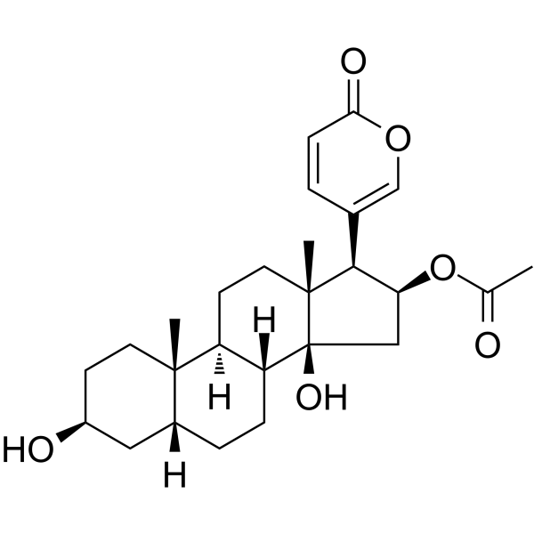
BufotalinApoptosis Inducer -
HY-A0154
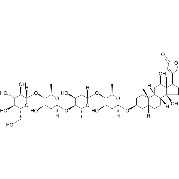
DeslanosideAnti-arrhythmia Drug -
HY-B1422
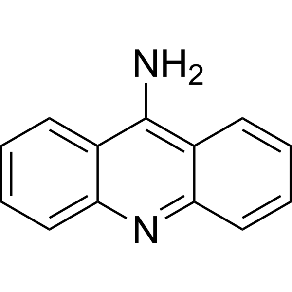
9-AminoacridineAntimicrobial -
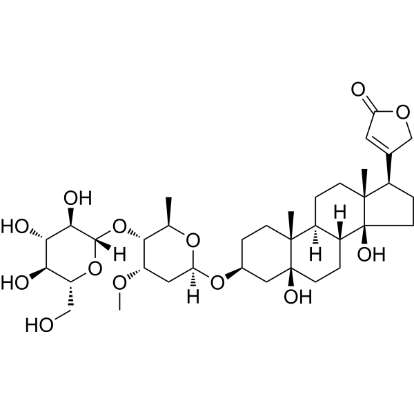
HY-N1381
PeriplocinAkt Inhibitor -
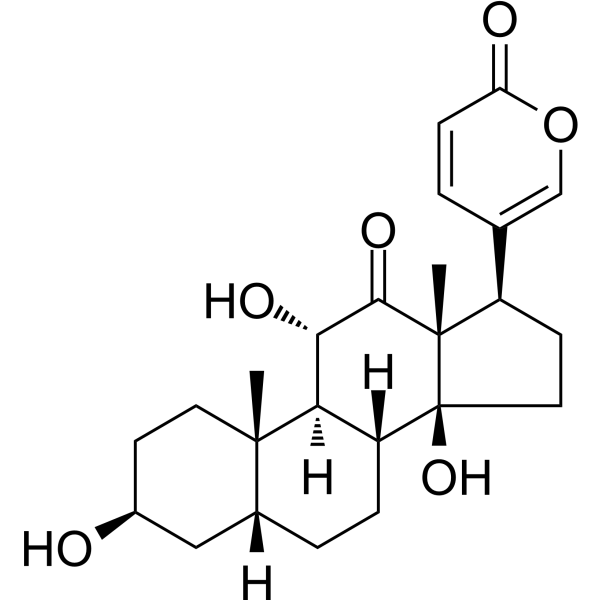
HY-N0876
ArenobufaginAnticancer Agent -

HY-N2414R
Periplogenin (Standard)Cardiac Glycoside -
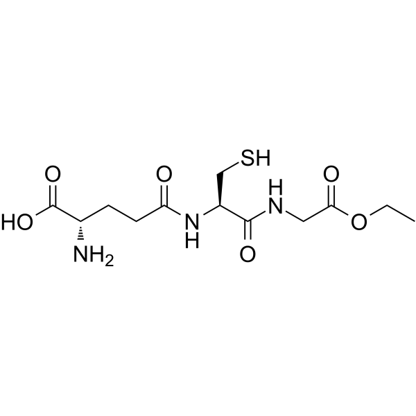
HY-134124
Glutathione ethyl esterGSH Donor -

HY-130743
Dieugenol
Publications Citing Use of MedChemExpress (MCE) Periplogenin
More Customer Validation & Images

-
Cell Proliferation/Viability Assay

-
Cell Migration/Invasion Assay

-
Cell Proliferation/Viability Assay

-
Flow Cytometry

-
WB

All Caspase Isoforms
More

Biological Activity
Description
IC50 & Target
[2]|
IL-1β |
IL-18 |
Caspase-1 |
NLRP3 |
Cellular Effect
|
Cell Line

|
Type | Value | Description | References |
|---|---|---|---|---|
| HT-1080 | IC50 |
810 nM
Compound: 11
|
Antiproliferative activity against human HT1080 cells after 72 hrs by MTT assay
Antiproliferative activity against human HT1080 cells after 72 hrs by MTT assay
|
[PMID: 14640513] |
In Vitro
Periplogenin (0.625-10 μg/mL 44 h) inhibits the viability of human HaCaT keratinocytes, with an IC50 of 1.56 μg/mL[1].
Periplogenin (2 μg/mL, 6-24 h) induces caspase-independent non-apoptotic and non-autophagic cell death in HaCaT cells[1].
Periplogenin (1-4 μg/mL, 12-48 h) increases ROS production and induces necroptosis in HaCaT cells[1].
Periplogenin (0.1-20 μg/mL, 24 h) inhibits the proliferation of rheumatoid arthritis fibroblast-like synoviocytes (RA-FLS), with an IC50 of 1.56 μg/mL[2].
Periplogenin (0.1-5 μg/mL, 24 h) inhibits the migration, invasion and pyroptosis in RA-FLS[2].
Periplogenin (0.1-5 μg/mL, 24 h) induces G0/G1 phase arrest in RA-FLS[2].
Periplogenin (0.1-5 μg/mL, 24 h) downregulates NLRP3, Cleaved-Caspase-1, GSDMD-N, IL-1β, and IL-18 expression in RA-FLS[2].
Periplogenin (0.02-33 μM, 72 h) inhibits the viability of DU145, DLD-1, U-87MG, and MCF-7 cancer cells, with IC50 values of 2.897 μM, 8.712 μM, 3.008 μM, and 20.025 μM respectively[3].
Periplogenin (0-100 μM) inhibits the enzymatic activity of ATP1A1 in wild-type DU145 cells[3].
MedChemExpress (MCE) has not independently confirmed the accuracy of these methods. They are for reference only. Further protocols information, click here.
-
Cell Line:HaCaT cells
-
Concentration:1, 2 and 4 μg/mL
-
Incubation Time:24 and 48 h
-
Result:Disrupted the membrane integrity.
Increased PI uptake and LDH release.
Revealed necrotic features (cytoplasmic swelling, reduced organelles, nuclear membrane disorder).
-
Cell Line:RA-FLS
-
Concentration:0.1, 0.5 and 5 μg/mL
-
Incubation Time:24 h
-
Result:Increased the proportion of G0/G1 phase cells.
Decreased the proportion of G2/M phase cells.
Showed no significant change in S phase cell proportion.
In Vivo
Periplogenin (10-20 mg/kg, i.g., once a day) inhibits tumor growth in DU145 wild-type xenograft mice models[3].
MedChemExpress (MCE) has not independently confirmed the accuracy of these methods. They are for reference only.
-
Animal Model:TPA-induced and IMQ (HY-B0180)-induced psoriasis-like mice models[1]
-
Dosage:2 μg per site
-
Administration:Topical application, daily for 11 days starting after 8 days of TPA induction
-
Result:Reduced ear thickness and weight.
Alleviated epidermal hyperplasia and inflammatory cell infiltration in dorsal skin and ears.
-
Animal Model:DU145 wild-type xenograft mice models[3]
-
Dosage:10 and 20 mg/kg
-
Administration:Orally gavage, once a day
-
Result:Reduced tumor volume and weight.
Had no significant change in body weight.


Chemical Information
-
CAS No. 514-39-6
-
Appearance Solid
-
Molecular Weight 390.51
-
Formula C23H34O5
-
Color White to off-white
-
SMILES
O[C@@]12[C@@]3([H])[C@@](CC[C@@]1([C@@H](C4=CC(OC4)=O)CC2)C)([H])[C@@]5([C@@](O)(C[C@@H](O)CC5)CC3)C
-
Structure Classification
-
Initial Source
-
Shipping
Room temperature in continental US; may vary elsewhere.
-
Storage
4°C, protect from light
* In solvent : -80°C, 6 months; -20°C, 1 month (protect from light)


Publications (1)
-
Journal Impact Factor
-
Most Recent
-
Int Immunopharmacol
Periplogenin inhibits pyroptosis of fibroblastic synoviocytes in rheumatoid arthritis through the NLRP3/Caspase-1/GSDMD signaling pathway. [Abstract]2024 May 30:133:112041. PMID: 38636373
Periplogenin purchased from MedChemExpress. Usage Cited in: Int Immunopharmacol. 2024 May 30:133:112041. [Abstract]
Effect of Periplogenin PPN on the cell proliferation of RA-FLS.
Periplogenin purchased from MedChemExpress. Usage Cited in: Int Immunopharmacol. 2024 May 30:133:112041. [Abstract]
Cell wound healing in the scratch test for 24 h treated with Periplogenin (PPN) (0.1, 0.5, 5 μg/mL).
Periplogenin purchased from MedChemExpress. Usage Cited in: Int Immunopharmacol. 2024 May 30:133:112041. [Abstract]
Transwell test cell invasion ability treated with Periplogenin (PPN) (0.1, 0.5, 5 μg/mL).
Periplogenin purchased from MedChemExpress. Usage Cited in: Int Immunopharmacol. 2024 May 30:133:112041. [Abstract]
Pyroptosis was determined by flow cytometry treated with Periplogenin (PPN) (0.1, 0.5, 5 μg/mL).
Periplogenin purchased from MedChemExpress. Usage Cited in: Int Immunopharmacol. 2024 May 30:133:112041. [Abstract]
Relative expression of NLRP3, GSDMD, Caspase-1, IL-1β, IL-18 treated with Periplogenin (PPN) (0.1, 0.5, 5 μg/mL).


Solvent & Solubility
In Vitro:
DMSO : 100 mg/mL (256.08 mM; Need ultrasonic; Hygroscopic DMSO has a significant impact on the solubility of product, please use newly opened DMSO)
Please refer to the solubility information to select the appropriate solvent. Once prepared, please aliquot and store the solution to prevent product inactivation from repeated freeze-thaw cycles.
Storage method and period of stock solution: -80°C, 6 months; -20°C, 1 month (protect from light). When stored at -80°C, please use it within 6 months. When stored at -20°C, please use it within 1 month.
Please refer to the solubility information to select the appropriate solvent. Once prepared, please aliquot and store the solution to prevent product inactivation from repeated freeze-thaw cycles.
Storage method and period of stock solution: -80°C, 6 months; -20°C, 1 month (protect from light). When stored at -80°C, please use it within 6 months. When stored at -20°C, please use it within 1 month.
Concentration (start) × Volume (start) = Concentration (final) × Volume (final)
In Vivo:
Select the appropriate dissolution method based on your experimental animal and administration route.
- For the following dissolution methods, please ensure to first prepare a clear stock solution using an In Vitro approach and then sequentially add co-solvents:
- To ensure reliable experimental results, the clarified stock solution can be appropriately stored based on storage conditions. As for the working solution for In Vivo experiments, it is recommended to prepare freshly and use it on the same day.
- The percentages shown for the solvents indicate their volumetric ratio in the final prepared solution. If precipitation or phase separation occurs during preparation, heat and/or sonication can be used to aid dissolution.
Add each solvent one by one: 10% DMSO 40% PEG300 5% Tween-80 45% Saline
Solubility: ≥ 2.5 mg/mL (6.40 mM); Clear solution
This protocol yields a clear solution of ≥ 2.5 mg/mL (saturation unknown).
Taking 1 mL working solution as an example, add 100 μL DMSO stock solution (25.0 mg/mL) to 400 μL PEG300, and mix evenly; then add 50 μL Tween-80 and mix evenly; then add 450 μL Saline to adjust the volume to 1 mL.
Preparation of Saline: Dissolve 0.9 g sodium chloride in ddH₂O and dilute to 100 mL to obtain a clear Saline solution.
Add each solvent one by one: 10% DMSO 90% (20% SBE-β-CD in Saline)
Solubility: ≥ 2.5 mg/mL (6.40 mM); Clear solution
This protocol yields a clear solution of ≥ 2.5 mg/mL (saturation unknown).
Taking 1 mL working solution as an example, add 100 μL DMSO stock solution (25.0 mg/mL) to 900 μL 20% SBE-β-CD in Saline, and mix evenly.
Preparation of 20% SBE-β-CD in Saline (4°C, storage for one week): 2 g SBE-β-CD powder is dissolved in 10 mL Saline, completely dissolve until clear.
In Vivo Dissolution Calculator
Please enter the basic information of animal experiments:
-
-
-
-
Recommended: Prepare an additional quantity of animals to account for potential losses during experiments.
Please enter your animal formula composition:
-
%DMSO +
Recommended: Keep the proportion of DMSO in working solution below 2% if your animal is weak.
-
%+
-
+%Tween-80 + +
-
%Saline +
The co-solvents required include: DMSO, . All of co-solvents are available by MedChemExpress (MCE). , Tween 80. All of co-solvents are available by MedChemExpress (MCE).
Working solution concentration: 0.22 mg/mL
Method for preparing stock solution: mg drug dissolved in μL DMSO. Stock solution concentration: mg/mL. * In solvent : -80°C, 6 months; -20°C, 1 month (protect from light)
1. Take μL DMSO stock solution;
2. Add μL .
μL , mix evenly;
3. Then add μL Tween 80, mix evenly;
4. Then add μL
Please ensure that the stock solution in the first step is dissolved to a clear state, and add co-solvents in sequence. You can use ultrasonic heating (ultrasonic cleaner, recommended frequency 20-40 kHz), vortexing, etc. to assist dissolution.

Protocols
-
Collagen-Induced Arthritis
Collagen-induced arthritis (CIA) is an autoimmune murine model of rheumatoid arthritis in which immunization with type II collagen (CII) emulsified in an adjuvant induces a T cell- and autoantibody-driven inflammatory arthritis characterized by synovial hyperplasia, immune cell infiltration, and joint destruction. The model typically relies on genetically susceptible mouse strains (e. g. , DBA/1) and reproduces key features of human rheumatoid arthritis, including anti-collagen immune responses and progressive joint inflammation. Disease onset generally occurs within ~3-4 weeks after immunization, depending on antigen/adjuvant combinations and protocol variation. The immunopathology is driven by adaptive immune activation against CII, leading to systemic and local joint inflammation mediated by pro-inflammatory cytokines and effector immune cells, making CIA a standard preclinical platform for evaluating immunomodulatory and anti-arthritic interventions.
-
Flow cytometric DNA-content cell-cycle staining
Flow cytometric DNA-content cell-cycle staining measures the fluorescence intensity of DNA-bound fluorochromes in single cells or nuclei to estimate DNA content distributions, allowing assignment of populations to G0/G1, S, and G2/M phases by DNA histogram deconvolution. Propidium iodide (PI) intercalates into DNA, and PI fluorescence is proportional to cellular DNA content when staining is performed under conditions that make DNA accessible and minimize non-DNA signal. Cells with G2/M DNA content are expected to show approximately twice the fluorescence intensity of G0/G1 cells, while S-phase cells occupy intermediate fluorescence values. PI-based DNA-content analysis can also detect cells with fractional DNA content, often reported as sub-G1, when DNA fragmentation and extraction during staining reduce retained DNA signal in apoptotic cells. DAPI is an alternative DNA fluorochrome for univariate DNA-content analysis, while bivariate approaches combining DNA content with proliferation
-
Imiquimod-Induced Psoriasiform Dermatitis
Imiquimod (IMQ)-induced psoriasiform dermatitis is a widely used murine model in which topical application of IMQ, a Toll-like receptor 7 (TLR7) agonist, triggers innate immune activation in the skin and induces a psoriasis-like inflammatory cascade characterized by epidermal hyperplasia, immune cell infiltration, and cytokine production dominated by the IL-23/IL-17 axis. This inflammatory response is mediated through activation of dendritic cells and downstream induction of IL-23, IL-17A, IL-22, and related pro-inflammatory mediators, recapitulating key features of human plaque psoriasis and enabling mechanistic and therapeutic studies. The model is commonly induced using Aldara (5% IMQ cream) applied topically to murine skin, resulting in rapid onset of erythema, scaling, and thickening that can be quantified as disease severity indices and validated histologically.
-
ROS/oxidative-stress fluorescent staining
ROS/oxidative-stress fluorescent staining uses cell-permeant fluorogenic probes that become fluorescent after oxidation inside cells or tissues; commonly used examples include DCFH-DA/DCFDA for broad cellular oxidant detection, DHE for superoxide-related signal detection, MitoSOX for mitochondrial superoxide-related signal detection, and CellROX probes for oxidative-stress-associated fluorescence readouts. The assay detects probe oxidation rather than a single ROS species unless the probe and analysis method have been chemically validated for that species. DCFH-DA enters cells, is deacetylated by intracellular esterases to DCFH, and produces fluorescent DCF after oxidation, so the readout is used as an operational measure of total cellular oxidative stress rather than a species-specific ROS measurement. DHE and MitoSOX can report superoxide-related oxidation, but red fluorescence alone can include non-specific ethidium-like oxidation products; HPLC or optimized spectral approaches are
-
Apoptosis Solutions
Apoptosis is a regulated, generally non-lytic cell-death pathway that removes unwanted, damaged, infected, or abnormal cells through coordinated morphological changes, caspase activation, DNA fragmentation, and membrane remodeling. The intrinsic apoptosis pathway is controlled mainly by mitochondrial outer membrane permeabilization, BCL-2 family proteins, cytochrome c release, apoptosome formation, caspase-9 activation, and downstream executioner caspase-3/7 activation. The extrinsic apoptosis pathway is initiated by death receptors such as Fas, TNFR, and TRAIL receptors, which recruit adaptor proteins and activate caspase-8 before engaging executioner caspases or mitochondrial amplification through BID cleavage. Apoptosis is linked to many phenotypes, including cancer cell killing, tissue homeostasis, immune regulation, neurodegeneration, infection response, and treatment-induced cytotoxicity; unresolved questions include how apoptosis interacts with necroptosis, pyroptosis, ferroptos
-
Protocol for Cell Cycle
Cell-cycle analysis by flow cytometry measures DNA content in single cells to estimate the fraction of cells in G0/G1, S, and G2/M phases. Propidium iodide intercalates into DNA, and after RNA removal with RNase, fluorescence intensity reflects cellular DNA content: 2N cells are assigned to G0/G1, cells between 2N and 4N to S phase, and 4N cells to G2/M. DNA-content analysis alone cannot reliably separate G0 from G1 or G2 from M. Ki-67 can distinguish quiescent G0 cells from cycling cells, EdU or BrdU incorporation marks active DNA synthesis in S phase, and phospho-histone H3 staining identifies mitotic cells within the 4N population.
-
Research Protocol for Inflammation-related Diseases
The NLRP3 inflammasome is a cytosolic innate immune signaling platform that integrates priming signals and danger-signal activation to promote caspase-1 activation, maturation of IL-1β and IL-18, and gasdermin D-mediated pyroptotic cell death. The core experimental logic is to determine whether inflammatory disease phenotypes are driven by increased NLRP3 expression, ASC-containing inflammasome assembly, caspase-1 cleavage, GSDMD cleavage, and extracellular release of IL-1β/IL-18 rather than by nonspecific cell injury alone. The pathway is strongly linked to inflammation-related disease phenotypes because monosodium urate crystals activate NALP3/NLRP3 inflammasome signaling in gout-like crystal inflammation, cholesterol crystals activate NLRP3 inflammasomes in atherogenesis models, and DSS-induced intestinal inflammation has been reported to involve NLRP3 inflammasome activity. However, experimental colitis studies also show context-dependent protective effects of NLRP3 inflammasome co
-
Pyroptosis Solutions
Pyroptosis is a lytic inflammatory cell-death pathway executed by gasdermin pores, most classically through inflammasome-mediated activation of caspase-1, cleavage of gasdermin D, membrane pore formation, LDH release, and secretion of IL-1β and IL-18. The canonical pathway is commonly modeled by priming cells with an inflammatory signal such as LPS to induce pro-IL-1β and inflammasome components, followed by an activation signal such as ATP or nigericin to activate NLRP3, ASC speck formation, caspase-1 cleavage, GSDMD cleavage, cytokine release, and pyroptotic membrane rupture. The non-canonical pathway is triggered when cytosolic LPS activates mouse caspase-11 or human caspase-4/5, leading to GSDMD cleavage and pyroptosis, and this can secondarily activate NLRP3-dependent IL-1β release. Pyroptosis is linked to inflammatory injury, infection, cancer, liver disease, ocular disease, placental inflammation, and other disease phenotypes, but unresolved questions include which gasdermin fam


Purity & Documentation
-
Data Sheet (286 KB)
-
SDS (251 KB)
- English - EN (251 KB)
- Français - FR (251 KB)
- Deutsch - DE (251 KB)
- Norwegian - NO (251 KB)
- Español - ES (251 KB)
- Swedish - SV (251 KB)
- Italian - IT (251 KB)
- Korean - KR (251 KB)
- Portuguese - PT (251 KB)
-
Handling Instructions (2659 KB)
References
[1]. Zhang WJ, et al. Periplogenin induces necroptotic cell death through oxidative stress in HaCaT cells and ameliorates skin lesions in the TPA- and IMQ-induced psoriasis-like mouse models. Biochem Pharmacol. 2016 Apr 1;105:66-79. [Content Brief]
[2]. Ma X, et al. Periplogenin inhibits pyroptosis of fibroblastic synoviocytes in rheumatoid arthritis through the NLRP3/Caspase-1/GSDMD signaling pathway. Int Immunopharmacol. 2024 May 30;133:112041. [Content Brief]
[3]. Zhang X, et al. The natural compound periplogenin suppresses the growth of prostate carcinoma cells by directly targeting ATP1A1. Sci Rep. 2024 Sep 3;14(1):20509. [Content Brief]


Complete Stock Solution Preparation Table
Please refer to the solubility information to select the appropriate solvent. Once prepared, please aliquot and store the solution to prevent product inactivation from repeated freeze-thaw cycles.
Storage method and period of stock solution: -80°C, 6 months; -20°C, 1 month (protect from light). When stored at -80°C, please use it within 6 months. When stored at -20°C, please use it within 1 month.
| Optional Solvent | Concentration Solvent Mass | 1 mg | 5 mg | 10 mg | 25 mg |
|---|---|---|---|---|---|
| DMSO | 1 mM | 2.5608 mL | 12.8038 mL | 25.6075 mL | 64.0188 mL |
| 5 mM | 0.5122 mL | 2.5608 mL | 5.1215 mL | 12.8038 mL | |
| 10 mM | 0.2561 mL | 1.2804 mL | 2.5608 mL | 6.4019 mL | |
| 15 mM | 0.1707 mL | 0.8536 mL | 1.7072 mL | 4.2679 mL | |
| 20 mM | 0.1280 mL | 0.6402 mL | 1.2804 mL | 3.2009 mL | |
| 25 mM | 0.1024 mL | 0.5122 mL | 1.0243 mL | 2.5608 mL | |
| 30 mM | 0.0854 mL | 0.4268 mL | 0.8536 mL | 2.1340 mL | |
| 40 mM | 0.0640 mL | 0.3201 mL | 0.6402 mL | 1.6005 mL | |
| 50 mM | 0.0512 mL | 0.2561 mL | 0.5122 mL | 1.2804 mL | |
| 60 mM | 0.0427 mL | 0.2134 mL | 0.4268 mL | 1.0670 mL | |
| 80 mM | 0.0320 mL | 0.1600 mL | 0.3201 mL | 0.8002 mL | |
| 100 mM | 0.0256 mL | 0.1280 mL | 0.2561 mL | 0.6402 mL |


Periplogenin Related Classifications
HY-N2414 Related Classifications
 Powered by Bioz