



Spicamycin
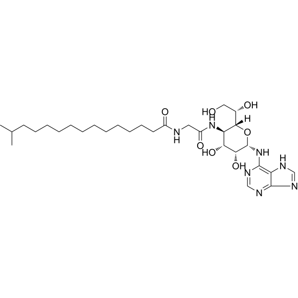
Spicamycin, an adenine nucleoside antibiotic with antifungal and antitumor activities. Spicamycin is also a potent inducer of differentiation of myeloid leukemia cells. Spicamycin induces apoptosis in NB4 cells via down-regulation of Bcl-2 expression and modulation of PML protein.
For research use only. We do not sell to patients.
- CAS No.: 87099-85-2
- Formula: C30H51N7O7
- Molecular Weight:621.77
-
Storage:
Please store the product under the recommended conditions in the Certificate of Analysis.

To place orders, for customer services and technical support, please contact: MedChemExpress USA
Tel: 609-228-6898 E-mail: [email protected] [email protected]


-
 Biological Activity
-
Chemical Information
- Protocols
- Purity & Documentation
- References
-
 Help & FAQs


All Antibiotic Isoforms
More

Biological Activity
Description
IC50 & Target
[1]|
Bcl-2 |
In Vitro
Spicamycin (10-160 ng/mL; 0-5 d) potently inhibits cell proliferation and viability of NB4, NKM-1, and HL-60 cells[1].
Spicamycin (20 ng/mL, 80 ng/mL; 48 h) induces apoptosis in NB4, HL-60 and NKM-1 cells[1].
Spicamycin (20 ng/mL, 40 ng/mL; 36 h) down-regulates the Bcl-2 expression in NB4 cells[1].
Spicamycin (2.5-640 ng/mL) shows anti-microbial activity against Saccharomyces cerevisiae ATCC 9763, Candida utilis IFO 0396, and Trichophyton mentagrophytes with MIC values of 25μg/mL, 25μg/mL, and 1.56 μg/mL, respectively[2].
MedChemExpress (MCE) has not independently confirmed the accuracy of these methods. They are for reference only.
-
Cell Line:NB4, HL-60, NKM-1, NOP-1 and Daudi cells
-
Concentration:10 ng/mL, 20 ng/mL, 40 ng/mL, 80 ng/mL, 160 ng/mL
-
Incubation Time:0, 1, 2, 3, 4, and 5 days or 72 hours
-
Result:Completely inhibited cell proliferation and viability of NB4 and NKM-1 at 40 ng/mL, of HL-60 at 80 ng/mL, but failed to inhibit NOP-1 and Daudi cells at higher dose of 160 ng/mL.
Inhibited cells viability of IC50s of 18.2 ng/mL, 28.6 ng/mL, 23.8 ng/mL, 74.9 ng/mL, and 37.4 ng/mL, respectively.
-
Cell Line:NB4 cells
-
Concentration:20 ng/mL, 40 ng/mL
-
Incubation Time:36 hours
-
Result:Reduced the Bcl-2 expression without affecting Bcl-xL and Bax expression.
In Vivo
Spicamycin shows an LD50 value of 40 mg/kg (i.p.) in mice[2].
MedChemExpress (MCE) has not independently confirmed the accuracy of these methods. They are for reference only.
-
Animal Model:P388 Mouse Leukemia model[2]
-
Dosage:0.125, 0.25, 0.5, 1.0, and 2.0 mg/kg
-
Administration:Intraperitoneal injection; once daily for 9 days
-
Result:Showed no biotoxicity at dose below 2.0 mg/kg.


Chemical Information
-
CAS No. 87099-85-2
-
Molecular Weight 621.77
-
Formula C30H51N7O7
-
SMILES
OC[C@H](O)[C@]([C@H](NC(CNC(CCCCCCCCCCCCC(C)C)=O)=O)[C@@H](O)[C@H]1O)([H])O[C@@H]1NC2=C3C(N=CN3)=NC=N2
-
Shipping
Room temperature in continental US; may vary elsewhere.
-
Storage
Please store the product under the recommended conditions in the Certificate of Analysis.


Purity & Documentation
References

Calculators
Concentration (start) × Volume (start) = Concentration (final) × Volume (final)


Spicamycin Related Classifications
HY-127130 Related Classifications
 Powered by Bioz