



Swertiamarin
Based on 1 publication(s) in Google Scholar
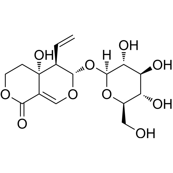
Swertiamarin is an orally active natural product with hypoglycemic, lipid-lowering, anti-rheumatic, and antioxidant activities. Swertiamarin can regulate the levels of pro-inflammatory cytokines, MMP, and NF-κB, and promote osteoblast proliferation. Swertiamarin has antioxidant and hepatoprotective effects against carbon tetrachloride induced rat liver toxicity through the Nrf2/HO-1 pathway. Swertiamarin can attenuate inflammatory mediators by regulating JAK2/STAT3 transcription factors in adjuvant induced arthritis rats. Swertiamarin can be used in the research of diabetes and arthritis.
Nos produits utilisent uniquement pour la recherche. Nous ne vendons pas aux patients.
- Pureté : 99.03%
- CAS No.: 17388-39-5
- Formule: C16H22O10
- Masse moléculaire:374.34
-
Stockage:
4°C, protect from light
* In solvent : -80°C, 2 years; -20°C, 1 year (protect from light)

To place orders, for customer services and technical support, please contact: MedChemExpress USA
Tel: 609-228-6898 E-mail: [email protected] [email protected]


-
 Activité biologique
-
Chemical Information
-
Solvant et solubilité
- Protocole
- Pureté et documentation
- Références
-
 Help & FAQs


-
HY-N0494
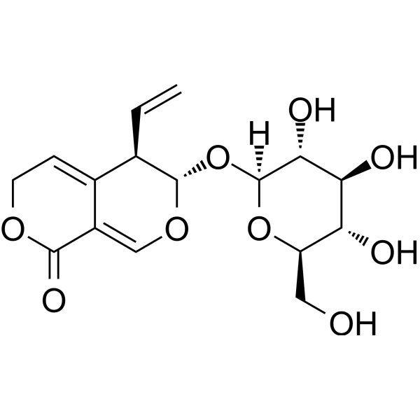
GentiopicrosideCytochrome P450 Inhibitor -
HY-N0806
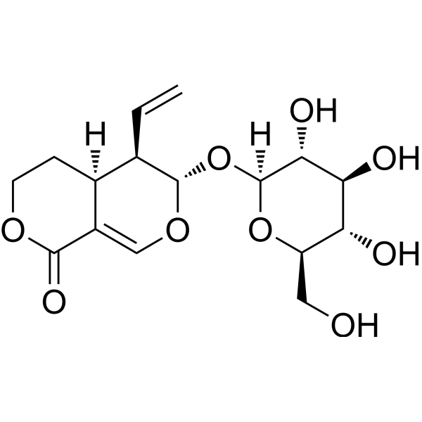
SwerosideKeap1/Nrf2 Activator -
HY-N0156
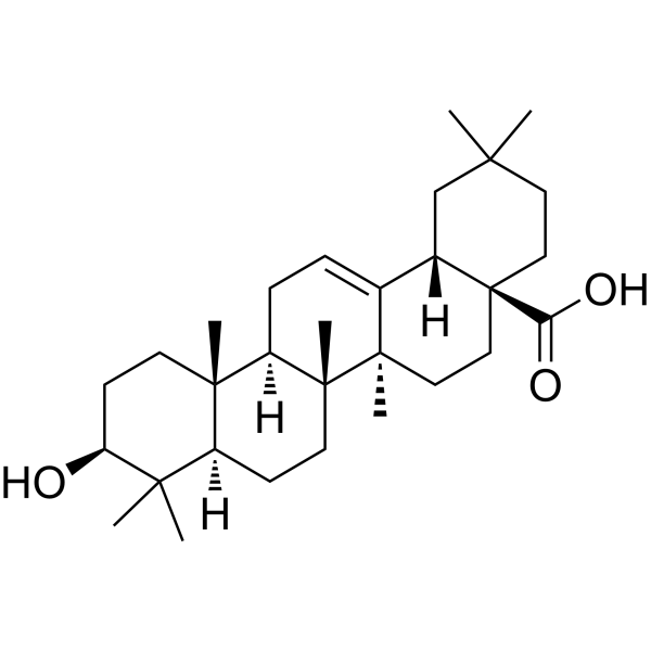
Oleanolic acidAutophagy Inducer -
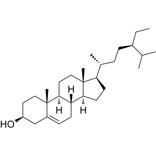
HY-N0171A
Beta-Sitosterol (purity>98%)Dietary Plant Sterols -

HY-N0807R
Swertiamarin (Standard)Natural Product -
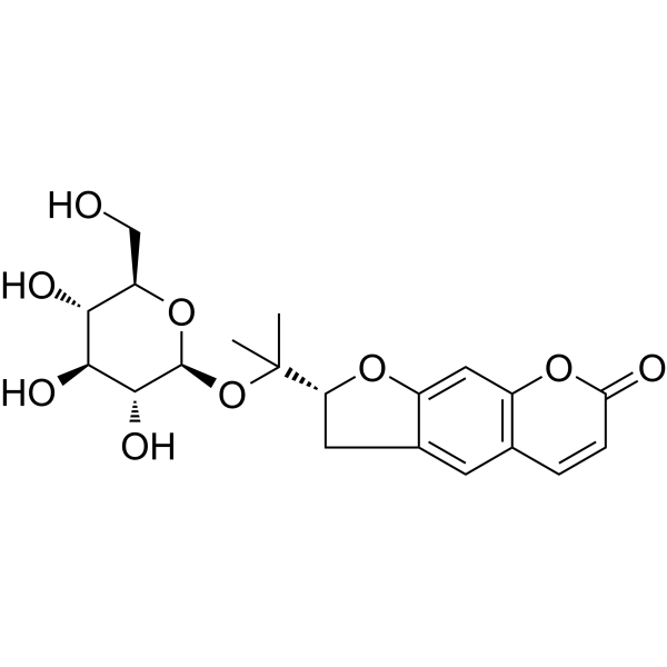
HY-N0825
NodakeninAChE Inhibitor -
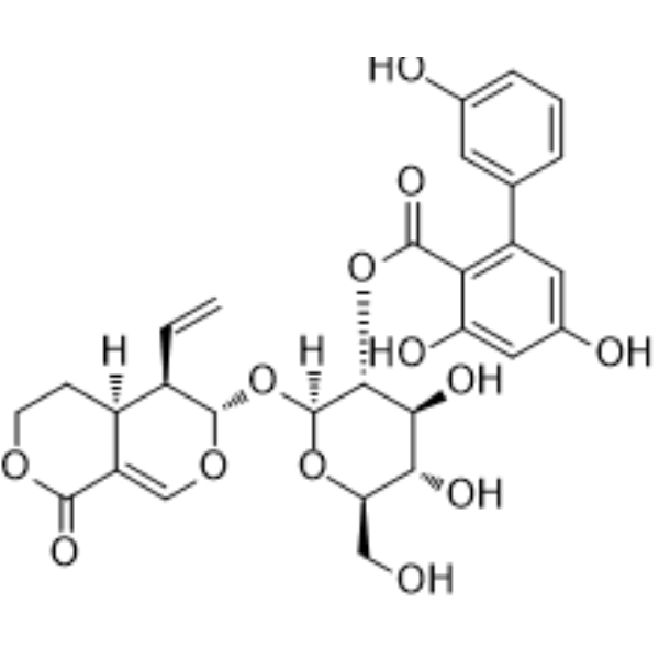
HY-N2447
AmarogentinAMPK Activator -
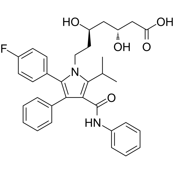
HY-B0589
AtorvastatinHMG-CoA Reductase Inhibitor -
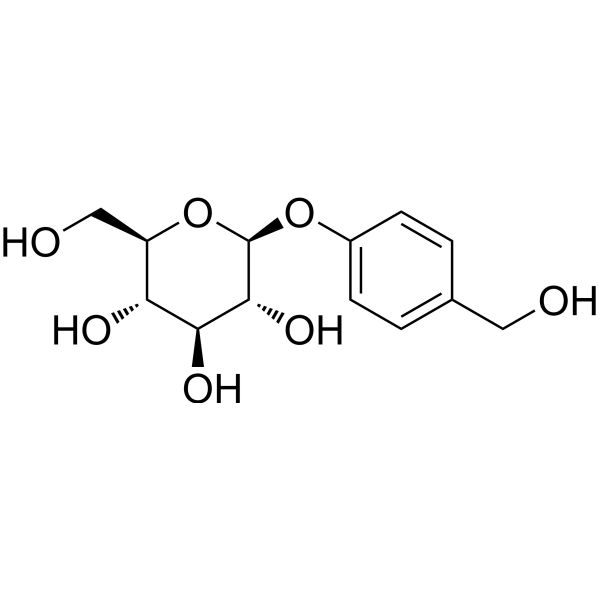
HY-N0115
GastrodinAnti-Inflammatory Agent -
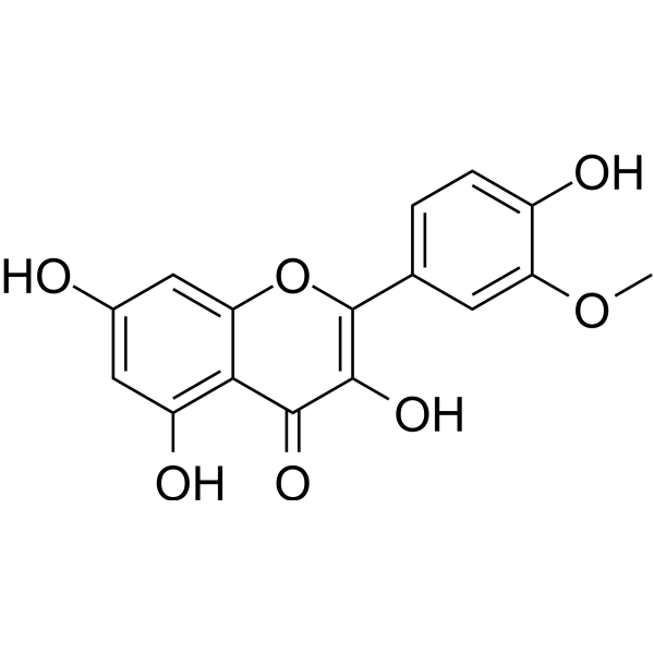
HY-N0776
IsorhamnetinEndogenous Metabolite -
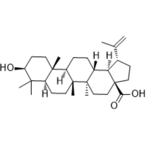
HY-10529
Betulinic acidApoptosis Inducer -
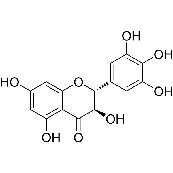
HY-N0112
DihydromyricetinAutophagy Inducer
Publications Citing Use of MedChemExpress (MCE) Swertiamarin
More

Activité biologique
Description
In Vitro
Swertiamarin (10-50 μg/mL, 48 h) can regulate the levels of pro-inflammatory cytokines, MMP, and NF-κB and promote the proliferation of osteoblasts[3]. Swertiamarin promotes 3T3-L1 adipocyte differentiation through its active metabolite gentianine, and induces PPAR-g, GLUT-4 and adiponectin mRNA expression, thus having anti diabetes activity[5].
MedChemExpress (MCE) has not independently confirmed the accuracy of these methods. They are for reference only.
-
Cell Line:Calvarial osteoblast cells
-
Concentration:10-50 μg/mL
-
Incubation Time:48 h
-
Result:Improved cell proliferation and ALP levels of osteoblasts.
In Vivo
MedChemExpress (MCE) has not independently confirmed the accuracy of these methods. They are for reference only.
-
Animal Model:Male Sprague Dawley (SD) rats with hypercholesterolemia induced by supplementing a diet rich in cholesterol[1].
-
Dosage:50, 75 mg/kg
-
Administration:Oral gavage (p.o.); once daily; 7 days
-
Result:Reduced serum total cholesterol, triglyceride concentration and atherosclerosis index.
-
Animal Model:Male Sprague Dawley (SD) rat model of liver injury induced by CCl4[2].
-
Dosage:100, 200 mg/kg
-
Administration:i.g. ; once daily; 8 weeks
-
Result:Reduced the levels of serum marker enzymes ALT, AST, and ALP representing liver damage, and restored antioxidant enzyme activity and GSH content in rat liver.
-
Animal Model:
-
Dosage:2, 5, 10 mg/kg
-
Administration:Oral gavage (p.o.); once daily; 2 weeks
-
Result:Reduced calcium and TRAP, ACP, and ALP levels in serum and urine of arthritis rats, and increased phosphorus and collagen levels[3]. Inhibited paw thickness, lysosomal enzyme levels, and increased body weight in rats[4].
- Collagen-Induced Arthritis
- Mitochondrial membrane-potential fluorescent assay
- LPS-Induced Endotoxemia/Systemic Inflammation
- Mitochondrial membrane-potential and mitochondrial mass staining
- Research Protocol for Inflammation-related Diseases
- Hepatotoxicity Study
- Research Protocol for Metabolic Diseases
- Cell Viability Determination by MTT Colorimetric Assay


Chemical Information
-
CAS No. 17388-39-5
-
Appearance Solid
-
Masse moléculaire 374.34
-
Formule C16H22O10
-
Color Off-white to light yellow
-
SMILES
C=C[C@@H]([C@@H]1O[C@]([C@@H]([C@@H](O)[C@@H]2O)O)([H])O[C@@H]2CO)[C@@](C3=CO1)(CCOC3=O)O
-
Structure Classification
-
Initial Source
-
Livraison
Room temperature in continental US; may vary elsewhere.
-
Stockage
4°C, protect from light
* In solvent : -80°C, 2 years; -20°C, 1 year (protect from light)


Publications (1)
-
Journal Impact Factor
-
Most Recent
-
PLoS One
Swertiamarin fails to induce cytotoxicity in colon cancer cell lines: Evidence against a direct anticancer effect. [Abstract]2026 Mar 16;21(3):e0344653. PMID: 41838704


Solvant et solubilité
In Vitro:
DMSO : 175 mg/mL (467.49 mM; Need ultrasonic; Hygroscopic DMSO has a significant impact on the solubility of product, please use newly opened DMSO)
Please refer to the solubility information to select the appropriate solvent. Once prepared, please aliquot and store the solution to prevent product inactivation from repeated freeze-thaw cycles.
Storage method and period of stock solution: -80°C, 2 years; -20°C, 1 year (protect from light). When stored at -80°C, please use it within 2 years. When stored at -20°C, please use it within 1 year.
Please refer to the solubility information to select the appropriate solvent. Once prepared, please aliquot and store the solution to prevent product inactivation from repeated freeze-thaw cycles.
Storage method and period of stock solution: -80°C, 2 years; -20°C, 1 year (protect from light). When stored at -80°C, please use it within 2 years. When stored at -20°C, please use it within 1 year.
Concentration (start) × Volume (start) = Concentration (final) × Volume (final)
In Vivo:
Select the appropriate dissolution method based on your experimental animal and administration route.
- For the following dissolution methods, please ensure to first prepare a clear stock solution using an In Vitro approach and then sequentially add co-solvents:
- To ensure reliable experimental results, the clarified stock solution can be appropriately stored based on storage conditions. As for the working solution for In Vivo experiments, it is recommended to prepare freshly and use it on the same day.
- The percentages shown for the solvents indicate their volumetric ratio in the final prepared solution. If precipitation or phase separation occurs during preparation, heat and/or sonication can be used to aid dissolution.
Add each solvent one by one: 10% DMSO 40% PEG300 5% Tween-80 45% Saline
Solubility: ≥ 2.17 mg/mL (5.80 mM); Clear solution
This protocol yields a clear solution of ≥ 2.17 mg/mL (saturation unknown).
Taking 1 mL working solution as an example, add 100 μL DMSO stock solution (21.7 mg/mL) to 400 μL PEG300, and mix evenly; then add 50 μL Tween-80 and mix evenly; then add 450 μL Saline to adjust the volume to 1 mL.
Preparation of Saline: Dissolve 0.9 g sodium chloride in ddH₂O and dilute to 100 mL to obtain a clear Saline solution.
Add each solvent one by one: 10% DMSO 90% (20% SBE-β-CD in Saline)
Solubility: ≥ 2.17 mg/mL (5.80 mM); Clear solution
This protocol yields a clear solution of ≥ 2.17 mg/mL (saturation unknown).
Taking 1 mL working solution as an example, add 100 μL DMSO stock solution (21.7 mg/mL) to 900 μL 20% SBE-β-CD in Saline, and mix evenly.
Preparation of 20% SBE-β-CD in Saline (4°C, storage for one week): 2 g SBE-β-CD powder is dissolved in 10 mL Saline, completely dissolve until clear.
In Vivo Dissolution Calculator
Please enter the basic information of animal experiments:
-
-
-
-
Recommended: Prepare an additional quantity of animals to account for potential losses during experiments.
Please enter your animal formula composition:
-
%DMSO +
Recommended: Keep the proportion of DMSO in working solution below 2% if your animal is weak.
-
%+
-
+%Tween-80 + +
-
%Saline +
The co-solvents required include: DMSO, . All of co-solvents are available by MedChemExpress (MCE). , Tween 80. All of co-solvents are available by MedChemExpress (MCE).
Working solution concentration: 0.22 mg/mL
Method for preparing stock solution: mg drug dissolved in μL DMSO. Stock solution concentration: mg/mL. * In solvent : -80°C, 2 years; -20°C, 1 year (protect from light)
1. Take μL DMSO stock solution;
2. Add μL .
μL , mix evenly;
3. Then add μL Tween 80, mix evenly;
4. Then add μL
Please ensure that the stock solution in the first step is dissolved to a clear state, and add co-solvents in sequence. You can use ultrasonic heating (ultrasonic cleaner, recommended frequency 20-40 kHz), vortexing, etc. to assist dissolution.


Pureté et documentation
-
Fiche technique (284 KB)
-
SDS (393 KB)
- English - EN (393 KB)
- Français - FR (393 KB)
- Deutsch - DE (393 KB)
- Norwegian - NO (393 KB)
- Español - ES (393 KB)
- Swedish - SV (393 KB)
- Italian - IT (393 KB)
- Korean - KR (393 KB)
- Portuguese - PT (393 KB)
-
Instruction de manipulation (2659 KB)
Références
[1]. Vaidya H, et al. Swertiamarin: a lead from Enicostemma littorale Blume. for anti-hyperlipidaemic effect. Eur J Pharmacol. 2009 Sep 1;617(1-3):108-12. [Content Brief]
[2]. Wu T, et al. Antioxidant and Hepatoprotective Effect of Swertiamarin on Carbon Tetrachloride-Induced Hepatotoxicity via the Nrf2/HO-1 Pathway. Cell Physiol Biochem. 2017;41(6):2242-2254. [Content Brief]
[3]. Hairul-Islam MI, et al. Swertiamarin, a natural steroid, prevent bone erosion by modulating RANKL/RANK/OPG signaling. Int Immunopharmacol. 2017 Dec;53:114-124. [Content Brief]
[4]. Saravanan S, et al. Swertiamarin attenuates inflammation mediators via modulating NF-κB/I κB and JAK2/STAT3 transcription factors in adjuvant induced arthritis. Eur J Pharm Sci. 2014 Jun 2;56:70-86. [Content Brief]
[5]. Vaidya H, et al. Anti-diabetic activity of swertiamarin is due to an active metabolite, gentianine, that upregulates PPAR-γ gene expression in 3T3-L1 cells. Phytother Res. 2013 Apr;27(4):624-7. [Content Brief]


Complete Stock Solution Preparation Table
Please refer to the solubility information to select the appropriate solvent. Once prepared, please aliquot and store the solution to prevent product inactivation from repeated freeze-thaw cycles.
Storage method and period of stock solution: -80°C, 2 years; -20°C, 1 year (protect from light). When stored at -80°C, please use it within 2 years. When stored at -20°C, please use it within 1 year.
| Optional Solvent | Concentration Solvent Mass | 1 mg | 5 mg | 10 mg | 25 mg |
|---|---|---|---|---|---|
| DMSO | 1 mM | 2.6714 mL | 13.3568 mL | 26.7137 mL | 66.7842 mL |
| 5 mM | 0.5343 mL | 2.6714 mL | 5.3427 mL | 13.3568 mL | |
| 10 mM | 0.2671 mL | 1.3357 mL | 2.6714 mL | 6.6784 mL | |
| 15 mM | 0.1781 mL | 0.8905 mL | 1.7809 mL | 4.4523 mL | |
| 20 mM | 0.1336 mL | 0.6678 mL | 1.3357 mL | 3.3392 mL | |
| 25 mM | 0.1069 mL | 0.5343 mL | 1.0685 mL | 2.6714 mL | |
| 30 mM | 0.0890 mL | 0.4452 mL | 0.8905 mL | 2.2261 mL | |
| 40 mM | 0.0668 mL | 0.3339 mL | 0.6678 mL | 1.6696 mL | |
| 50 mM | 0.0534 mL | 0.2671 mL | 0.5343 mL | 1.3357 mL | |
| 60 mM | 0.0445 mL | 0.2226 mL | 0.4452 mL | 1.1131 mL | |
| 80 mM | 0.0334 mL | 0.1670 mL | 0.3339 mL | 0.8348 mL | |
| 100 mM | 0.0267 mL | 0.1336 mL | 0.2671 mL | 0.6678 mL |


Swertiamarin Related Classifications
HY-N0807 Related Classifications
 Powered by Bioz