



Ibuprofen
Based on 23 publication(s) in Google Scholar
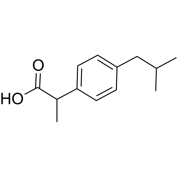
Ibuprofen ((±)-Ibuprofen) is a potent, orally active, selective COX-1 inhibitor with an IC50 value of 13 μM. Ibuprofen inhibits cell proliferation, angiogenesis, and induces cell apoptosis. Ibuprofen is a nonsteroidal anti-inflammatory agent and a nitric oxide (NO) donor. Ibuprofen ((±)-Ibuprofen) can be used in the research of pain, swelling, inflammation, infection, immunology, cancers.
For research use only. We do not sell to patients.
- Purity : 99.97%
- CAS No.: 15687-27-1
- Formula: C13H18O2
- Molecular Weight:206.28
-
Storage:
Store at room temperature 3 years.
In solvent -80°C, 2 years , -20°C, 1 year

To place orders, for customer services and technical support, please contact: MedChemExpress USA
Tel: 609-228-6898 E-mail: [email protected] [email protected]


-
 Biological Activity
-
Chemical Information
-
Solvent & Solubility
- Protocols
- Purity & Documentation
- References
-
 Help & FAQs


-
HY-14397
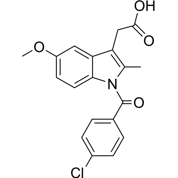
IndomethacinCOX1/2 Inhibitor -
HY-14398
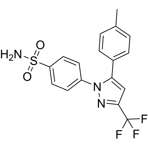
CelecoxibCOX-2inhibitor -
HY-15030
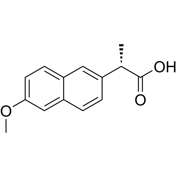
NaproxenCOX Inhibitor -
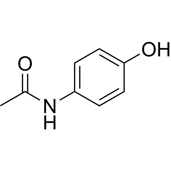
HY-66005
AcetaminophenCOX-2 Inhibitor -
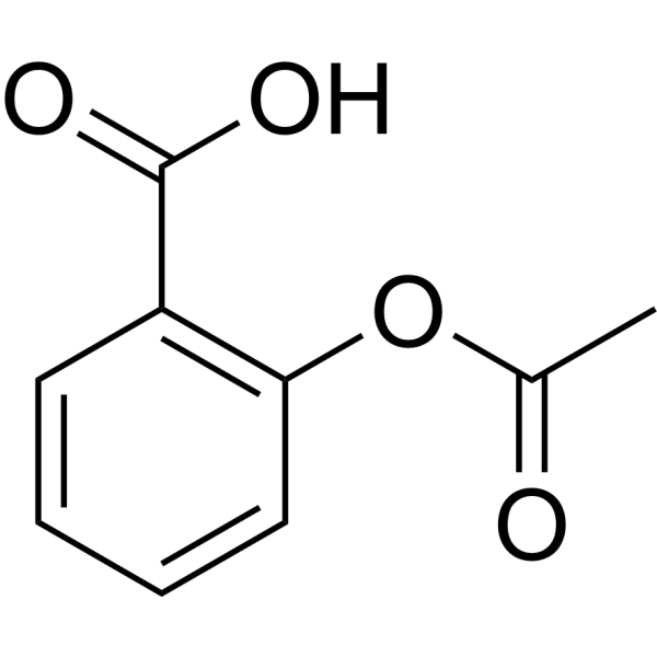
HY-14654
AspirinCOX Inhibitor -
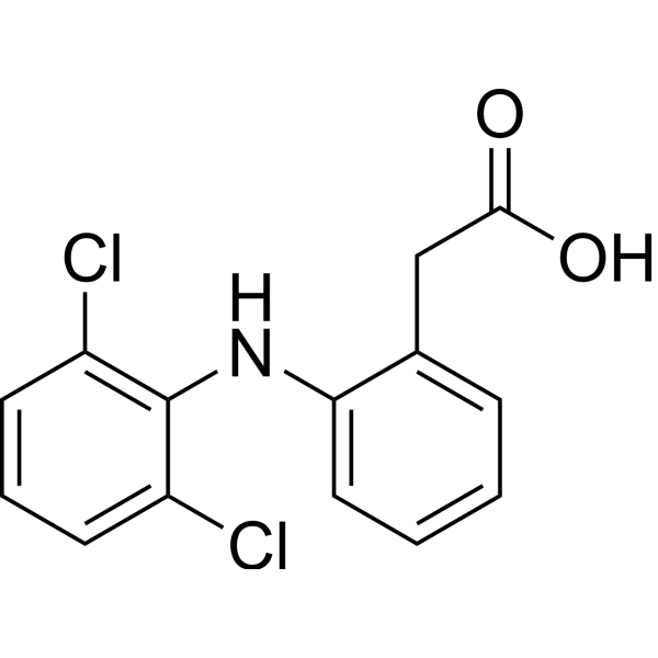
HY-15036
DiclofenacCOX Inhibitor -
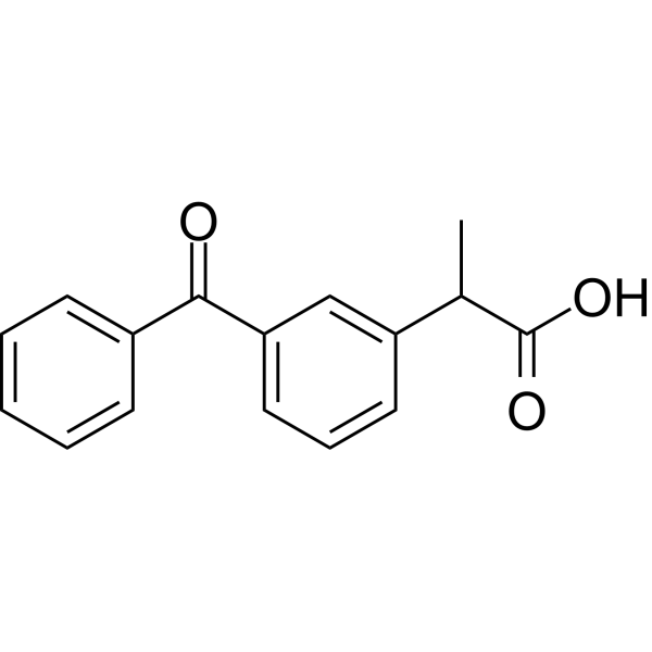
HY-B0227
KetoprofenAnti-inflammatory agent -
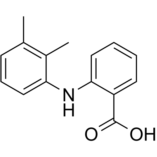
HY-B0574
Mefenamic acidCOX Inhibitor -
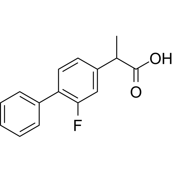
HY-10582
FlurbiprofenCOX Inhibitor -
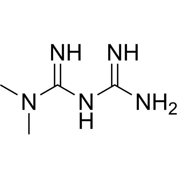
HY-B0627
MetforminAntihyperglycemic Agent -
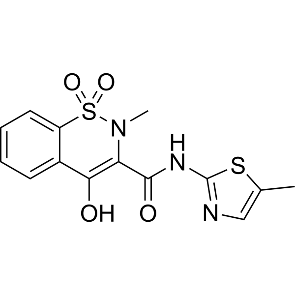
HY-B0261
MeloxicamCOX Inhibitor -
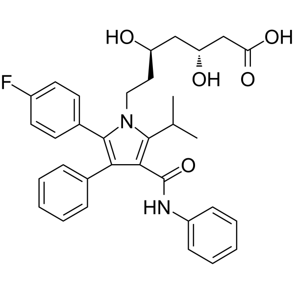
HY-B0589
AtorvastatinHMG-CoA Reductase Inhibitor
Publications Citing Use of MedChemExpress (MCE) Ibuprofen
More- Biomaterials. 2025 Nov:322:123402. [Abstract]
- Phytomedicine. 2022 Nov:106:154427. [Abstract]
- J Hazard Mater Lett. 2025 Nov.
- Int J Biol Macromol. 2025 Dec 9;337(Pt 2):149613. [Abstract]
- Plant Physiol. 2026 Mar 2;200(3):kiag108. [Abstract]
- Cell Rep. 2025 Apr 2;44(4):115489. [Abstract]
- Cell Rep. 2019 Dec 17;29(12):3847-3858.e5. [Abstract]
- Biochem Pharmacol. 2024 Nov:229:116478. [Abstract]
- Inflammopharmacology. 2024 Feb;32(1):733-745. [Abstract]
- Cells. 2022 Jun 9;11(12):1870. [Abstract]
- EMBO Rep. 2022 Jun 7;23(6):e53932. [Abstract]
- Cell Rep Methods. 2023 Oct 23;3(10):100599. [Abstract]
- Int Immunopharmacol. 2026 Aug 15:183:116873. [Abstract]
- Int J Mol Sci. 2021 Nov 22;22(22):12597. [Abstract]
- Bioresour Technol Rep. 2023 Nov 10, 101696.
- Bioresour Technol Rep. 2023 Dec, 24, 101619.
- Sci Rep. 2020 Oct 2;10(1):16383. [Abstract]
- Neuropharmacology. 2022 Oct 1:217:109191. [Abstract]
- ACS Chem Neurosci. 2025 Sep 3;16(17):3257-3266. [Abstract]
- J Neurosci. 2024 Aug 21;44(34):e0740242024. [Abstract]
- Reprod Toxicol. 2025 May:134:108895. [Abstract]
- J Appl Toxicol. 2024 Oct;44(10):1528-1539. [Abstract]
- Chemosphere. 2019 Jun:225:378-387. [Abstract]
 Customer Validation & Images

-
Cell Proliferation/Viability Assay
-
Cell Migration/Invasion Assay
-
Cell Migration/Invasion Assay
-
Flow Cytometry
-
Bio/Physico-chemical Assay
All Parasite Isoforms
More

Biological Activity
Description
IC50 & Target
[1]|
COX-1 13 μM (IC50) |
COX-2 370 μM (IC50) |
Cellular Effect
|
Cell Line

|
Type | Value | Description | References |
|---|---|---|---|---|
| A549 | IC50 |
>50 μM
Compound: Ibuprofen
|
Cytotoxicity against human A549 cells measured after 24 to 72 hrs by MTT assay
Cytotoxicity against human A549 cells measured after 24 to 72 hrs by MTT assay
|
[PMID: 37482018] |
| Bel-7402 | IC50 |
>100 μM
Compound: Ibuprofen
|
Antiproliferative activity against human Bel7402 cells after 72 hrs by CCK-8 assay
Antiproliferative activity against human Bel7402 cells after 72 hrs by CCK-8 assay
|
[PMID: 28301815] |
| Bel7402/5-FU | IC50 |
>100 μM
Compound: Ibuprofen
|
Antiproliferative activity against human Bel7402/5-FU cells after 72 hrs by CCK-8 assay
Antiproliferative activity against human Bel7402/5-FU cells after 72 hrs by CCK-8 assay
|
[PMID: 28301815] |
| BGC-823 | IC50 |
>50 μM
Compound: Ibuprofen
|
Cytotoxicity against human BGC-823 cells assessed as reduction in cell viability measured upto 72 hrs by MTT assay
Cytotoxicity against human BGC-823 cells assessed as reduction in cell viability measured upto 72 hrs by MTT assay
|
[PMID: 33799070] |
| BGC-823 | IC50 |
>50 μM
Compound: Ibuprofen
|
Cytotoxicity against human BGC-823 cells measured after 24 to 72 hrs by MTT assay
Cytotoxicity against human BGC-823 cells measured after 24 to 72 hrs by MTT assay
|
[PMID: 37482018] |
| Caco-2 | IC50 |
>50 μM
Compound: Ibuprofen
|
Cytotoxicity against human Caco2 cells assessed as reduction in cell viability measured upto 72 hrs by MTT assay
Cytotoxicity against human Caco2 cells assessed as reduction in cell viability measured upto 72 hrs by MTT assay
|
[PMID: 33799070] |
| CHO | IC50 |
8 μM
Compound: Ibuprofen
|
TP_TRANSPORTER: inhibition of Adefovir uptake in OAT1-expressing CHO cells
TP_TRANSPORTER: inhibition of Adefovir uptake in OAT1-expressing CHO cells
|
[PMID: 10991954] |
| CHO | IC50 |
200 μM
Compound: 1
|
Modulation of gamma-secretase-mediated cleavage of human APP expressed in CHO cells with human presenilin-1 assessed as inhibition of amyloid beta42 production after 24 hrs by ELISA
Modulation of gamma-secretase-mediated cleavage of human APP expressed in CHO cells with human presenilin-1 assessed as inhibition of amyloid beta42 production after 24 hrs by ELISA
|
[PMID: 20503989] |
| CHO | IC50 |
200 μM
Compound: 1
|
Modulation of gamma-secretase expressed in CHO cells co-expressing human APP and wild type human presenilin-1 assessed as inhibition of amyloid beta42 production by ELISA
Modulation of gamma-secretase expressed in CHO cells co-expressing human APP and wild type human presenilin-1 assessed as inhibition of amyloid beta42 production by ELISA
|
[PMID: 21873070] |
| COS-7 | IC50 |
>1000 μM
Compound: Ibuprofen
|
Cytotoxicity against African green monkey COS7 cells assessed as cell growth inhibition measured after 48 hrs by MTT assay
Cytotoxicity against African green monkey COS7 cells assessed as cell growth inhibition measured after 48 hrs by MTT assay
|
[PMID: 32527536] |
| HCT-116 | IC50 |
448 μM
Compound: IB
|
Cytotoxicity against human HCT116 cells after 72 hrs by MTT assay
Cytotoxicity against human HCT116 cells after 72 hrs by MTT assay
|
[PMID: 20171760] |
| HCT-15 | IC50 |
>1000 μM
Compound: Ibuprofen
|
Anticancer activity against human HCT15 cells assessed as cell growth inhibition measured after 48 hrs by MTT assay
Anticancer activity against human HCT15 cells assessed as cell growth inhibition measured after 48 hrs by MTT assay
|
[PMID: 32527536] |
| HeLa | IC50 |
>50 μM
Compound: Ibuprofen
|
Cytotoxicity against human HeLa cells measured after 24 to 72 hrs by MTT assay
Cytotoxicity against human HeLa cells measured after 24 to 72 hrs by MTT assay
|
[PMID: 37482018] |
| HT-29 | IC50 |
552 μM
Compound: IB
|
Cytotoxicity against human HT-29 cells after 72 hrs by MTT assay
Cytotoxicity against human HT-29 cells after 72 hrs by MTT assay
|
[PMID: 20171760] |
| HT-29 | IC50 |
>50 μM
Compound: Ibu
|
Antiproliferative activity against human HT-29 cells assessed as reduction in cell viability after 24 hrs by MTT assay
Antiproliferative activity against human HT-29 cells assessed as reduction in cell viability after 24 hrs by MTT assay
|
[PMID: 26750401] |
| HT-29 | IC50 |
>50 μM
Compound: Ibu
|
Antiproliferative activity against human HT-29 cells assessed as reduction in cell viability after 48 hrs by MTT assay
Antiproliferative activity against human HT-29 cells assessed as reduction in cell viability after 48 hrs by MTT assay
|
[PMID: 26750401] |
| HT-29 | IC50 |
>50 μM
Compound: Ibu
|
Antiproliferative activity against human HT-29 cells assessed as reduction in cell viability after 72 hrs by MTT assay
Antiproliferative activity against human HT-29 cells assessed as reduction in cell viability after 72 hrs by MTT assay
|
[PMID: 26750401] |
| Huh-7 | IC50 |
37.2 μM
Compound: Ibu
|
Cytotoxicity against human HuH7 cells assessed as growth inhibition after 72 hrs by sulforhodamine B assay
Cytotoxicity against human HuH7 cells assessed as growth inhibition after 72 hrs by sulforhodamine B assay
|
[PMID: 26810835] |
| K562 | IC50 |
>1000 μM
Compound: Ibuprofen
|
Anticancer activity against human K562 cells assessed as cell growth inhibition measured after 48 hrs by MTT assay
Anticancer activity against human K562 cells assessed as cell growth inhibition measured after 48 hrs by MTT assay
|
[PMID: 32527536] |
| L02 | IC50 |
>100 μM
Compound: Ibuprofen
|
Antiproliferative activity against human LO2 cells after 72 hrs by CCK-8 assay
Antiproliferative activity against human LO2 cells after 72 hrs by CCK-8 assay
|
[PMID: 28301815] |
| MCF7 | IC50 |
92.14 μM
Compound: Ibuprofen
|
Anticancer activity against human MCF7 cells assessed as cell growth inhibition measured after 48 hrs by MTT assay
Anticancer activity against human MCF7 cells assessed as cell growth inhibition measured after 48 hrs by MTT assay
|
[PMID: 32527536] |
| MCF7 | IC50 |
>50 μM
Compound: Ibuprofen
|
Cytotoxicity against human MCF7 cells assessed as reduction in cell viability measured upto 72 hrs by MTT assay
Cytotoxicity against human MCF7 cells assessed as reduction in cell viability measured upto 72 hrs by MTT assay
|
[PMID: 33799070] |
| MCF7 | IC50 |
>50 μM
Compound: 6
|
Cytotoxicity against human MCF7 cells assessed as reduction in cell viability measured after 24 hrs by MTT assay
Cytotoxicity against human MCF7 cells assessed as reduction in cell viability measured after 24 hrs by MTT assay
|
[PMID: 35468536] |
| MCF7 | IC50 |
>50 μM
Compound: Ibuprofen
|
Cytotoxicity against human MCF7 cells measured after 24 to 72 hrs by MTT assay
Cytotoxicity against human MCF7 cells measured after 24 to 72 hrs by MTT assay
|
[PMID: 37482018] |
| MDA-MB-231 | IC50 |
>50 μM
Compound: Ibu
|
Antiproliferative activity against human MDA-MB-231 cells assessed as reduction in cell viability after 24 hrs by MTT assay
Antiproliferative activity against human MDA-MB-231 cells assessed as reduction in cell viability after 24 hrs by MTT assay
|
[PMID: 26750401] |
| MDA-MB-231 | IC50 |
>50 μM
Compound: Ibu
|
Antiproliferative activity against human MDA-MB-231 cells assessed as reduction in cell viability after 48 hrs by MTT assay
Antiproliferative activity against human MDA-MB-231 cells assessed as reduction in cell viability after 48 hrs by MTT assay
|
[PMID: 26750401] |
| MDA-MB-231 | IC50 |
>50 μM
Compound: Ibu
|
Antiproliferative activity against human MDA-MB-231 cells assessed as reduction in cell viability after 72 hrs by MTT assay
Antiproliferative activity against human MDA-MB-231 cells assessed as reduction in cell viability after 72 hrs by MTT assay
|
[PMID: 26750401] |
| Neutrophil | IC50 |
27.5 μM
Compound: ibuprofen
|
Inhibition of fMLP-induced superoxide production in human neutrophils
Inhibition of fMLP-induced superoxide production in human neutrophils
|
[PMID: 17559265] |
| Neutrophil | IC50 |
32.55 μM
Compound: ibuprofen
|
Antiinflammatory activity in human neutrophils assessed as fMLP-induced superoxide release after 5 mins by spectrometry
Antiinflammatory activity in human neutrophils assessed as fMLP-induced superoxide release after 5 mins by spectrometry
|
[PMID: 17822293] |
| Neutrophil | IC50 |
12.1 μM
Compound: Ibuprofen
|
Immunomodulatory activity in polymorphoneutrophils assessed as inhibition of luminol-induced oxidative burst by chemiluminescence assay
Immunomodulatory activity in polymorphoneutrophils assessed as inhibition of luminol-induced oxidative burst by chemiluminescence assay
|
[PMID: 18950230] |
| Neutrophil | IC50 |
27.53 μM
Compound: ibuprofen
|
Antiinflammatory activity in human neutrophils assessed as inhibition of fMLP-indcued superoxide production after 5 mins
Antiinflammatory activity in human neutrophils assessed as inhibition of fMLP-indcued superoxide production after 5 mins
|
[PMID: 20839880] |
| PANC-1 | IC50 |
>50 μM
Compound: Ibu
|
Antiproliferative activity against human PANC1 cells assessed as reduction in cell viability after 24 hrs by MTT assay
Antiproliferative activity against human PANC1 cells assessed as reduction in cell viability after 24 hrs by MTT assay
|
[PMID: 26750401] |
| PANC-1 | IC50 |
>50 μM
Compound: Ibu
|
Antiproliferative activity against human PANC1 cells assessed as reduction in cell viability after 48 hrs by MTT assay
Antiproliferative activity against human PANC1 cells assessed as reduction in cell viability after 48 hrs by MTT assay
|
[PMID: 26750401] |
| PANC-1 | IC50 |
>50 μM
Compound: Ibu
|
Antiproliferative activity against human PANC1 cells assessed as reduction in cell viability after 72 hrs by MTT assay
Antiproliferative activity against human PANC1 cells assessed as reduction in cell viability after 72 hrs by MTT assay
|
[PMID: 26750401] |
| PC-3 | IC50 |
>1000 μM
Compound: Ibuprofen
|
Anticancer activity against human PC3 cells assessed as cell growth inhibition measured after 48 hrs by MTT assay
Anticancer activity against human PC3 cells assessed as cell growth inhibition measured after 48 hrs by MTT assay
|
[PMID: 32527536] |
| PC-3 | IC50 |
>50 μM
Compound: Ibuprofen
|
Cytotoxicity against human PC-3 cells assessed as reduction in cell viability measured upto 72 hrs by MTT assay
Cytotoxicity against human PC-3 cells assessed as reduction in cell viability measured upto 72 hrs by MTT assay
|
[PMID: 33799070] |
| RAW264.7 | IC50 |
0.86 μM
Compound: Ibuprofen
|
Inhibition of COX-2 in mouse RAW264.7 cells assessed as decrease in LPS-induced PGE2 production treated prior to LPS challenge by enzyme immunoassay
Inhibition of COX-2 in mouse RAW264.7 cells assessed as decrease in LPS-induced PGE2 production treated prior to LPS challenge by enzyme immunoassay
|
[PMID: 24656662] |
| RAW264.7 | IC50 |
0.39 μM
Compound: Ibuprofen
|
Antiinflammatory activity in mouse RAW264.7 cells assessed as inhibition of LPS-stimulated PGE2 production by ELISA
Antiinflammatory activity in mouse RAW264.7 cells assessed as inhibition of LPS-stimulated PGE2 production by ELISA
|
[PMID: 28408221] |
| RAW264.7 | IC50 |
1483.87 μM
Compound: Ibuprofen
|
Antiinflammatory activity in mouse RAW264.7 cells assessed as inhibition of LPS-stimulated NO production by ELISA
Antiinflammatory activity in mouse RAW264.7 cells assessed as inhibition of LPS-stimulated NO production by ELISA
|
[PMID: 28408221] |
| RAW264.7 | IC50 |
3309.84 μM
Compound: Ibuprofen
|
Cytotoxicity against mouse RAW264.7 cells by MTT assay
Cytotoxicity against mouse RAW264.7 cells by MTT assay
|
[PMID: 28408221] |
| RAW264.7 | IC50 |
54.5 μM
Compound: Ibuprofen
|
Antiinflammatory activity in mouse RAW264.7 cells assessed as inhibition of LPS-induced nitric oxide production by measuring nitrite accumulation by Griess method
Antiinflammatory activity in mouse RAW264.7 cells assessed as inhibition of LPS-induced nitric oxide production by measuring nitrite accumulation by Griess method
|
[PMID: 28561586] |
| RAW264.7 | IC50 |
14.4 μM
Compound: Ibuprofen
|
Anti-inflammatory activity against LPS-stimulated mouse RAW264.7 cells assessed as reduction in IL1beta protein expression level after 24 hrs by ELISA
Anti-inflammatory activity against LPS-stimulated mouse RAW264.7 cells assessed as reduction in IL1beta protein expression level after 24 hrs by ELISA
|
[PMID: 34890996] |
| RAW264.7 | IC50 |
2.1 μM
Compound: Ibuprofen
|
Anti-inflammatory activity against LPS-stimulated mouse RAW264.7 cells assessed as reduction in TNFalpha protein expression level after 24 hrs by ELISA
Anti-inflammatory activity against LPS-stimulated mouse RAW264.7 cells assessed as reduction in TNFalpha protein expression level after 24 hrs by ELISA
|
[PMID: 34890996] |
| RAW264.7 | EC50 |
26.74 μM
Compound: IBP
|
Anti-inflammatory activity in mouse RAW264.7 cells assessed as inhibition of LPS-stimulated NO production
Anti-inflammatory activity in mouse RAW264.7 cells assessed as inhibition of LPS-stimulated NO production
|
[PMID: 37925088] |
| RAW264.7 | IC50 |
>1000 μM
Compound: IBP
|
Cytotoxicity against mouse RAW264.7 cells assessed as cell growth inhibition by CCK-8 colorimetric assay
Cytotoxicity against mouse RAW264.7 cells assessed as cell growth inhibition by CCK-8 colorimetric assay
|
[PMID: 37925088] |
| RBL-1 | IC50 |
2 μM
Compound: 10
|
Inhibition of Prostaglandin G/H synthase in intact RBL-1 cell line
Inhibition of Prostaglandin G/H synthase in intact RBL-1 cell line
|
[PMID: 2115586] |
| Sf21 | IC50 |
290.9 μM
Compound: Ibuprofen
|
Inhibition of Sprague-Dawley rat Bsep expressed in plasma membrane vesicles of Sf21 cells assessed as inhibition of ATP-dependent [3H]taurocholate uptake
Inhibition of Sprague-Dawley rat Bsep expressed in plasma membrane vesicles of Sf21 cells assessed as inhibition of ATP-dependent [3H]taurocholate uptake
|
[PMID: 21965623] |
| Sf21 | IC50 |
598.6 μM
Compound: Ibuprofen
|
Inhibition of human BSEP expressed in plasma membrane vesicles of Sf21 cells assessed as inhibition of ATP-dependent [3H]taurocholate uptake
Inhibition of human BSEP expressed in plasma membrane vesicles of Sf21 cells assessed as inhibition of ATP-dependent [3H]taurocholate uptake
|
[PMID: 21965623] |
| SK-LU-1 | IC50 |
90.61 μM
Compound: Ibuprofen
|
Anticancer activity against human SKLU1 cells assessed as cell growth inhibition measured after 48 hrs by MTT assay
Anticancer activity against human SKLU1 cells assessed as cell growth inhibition measured after 48 hrs by MTT assay
|
[PMID: 32527536] |
| SW480 | IC50 |
>50 μM
Compound: Ibuprofen
|
Cytotoxicity against human SW480 cells measured after 24 to 72 hrs by MTT assay
Cytotoxicity against human SW480 cells measured after 24 to 72 hrs by MTT assay
|
[PMID: 37482018] |
| TERT-RPE1 | IC50 |
>40 μM
Compound: 6
|
Cytotoxicity against human RPE-1 cells assessed as reduction in cell viability measured after 24 hrs by MTT assay
Cytotoxicity against human RPE-1 cells assessed as reduction in cell viability measured after 24 hrs by MTT assay
|
[PMID: 35468536] |
| U-251 | IC50 |
>1000 μM
Compound: Ibuprofen
|
Anticancer activity against human U251 cells assessed as cell growth inhibition measured after 48 hrs by MTT assay
Anticancer activity against human U251 cells assessed as cell growth inhibition measured after 48 hrs by MTT assay
|
[PMID: 32527536] |
| UACC-903 | IC50 |
>50 μM
Compound: Ibu
|
Antiproliferative activity against human UACC-903 cells assessed as reduction in cell viability after 24 hrs by MTT assay
Antiproliferative activity against human UACC-903 cells assessed as reduction in cell viability after 24 hrs by MTT assay
|
[PMID: 26750401] |
| UACC-903 | IC50 |
>50 μM
Compound: Ibu
|
Antiproliferative activity against human UACC-903 cells assessed as reduction in cell viability after 48 hrs by MTT assay
Antiproliferative activity against human UACC-903 cells assessed as reduction in cell viability after 48 hrs by MTT assay
|
[PMID: 26750401] |
| UACC-903 | IC50 |
>50 μM
Compound: Ibu
|
Antiproliferative activity against human UACC-903 cells assessed as reduction in cell viability after 72 hrs by MTT assay
Antiproliferative activity against human UACC-903 cells assessed as reduction in cell viability after 72 hrs by MTT assay
|
[PMID: 26750401] |
In Vitro
Ibuprofen (24 h) inhibits COX-1 and COX-2 activity with IC50 values of 13 μM and 370 μM[1].
Ibuprofen (500 μM, 48 h) inhibits cell proliferation and angiogenesis, and induces apoptosis in AGS cells (Adenocarcinoma gastric cell line)[2].
Ibuprofen (500 μM, 48 h) downregulates transcription of Akt, VEGF-A, PCNA, Bcl2, OCT3/4 and CD44 genes, but upregulates RNA levels of wild type P53 and Bax genes in AGS cell[2].
Ibuprofen (500 μM, 24 h) restores microtubule reformation, microtubule-dependent intracellular cholesterol transport, and induces extension of microtubules to the cell periphery in both cystic fibrosis (CF) cell models and primary CF nasal epithelial cells[3].
Ibuprofen (500 μM, 24 h) enhances UV-induced cell death in MCF-7 cells and MDA-MB-231 cells by a photosensitization process[4].
MedChemExpress (MCE) has not independently confirmed the accuracy of these methods. They are for reference only. Further protocols information, click here.
-
Cell Line:AGS cells
-
Concentration:100-1000 μM
-
Incubation Time:24 h, 48 h
-
Result:Inhibited AGS cell viability with IC50 values of 630 μM (trypan blue staining, 24 h), 456 μM (neutral red assay, 24 h), 549 μM (trypan blue staining, 48 h) and 408 μM (neutral red assay, 48 h).
In Vivo
Ibuprofen (60 mg/kg; i.h.; every second day for 15 days) reduces the risk of neuropathy in a rat model of chronic Oxaliplatin induced peripheral neuropathy[6].
Ibuprofen (20 mg/kg; p.o.; every 12 hours, 5 doses total) decreases muscle growth (average muscle fiber cross-sectional area) without affecting regulation of supraspinatus tendon adaptions to exercise[7].
Ibuprofen (35 mg/kg; p.o.; twice daily) attenuates the Inflammatory response to pseudomonas aeruginosa in a rat model of chronic pulmonary infection[8].
MedChemExpress (MCE) has not independently confirmed the accuracy of these methods. They are for reference only.
-
Animal Model:Syngeneic (D2A1) orthotopic Balb/c mouse model of PPBC (postpartum)[5]
-
Dosage:300 mg/kg, daily for 14 days
-
Administration:Fed in animal feedings (added to pulverized standard chow and mixed dry, then mixed with water, made into chow pellets and dried thoroughly)
-
Result:Suppresed tumor growth, reduced presence of immature monocytes and increased numbers of T cells.
Enhanced Th1 associated cytokines as well as promoted tumor border accumulation of T cells.
-
Animal Model:Oxaliplatin‑induced peripheral neuropathy[6]
-
Dosage:60 mg/kg, every second day for 15 days
-
Administration:Subcutaneous injection
-
Result:Lowered sensory nerve conduction velocity (SNCV).


Chemical Information
-
CAS No. 15687-27-1
-
Appearance Solid
-
Molecular Weight 206.28
-
Formula C13H18O2
-
Color White to off-white
-
SMILES
OC(C(C1=CC=C(CC(C)C)C=C1)C)=O
-
Synonyms
(±)-Ibuprofen
-
Shipping
Room temperature in continental US; may vary elsewhere.
-
Storage
Store at room temperature 3 years
In solvent -80°C 2 years -20°C 1 year


Publications (23)
-
Journal Impact Factor
-
Most Recent
-
Biomaterials
GSH-activable and cytolytic iPep-coupled immune nanoagonist for cancer synergetic therapy. [Abstract]2025 Nov:322:123402. PMID: 40373515 -
Phytomedicine
Prim-O-glucosycimifugin attenuates liver injury in septic mice by inhibiting NLRP3 inflammasome/caspase-1 signaling cascades in macrophages. [Abstract]2022 Nov:106:154427. PMID: 36088791 -
-
Int J Biol Macromol
Molecular interaction of abemaciclib with human serum albumin: Insights from spectroscopy, microscopy, and computational approaches. [Abstract]2025 Dec 9;337(Pt 2):149613. PMID: 41380878 -
Plant Physiol
The MdSP1-MdANK immune module activated by effector SyCD1 enhances Malus domestica resistance to Valsa mali. [Abstract]2026 Mar 2;200(3):kiag108. PMID: 41757702 -
Cell Rep
LAMP2A-mediated neuronal hyperexcitability by enhancing NKAβ1 degradation underlies depression-induced allodynia. [Abstract]2025 Apr 2;44(4):115489. PMID: 40178973 -
Cell Rep
A Central Amygdala Input to the Parafascicular Nucleus Controls Comorbid Pain in Depression. [Abstract]2019 Dec 17;29(12):3847-3858.e5. PMID: 31851918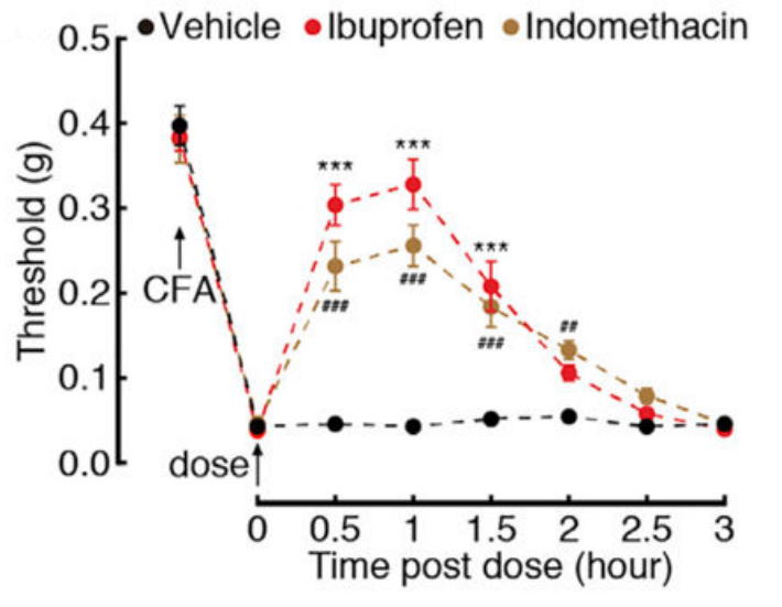
Ibuprofen purchased from MedChemExpress. Usage Cited in: Cell Rep. 2019 Dec 17;29(12):3847-3858.e5. [Abstract]
Effects of Ibuprofen (10 mg/kg, i.p.) or indomethacin (10 mg/kg, i.p.) on pain thresholds at day 3 after CFA treatment.
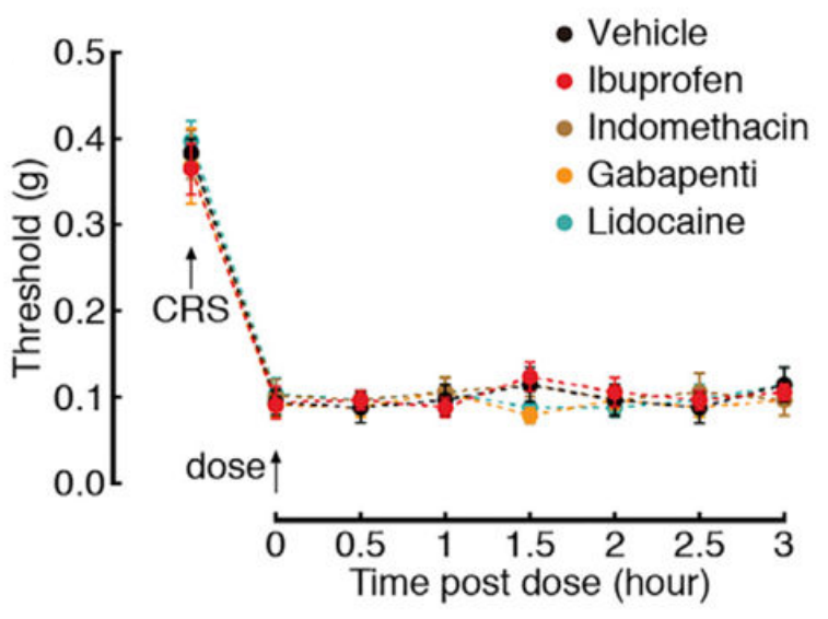
Ibuprofen purchased from MedChemExpress. Usage Cited in: Cell Rep. 2019 Dec 17;29(12):3847-3858.e5. [Abstract]
Effects of Ibuprofen (10 mg/kg, i.p.), Indomethacin, Gabapentin, or Lidocaine on pain thresholds of CRS 3W mice.
-
Biochem Pharmacol
The 'ABC' of split-nanoluciferase HIF heterodimerization bioassays: Applications, Benefits & Considerations. [Abstract]2024 Nov:229:116478. PMID: 39128589 -
Inflammopharmacology
Ibuprofen inhibits anaplastic thyroid cells in vivo and in vitro by triggering NLRP3-ASC-GSDMD-dependent pyroptosis. [Abstract]2024 Feb;32(1):733-745. PMID: 37999895
Ibuprofen purchased from MedChemExpress. Usage Cited in: Inflammopharmacology. 2024 Feb;32(1):733-745. [Abstract]
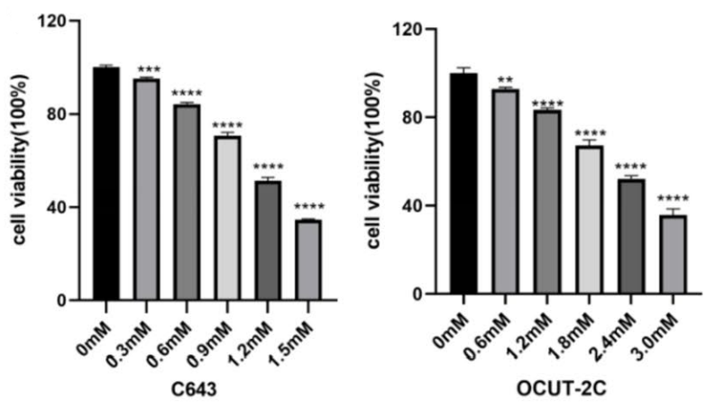
The inhibitory effect of Ibuprofen (0-3 mM, 48 h) on the proliferation of C643 and OCUT-2C cells was detected by CCK8 assay.
Ibuprofen purchased from MedChemExpress. Usage Cited in: Inflammopharmacology. 2024 Feb;32(1):733-745. [Abstract]
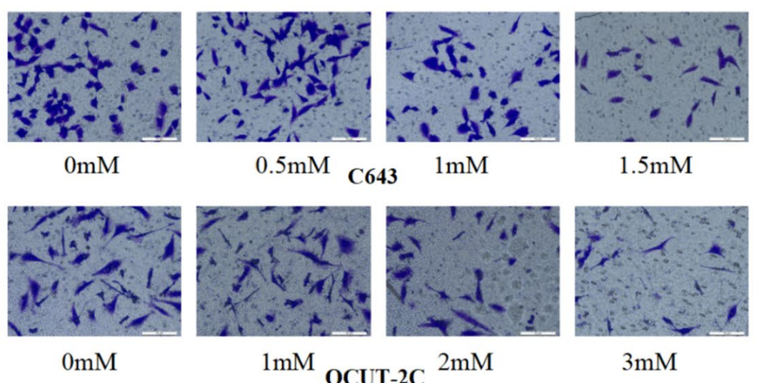
Transwell assay, cells were treated with various concentrations of Ibuprofen (C643 at 0, 0.5, 1, and 1.5 mM; OCUT-2C at 0, 1, 2, and 3 mM) for 48 h view cell invasion abilities.
Ibuprofen purchased from MedChemExpress. Usage Cited in: Inflammopharmacology. 2024 Feb;32(1):733-745. [Abstract]
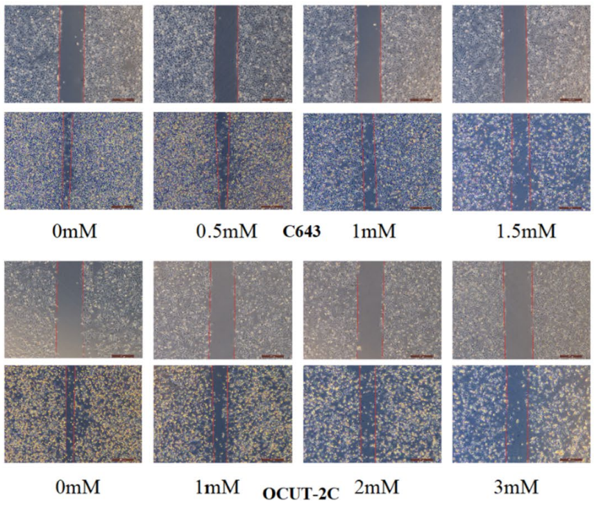
Wound healing assay, cells were treated with different concentrations of Ibuprofen (C643 at 0, 0.5, 1, and 1.5 mM; OCUT-2C at 0, 1, 2, and 3 mM) for 48 h view cell migration abilities.
Ibuprofen purchased from MedChemExpress. Usage Cited in: Inflammopharmacology. 2024 Feb;32(1):733-745. [Abstract]
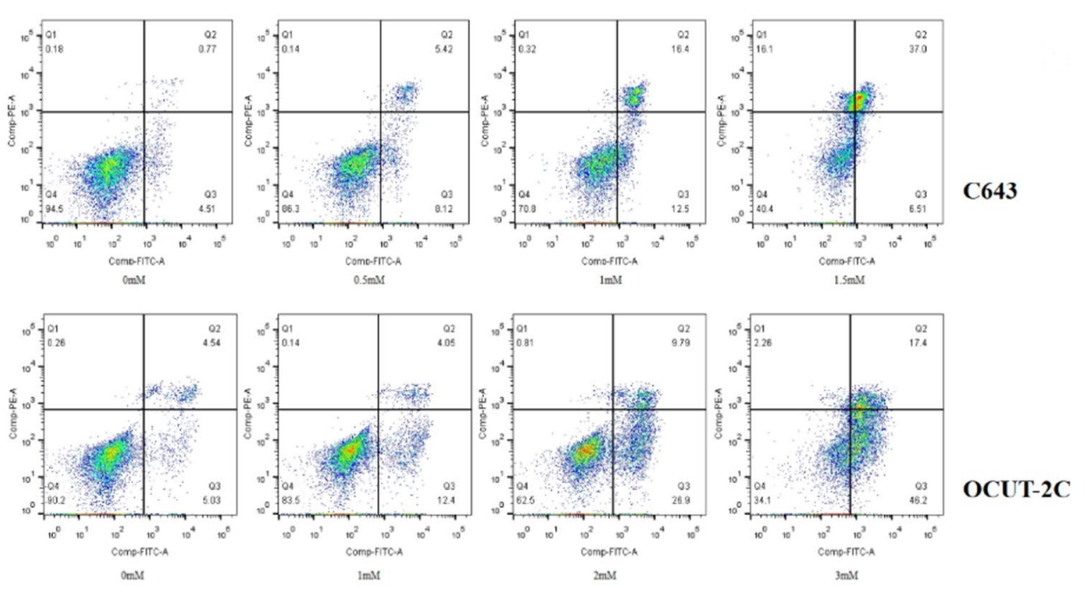
Flow cytometry analysis of ATC cells treated with different Ibuprofen (0-3 mM) concentrations for 48 h stained with Annexin V-FITC and PI.
Ibuprofen purchased from MedChemExpress. Usage Cited in: Inflammopharmacology. 2024 Feb;32(1):733-745. [Abstract]
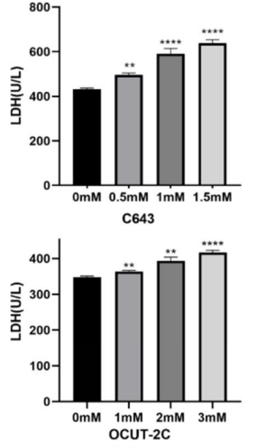
The effect of Ibuprofen (0-1.5 mM) on LDH activity in C643 and OCUT-2C cells.
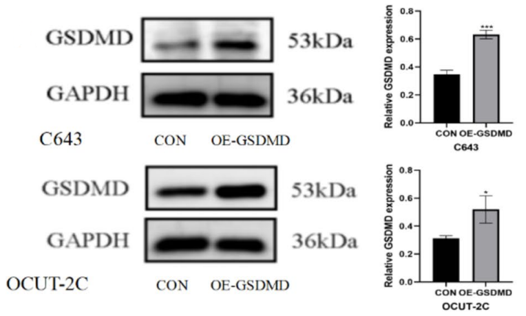
Ibuprofen purchased from MedChemExpress. Usage Cited in: Inflammopharmacology. 2024 Feb;32(1):733-745. [Abstract]
Ibuprofen-induced expression of GSDMD protein in C643 and OCUT-2C cells.
-
Cells
Prostaglandin 2α Promotes Autophagy and Mitochondrial Energy Production in Fish Hepatocytes. [Abstract]2022 Jun 9;11(12):1870. PMID: 35740999 -
EMBO Rep
2022 Jun 7;23(6):e53932. PMID: 35403787 -
Cell Rep Methods
RECOVER identifies synergistic drug combinations in vitro through sequential model optimization. [Abstract]2023 Oct 23;3(10):100599. PMID: 37797618 -
Int Immunopharmacol
Oxyphenbutazone suppresses non-canonical inflammasome activation and protects against LPS-induced sepsis. [Abstract]2026 Aug 15:183:116873. PMID: 42150290 -
Int J Mol Sci
Singapore Grouper Iridovirus Disturbed Glycerophospholipids Homeostasis: Cytosolic Phospholipase A2 Was Essential for Virus Replication. [Abstract]2021 Nov 22;22(22):12597. PMID: 34830477 -
-
-
Sci Rep
Characterization of five novel vasopressin V2 receptor mutants causing nephrogenic diabetes insipidus reveals a role of tolvaptan for M272R-V2R mutation. [Abstract]2020 Oct 2;10(1):16383. PMID: 33009446 -
Neuropharmacology
Malfunction of astrocyte and cholinergic input is involved in postoperative impairment of hippocampal synaptic plasticity and cognitive function. [Abstract]2022 Oct 1:217:109191. PMID: 35835213 -
ACS Chem Neurosci
Identification of a Nonelectrophilic and Selective TRPA1 Agonist for Alleviation of Inflammatory Pain through Channel Desensitization. [Abstract]2025 Sep 3;16(17):3257-3266. PMID: 40846466 -
J Neurosci
Acute Ongoing Nociception Delays Recovery of Consciousness from Sevoflurane Anesthesia via a Midbrain Circuit. [Abstract]2024 Aug 21;44(34):e0740242024. PMID: 39019613 -
Reprod Toxicol
2025 May:134:108895. PMID: 40097051 -
J Appl Toxicol
Differential impacts of nonsteroidal anti-inflammatory drugs on lifespan and healthspan in aged Caenorhabditis elegans. [Abstract]2024 Oct;44(10):1528-1539. PMID: 38840409 -
Chemosphere
Mass-balance-model-based evaluation of sewage treatment plant contribution to residual pharmaceuticals in environmental waters. [Abstract]2019 Jun:225:378-387. PMID: 30884299


Solvent & Solubility
In Vitro:
DMSO : 100 mg/mL (484.78 mM; Need ultrasonic; Hygroscopic DMSO has a significant impact on the solubility of product, please use newly opened DMSO)
H2O : < 0.1 mg/mL (insoluble)
Please refer to the solubility information to select the appropriate solvent. Once prepared, please aliquot and store the solution to prevent product inactivation from repeated freeze-thaw cycles.
Storage method and period of stock solution: -80°C, 2 years; -20°C, 1 year. When stored at -80°C, please use it within 2 years. When stored at -20°C, please use it within 1 year.
Please refer to the solubility information to select the appropriate solvent. Once prepared, please aliquot and store the solution to prevent product inactivation from repeated freeze-thaw cycles.
Storage method and period of stock solution: -80°C, 2 years; -20°C, 1 year. When stored at -80°C, please use it within 2 years. When stored at -20°C, please use it within 1 year.
Concentration (start) × Volume (start) = Concentration (final) × Volume (final)
In Vivo:
Select the appropriate dissolution method based on your experimental animal and administration route.
- For the following dissolution methods, please ensure to first prepare a clear stock solution using an In Vitro approach and then sequentially add co-solvents:
- To ensure reliable experimental results, the clarified stock solution can be appropriately stored based on storage conditions. As for the working solution for In Vivo experiments, it is recommended to prepare freshly and use it on the same day.
- The percentages shown for the solvents indicate their volumetric ratio in the final prepared solution. If precipitation or phase separation occurs during preparation, heat and/or sonication can be used to aid dissolution.
Add each solvent one by one: 10% DMSO 40% PEG300 5% Tween-80 45% Saline
Solubility: ≥ 2.5 mg/mL (12.12 mM); Clear solution
This protocol yields a clear solution of ≥ 2.5 mg/mL (saturation unknown).
Taking 1 mL working solution as an example, add 100 μL DMSO stock solution (25.0 mg/mL) to 400 μL PEG300, and mix evenly; then add 50 μL Tween-80 and mix evenly; then add 450 μL Saline to adjust the volume to 1 mL.
Preparation of Saline: Dissolve 0.9 g sodium chloride in ddH₂O and dilute to 100 mL to obtain a clear Saline solution.
Add each solvent one by one: 10% DMSO 90% (20% SBE-β-CD in Saline)
Solubility: ≥ 2.5 mg/mL (12.12 mM); Clear solution
This protocol yields a clear solution of ≥ 2.5 mg/mL (saturation unknown).
Taking 1 mL working solution as an example, add 100 μL DMSO stock solution (25.0 mg/mL) to 900 μL 20% SBE-β-CD in Saline, and mix evenly.
Preparation of 20% SBE-β-CD in Saline (4°C, storage for one week): 2 g SBE-β-CD powder is dissolved in 10 mL Saline, completely dissolve until clear.
In Vivo Dissolution Calculator
Please enter the basic information of animal experiments:
-
-
-
-
Recommended: Prepare an additional quantity of animals to account for potential losses during experiments.
Please enter your animal formula composition:
-
%DMSO +
Recommended: Keep the proportion of DMSO in working solution below 2% if your animal is weak.
-
%+
-
+%Tween-80 + +
-
%Saline +
The co-solvents required include: DMSO, . All of co-solvents are available by MedChemExpress (MCE). , Tween 80. All of co-solvents are available by MedChemExpress (MCE).
Working solution concentration: 0.22 mg/mL
Method for preparing stock solution: mg drug dissolved in μL DMSO. Stock solution concentration: mg/mL.
1. Take μL DMSO stock solution;
2. Add μL .
μL , mix evenly;
3. Then add μL Tween 80, mix evenly;
4. Then add μL
Please ensure that the stock solution in the first step is dissolved to a clear state, and add co-solvents in sequence. You can use ultrasonic heating (ultrasonic cleaner, recommended frequency 20-40 kHz), vortexing, etc. to assist dissolution.

Protocols
-
Human pluripotent stem cell endothelial-cell differentiation
Human pluripotent stem cell endothelial differentiation is based on stepwise developmental patterning: early activation of WNT/GSK3β inhibition promotes mesodermal or vascular progenitor entry, followed by endothelial specification using VEGF-related signaling, BMP4, FGF2, Notch modulation, or cAMP depending on the published protocol. Endothelial differentiation is read out by acquisition of CD31, CD34, VE-cadherin/CD144, KDR/VEGFR2, vWF, Tie2, NOS3, acetylated LDL uptake, tube/network formation, barrier function, and in vivo vessel-forming capacity where tested.
-
Somatic Cell Culture
A method of simulating the in vivo environment in vitro to maintain the cell growth, differentation and main functions.
-
Apoptosis
Apoptosis, also called programmed cell death, is generally characterized by distinct morphological characteristics.
-
TUNEL staining for apoptotic DNA fragmentation
TUNEL staining detects DNA strand breaks by using terminal deoxynucleotidyl transferase to add labeled nucleotides to exposed 3′-OH DNA termini, generating either microscopic staining in fixed cells or tissue sections, or fluorescence/cytometric signal in cell suspensions. TUNEL positivity reflects DNA fragmentation but should not be interpreted alone as definitive apoptosis, because TUNEL can also label necrotic, autolytic, mechanically damaged, or DNA-repair-associated DNA breaks.
-
Cell Cytotoxicity Assay
Cytotoxicity assays are usually based on the assessment of cell membrane damage, which can also be indirectly detected by measuring cell viability. Detection methods include MTT assay, CKK-8 assay, LDH assay and ATP assay, etc.
-
Endothelial Tube Formation Assay
Endothelial tube formation assay evaluates the ability of endothelial cells to attach, migrate, align, and organize into capillary-like networks when cultured on gelled basement membrane extract or Matrigel; the readout is the morphology and quantity of tube-like networks, which reflects an in vitro endothelial morphogenesis step related to angiogenesis. Basement membrane extract/Matrigel provides laminin-rich extracellular matrix cues that support endothelial differentiation into capillary-like structures, but it can contain biologically active growth factors, so growth-factor-reduced matrix is preferred when testing defined angiogenic stimulators or inhibitors.
-
Apoptosis Solutions
Apoptosis is a regulated, generally non-lytic cell-death pathway that removes unwanted, damaged, infected, or abnormal cells through coordinated morphological changes, caspase activation, DNA fragmentation, and membrane remodeling. The intrinsic apoptosis pathway is controlled mainly by mitochondrial outer membrane permeabilization, BCL-2 family proteins, cytochrome c release, apoptosome formation, caspase-9 activation, and downstream executioner caspase-3/7 activation. The extrinsic apoptosis pathway is initiated by death receptors such as Fas, TNFR, and TRAIL receptors, which recruit adaptor proteins and activate caspase-8 before engaging executioner caspases or mitochondrial amplification through BID cleavage. Apoptosis is linked to many phenotypes, including cancer cell killing, tissue homeostasis, immune regulation, neurodegeneration, infection response, and treatment-induced cytotoxicity; unresolved questions include how apoptosis interacts with necroptosis, pyroptosis, ferroptos
-
MTT Cell Proliferation Assay
The MTT assay is a colorimetric endpoint assay for estimating viable cell number, cell growth, cytotoxicity, or cell activation in cultured mammalian cells. Living cells reduce the yellow tetrazolium salt MTT into purple/blue formazan, while dead cells do not generate the same signal; the resulting color can be quantified with a multiwell spectrophotometer. MTT reduction is commonly interpreted as a readout of metabolic activity that often correlates with viable cell number, but it should not be treated as a direct cell-counting method unless the assay is optimized for the cell type and experimental condition. Studies show that MTT reduction can involve mitochondrial and non-mitochondrial reducing systems, and formazan may accumulate in intracellular lipid droplets rather than simply marking mitochondria.
-
Endothelial Cell Migration/Angiogenic Sprouting Assay
Endothelial cell migration and angiogenic sprouting assays are in vitro (and partially ex vivo-adapted) functional models that quantify the ability of endothelial cells to undergo coordinated migration, extracellular matrix invasion, and multicellular organization into capillary-like sprouts in response to pro-angiogenic stimuli such as VEGF, bFGF, or conditioned microenvironments. These assays are used to model early angiogenic events including tip-cell formation, directional migration, and lumen-like sprout extension, which collectively reflect angiogenic activation and vascular morphogenesis processes observed in vivo.
-
Research Protocol for Infectious Diseases
Infectious-disease experiments test how pathogens interact with host barriers, innate immune receptors, inflammatory signaling, pathogen replication, and tissue injury; pattern-recognition receptors such as TLRs, RIG-I-like receptors, NOD-like receptors, and inflammasomes detect microbial molecules and activate NF-κB, interferon, and cytokine responses. The central hypothesis is that infection severity reflects the balance between pathogen burden and host response: protective inflammation restricts pathogen growth, whereas excessive or mislocalized inflammation contributes to tissue damage and disease phenotype. Unresolved questions include which host pathways are protective versus pathogenic, why some infection models fail to translate to human disease, and which combined readouts best predict clinically relevant infection outcomes.
-
Vascular/Branching Fractal Analysis
Vascular/branching fractal analysis quantifies the geometric complexity of vessel trees or vascular networks from segmented 2D images, commonly by converting vessels into binary and/or skeletonized maps and estimating fractal dimension using box-counting or related approaches. Fractal dimension is interpreted as an image-derived readout of vascular branching complexity, space filling, or density, and has been applied to retinal photographs, fluorescein angiography, OCT angiography, capillary perfusion maps, and in vitro Matrigel angiogenesis networks. The assay readout is generated from vessel-positive pixels after image preprocessing, vessel segmentation, binarization, and optional skeletonization; reported outputs include fractal dimension, vessel density, branchpoint density, endpoint density, vessel length density, tortuosity, and generation-based branching metrics when VESGEN-style analysis is used. The biological interpretation is limited to quantitative vascular patterning and s
-
Annexin V plus membrane-impermeant dye apoptosis staining
Annexin V-based apoptosis assays rely on the detection of phosphatidylserine (PS) externalization from the inner leaflet of the plasma membrane to the outer leaflet, an early biochemical hallmark of apoptosis. Fluorescently labeled Annexin V binds PS in a calcium-dependent manner, enabling identification of early apoptotic cells by flow cytometry or fluorescence microscopy. When combined with a membrane-impermeant DNA-binding dye (e. g. , propidium iodide), this approach allows discrimination between viable (Annexin V−/dye−), early apoptotic (Annexin V+/dye−), and late apoptotic or necrotic (Annexin V+/dye+) cell populations by assessing membrane integrity and PS exposure.
-
BrdU Incorporation Assay
Bromodeoxyuridine (BrdU) incorporation assay is based on the principle that BrdU, a thymidine analog, is incorporated into newly synthesized DNA during the S phase of the cell cycle, thereby serving as a marker of DNA replication and cellular proliferation. Incorporated BrdU can be detected using anti-BrdU antibodies following DNA denaturation, enabling visualization or quantification of proliferating cells through immunochemical detection methods such as immunofluorescence or immunohistochemistry.
-
CFSE Dye Dilution Proliferation Assay
The CFSE (carboxyfluorescein diacetate succinimidyl ester) dye dilution proliferation assay is based on the covalent labeling of intracellular proteins by a cell-permeant fluorescent dye that becomes fluorescent upon intracellular ester cleavage and then is stably retained within cells. As labeled cells divide, the dye is partitioned equally between daughter cells, resulting in a stepwise halving of fluorescence intensity that can be quantified by flow cytometry to determine the number of cell divisions undergone by each cell population. This fluorescence dilution approach enables quantitative tracking of lymphocyte proliferation at the single-cell level over multiple rounds of division. CFSE-based proliferation analysis has been widely applied to measure antigen-driven lymphocyte expansion in vitro, where discrete fluorescence peaks correspond to successive cell divisions and allow reconstruction of proliferative history within heterogeneous populations.
-
Dye-dilution cell tracking and proliferation staining
Dye-dilution cell tracking assays quantify cell proliferation by covalently labeling intracellular proteins with a stable fluorescent dye that is equally partitioned between daughter cells during mitosis, resulting in stepwise halving of fluorescence intensity with each cell division as measured by flow cytometry histograms. Carboxyfluorescein diacetate succinimidyl ester (CFSE) is a prototypical dye that diffuses into cells, is enzymatically converted into a fluorescent compound, and then covalently binds intracellular amine groups, producing long-lived fluorescence suitable for tracking multiple rounds of division in vitro and in vivo. Successive generations of dividing cells form discrete peaks of decreasing fluorescence intensity, enabling estimation of proliferation history, precursor frequency, and division index within heterogeneous populations. Alternative dyes such as CellTrace Violet (CTV) and far-red membrane dyes (e. g. , PKH26) follow the same dilution principle but differ
-
CCK-8/WST-8 Cell Proliferation Assay
The CCK-8/WST-8 assay is based on the reduction of the water-soluble tetrazolium salt WST-8 to a water-soluble formazan product by cellular dehydrogenases in metabolically active cells, where the generated formazan amount is proportional to the number of living cells and is quantified by measuring absorbance in the visible range, providing a colorimetric readout for cell viability and proliferation assessment. This class of tetrazolium-based assays improves upon earlier MTT-based systems by producing a water-soluble formazan, eliminating the need for organic solubilization steps and enabling direct spectrophotometric measurement in culture medium.
-
Protocol for Cell Cycle
Cell-cycle analysis by flow cytometry measures DNA content in single cells to estimate the fraction of cells in G0/G1, S, and G2/M phases. Propidium iodide intercalates into DNA, and after RNA removal with RNase, fluorescence intensity reflects cellular DNA content: 2N cells are assigned to G0/G1, cells between 2N and 4N to S phase, and 4N cells to G2/M. DNA-content analysis alone cannot reliably separate G0 from G1 or G2 from M. Ki-67 can distinguish quiescent G0 cells from cycling cells, EdU or BrdU incorporation marks active DNA synthesis in S phase, and phospho-histone H3 staining identifies mitotic cells within the 4N population.
-
Cell Counting-Based Growth Curve Assay
Cell counting-based growth curve assays quantify cell proliferation by directly measuring changes in viable cell number over time using manual or automated counting methods such as hemocytometer-based counting or instrument-assisted cell enumeration, enabling construction of growth curves that reflect population expansion dynamics in response to culture conditions. A widely used approach is trypan blue exclusion with hemocytometer counting, where membrane-compromised (non-viable) cells take up the dye, allowing discrimination between viable and non-viable cells while simultaneously enabling total cell number quantification. Repeated sampling across time points allows estimation of proliferation rate, growth phases, and comparative growth kinetics between experimental conditions.
-
Research Protocol for Inflammation-related Diseases
The NLRP3 inflammasome is a cytosolic innate immune signaling platform that integrates priming signals and danger-signal activation to promote caspase-1 activation, maturation of IL-1β and IL-18, and gasdermin D-mediated pyroptotic cell death. The core experimental logic is to determine whether inflammatory disease phenotypes are driven by increased NLRP3 expression, ASC-containing inflammasome assembly, caspase-1 cleavage, GSDMD cleavage, and extracellular release of IL-1β/IL-18 rather than by nonspecific cell injury alone. The pathway is strongly linked to inflammation-related disease phenotypes because monosodium urate crystals activate NALP3/NLRP3 inflammasome signaling in gout-like crystal inflammation, cholesterol crystals activate NLRP3 inflammasomes in atherogenesis models, and DSS-induced intestinal inflammation has been reported to involve NLRP3 inflammasome activity. However, experimental colitis studies also show context-dependent protective effects of NLRP3 inflammasome co
-
Colony Formation (Clonogenic) Assay
The clonogenic (colony formation) assay measures the ability of a single cell to retain reproductive viability and form a macroscopic colony, typically defined as a cluster derived from one progenitor cell after a defined growth period. This assay is widely used to evaluate cell survival after exposure to ionizing radiation or cytotoxic treatments and is considered a standard method in radiation biology for generating dose-response relationships of reproductive cell death. Colony formation reflects long-term proliferative capacity rather than short-term metabolic activity, and survival is quantified by comparing treated versus untreated conditions based on colony number and derived survival fractions.
-
Cell Viability Determination by MTT Colorimetric Assay
The following protocol uses the MTT colorimetric assay as a classic literature-established method for assessing cell viability/metabolic activity in cultured mammalian cells. MTT[3-(4,5-dimethylthiazol-2-yl)-2,5-diphenyltetrazolium bromide] is reduced by metabolically active cells to a colored formazan product; the amount of formazan is quantified spectrophotometrically and provides an indirect measure of metabolically active viable cells. Importantly, MTT reduction reflects cellular oxidoreductase/metabolic activity rather than an absolute direct count of living cells, so changes in cellular metabolism can alter the signal independently of cell number.
-
EdU Incorporation Assay (Click Chemistry-Based DNA Synthesis Measurement)
The EdU incorporation assay measures DNA synthesis by adding the thymidine analog 5-ethynyl-2′-deoxyuridine to cells or tissues, where it is incorporated into newly synthesized DNA during S phase. Incorporated EdU is detected by copper-catalyzed azide-alkyne cycloaddition, in which a fluorescent azide covalently reacts with the ethynyl group on EdU, allowing S-phase cells to be detected by fluorescence microscopy, flow cytometry, or high-content imaging. EdU detection does not require DNA denaturation or anti-BrdU antibody access, which preserves sample structure and improves compatibility with immunostaining and multiparameter cytometry compared with BrdU-based detection. EdU can be cytotoxic in a cell-type- and exposure-dependent manner, so pulse duration, concentration, and continuous-labeling designs should be validated for each cell type.
-
Ki-67 Immunostaining Proliferation Assay
Ki-67 immunostaining measures the growth fraction of a cell population by detecting Ki-67, a nuclear antigen present in proliferating cells and absent in quiescent G0 cells. The readout is the percentage of Ki-67-positive nuclei among total counted cells, commonly called the Ki-67 labeling index or proliferation index.
-
PCNA Immunodetection Proliferation Assay
PCNA immunodetection measures proliferative activity by detecting proliferating cell nuclear antigen, a nuclear protein associated with DNA polymerase δ function and DNA replication. The assay readout is the proportion of PCNA-positive nuclei among total counted cells, but PCNA labeling is not identical to BrdU labeling because PCNA can mark late G1/early S-associated replication competence and may persist beyond active DNA synthesis depending on fixation and extraction conditions.
-
Protocol for Cell Counting and Cell Density Analysis
Cell counting and cell-density analysis estimate the number of cells in a known volume or field area. Manual hemocytometer counting uses a chamber of defined geometry to convert counted cells into cells/mL, while automated counters and image-analysis workflows detect cell objects from optical, brightfield, fluorescence, impedance, or digital-image features. Trypan blue viability counting is based on dye exclusion: viable cells with intact membranes exclude dye, while non-viable cells with compromised membranes stain blue. The readout is total cell density, viable-cell density, dead-cell density, and percent viability. Cell density can also be estimated from microscopy images by counting objects per image area, from flow cytometry using calibrated volume or reference particles, or from in situ microscopy in bioreactors after calibration against reference methods such as hemocytometer or flow cytometry.


Purity & Documentation
-
Data Sheet (280 KB)
-
SDS (394 KB)
- English - EN (394 KB)
- Français - FR (394 KB)
- Deutsch - DE (394 KB)
- Norwegian - NO (394 KB)
- Español - ES (394 KB)
- Swedish - SV (394 KB)
- Italian - IT (394 KB)
- Korean - KR (394 KB)
- Portuguese - PT (394 KB)
-
Handling Instructions (2659 KB)
References
[1]. Noreen Y, et al. Development of a radiochemical cyclooxygenase-1 and -2 in vitro assay for identification of natural products as inhibitors of prostaglandin biosynthesis. J Nat Prod. 1998 Jan;61(1):2-7. [Content Brief]
[2]. Hassan Akrami, et al. Inhibitory effect of ibuprofen on tumor survival and angiogenesis in gastric cancer cell. Tumour Biol. 2015 May;36(5):3237-43. [Content Brief]
[3]. Sharon M Rymut, et al. Ibuprofen regulation of microtubule dynamics in cystic fibrosis epithelial cells. Am J Physiol Lung Cell Mol Physiol. 2016 Aug 1;311(2):L317-27. [Content Brief]
[4]. Emmanuelle Bignon, et al. Ibuprofen and ketoprofen potentiate UVA-induced cell death by a photosensitization process. Sci Rep. 2017 Aug 21;7(1):8885. [Content Brief]
[5]. Nathan D Pennock, et al. Ibuprofen supports macrophage differentiation, T cell recruitment, and tumor suppression in a model of postpartum breast cancer. J Immunother Cancer. 2018 Oct 1;6(1):98. [Content Brief]
[6]. Thomas Krøigård, et al. Protective effect of ibuprofen in a rat model of chronic oxaliplatin-induced peripheral neuropathy. Exp Brain Res. 2019 Oct;237(10):2645-2651. [Content Brief]
[7]. Sarah Ilkhanipour Rooney, et al. Ibuprofen Differentially Affects Supraspinatus Muscle and Tendon Adaptations to Exercise in a Rat Model. Am J Sports Med. 2016 Sep;44(9):2237-45. [Content Brief]
[8]. M W Konstan, et al. Ibuprofen attenuates the inflammatory response to Pseudomonas aeruginosa in a rat model of chronic pulmonary infection. Implications for antiinflammatory therapy in cystic fibrosis. Am Rev Respir Dis. 1990 Jan;141(1):186-92. [Content Brief]


Complete Stock Solution Preparation Table
Please refer to the solubility information to select the appropriate solvent. Once prepared, please aliquot and store the solution to prevent product inactivation from repeated freeze-thaw cycles.
Storage method and period of stock solution: -80°C, 2 years; -20°C, 1 year. When stored at -80°C, please use it within 2 years. When stored at -20°C, please use it within 1 year.
| Optional Solvent | Concentration Solvent Mass | 1 mg | 5 mg | 10 mg | 25 mg |
|---|---|---|---|---|---|
| DMSO | 1 mM | 4.8478 mL | 24.2389 mL | 48.4778 mL | 121.1945 mL |
| 5 mM | 0.9696 mL | 4.8478 mL | 9.6956 mL | 24.2389 mL | |
| 10 mM | 0.4848 mL | 2.4239 mL | 4.8478 mL | 12.1194 mL | |
| 15 mM | 0.3232 mL | 1.6159 mL | 3.2319 mL | 8.0796 mL | |
| 20 mM | 0.2424 mL | 1.2119 mL | 2.4239 mL | 6.0597 mL | |
| 25 mM | 0.1939 mL | 0.9696 mL | 1.9391 mL | 4.8478 mL | |
| 30 mM | 0.1616 mL | 0.8080 mL | 1.6159 mL | 4.0398 mL | |
| 40 mM | 0.1212 mL | 0.6060 mL | 1.2119 mL | 3.0299 mL | |
| 50 mM | 0.0970 mL | 0.4848 mL | 0.9696 mL | 2.4239 mL | |
| 60 mM | 0.0808 mL | 0.4040 mL | 0.8080 mL | 2.0199 mL | |
| 80 mM | 0.0606 mL | 0.3030 mL | 0.6060 mL | 1.5149 mL | |
| 100 mM | 0.0485 mL | 0.2424 mL | 0.4848 mL | 1.2119 mL |


Ibuprofen Related Classifications
HY-78131 Related Classifications
 Powered by Bioz