



SB 220025

Based on 1 Customer Validation
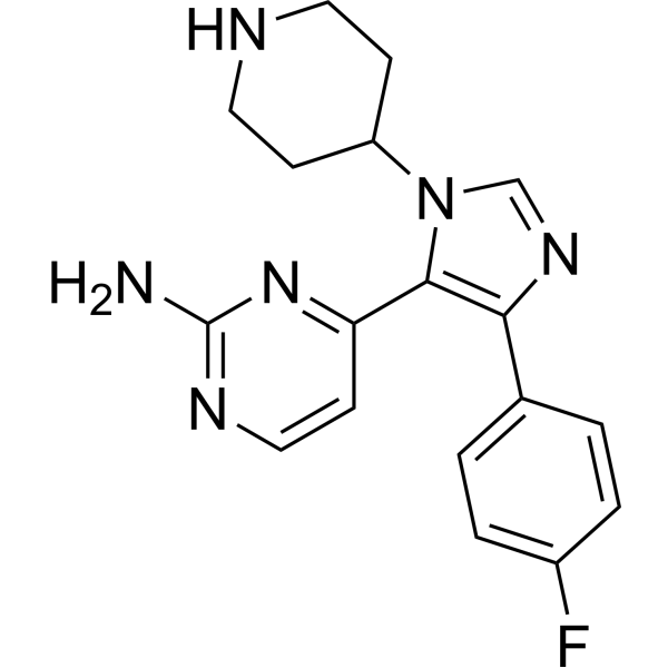
SB 220025 is a reversible, orally active, cell-permeable, ATP-competitive and selective human p38 MAPK inhibitor (IC50 = 60 nM). SB 220025 also inhibits p56Lck and PKC with IC50 values of 3.5 and 2.89 µM, respectively. SB 220025 inhibits the expression of IL-8 gene in response to globular adiponectin (gAd), reduces inflammatory cytokine production and inhibits angiogenesis. SB 220025 effectively prevents the progression of arthritis in a chronic inflammatory disease model and can be used in the study of inflammation.
For research use only. We do not sell to patients.
- Purity : 99.90%
- CAS No.: 165806-53-1
- Formula: C18H19FN6
- Molecular Weight:338.38
-
Storage:Powder -20°C, 3 years , 4°C, 2 years ; In solvent -80°C, 6 months , -20°C, 1 month

To place orders, for customer services and technical support, please contact: MedChemExpress USA
Tel: 609-228-6898 E-mail: [email protected] [email protected]


-
 Biological Activity
-
Chemical Information
-
Solvent & Solubility
- Protocols
- Purity & Documentation
- References
-
 Help & FAQs




Biological Activity
Description
IC50 & Target
[1]|
p38 60 nM (IC50) |
p56-Lck 3.5 μM (IC50) |
PKC 2.89 μM (IC50) |
In Vitro
SB 220025 (20 μM; 6 h) markedly reduces IL-8 gene expression in response to globular adiponectin (gAd) in HUVEC cells[1].
MedChemExpress (MCE) has not independently confirmed the accuracy of these methods. They are for reference only.
-
Cell Line:HUVEC cells
-
Concentration:20 μM
-
Incubation Time:6 h
-
Result:Inhibited MCP-1 gene expression.
In Vivo
SB 220025 (5, 30, 50 mg/kg; i.p.; b.i.d.) inhibits angiogenesis in the murine air pouch granuloma model[2].
SB 220025 (30 mg/kg; p.o.; twice a day for 3, 5, 7 or 14 days) prevents the increase in angiogenesis that occurs after day 3 in murine air pouch angiogenesis model[2].
SB 220025 (50 mg/kg; p.o.; b.i.d.; 10 days) effectively blocks the progression of arthritis in a chronic inflammatory disease model[2].
MedChemExpress (MCE) has not independently confirmed the accuracy of these methods. They are for reference only.
-
Animal Model:Acute model of LPS-induced TNF-a expression[2].
-
Dosage:3-50 mg/kg
-
Administration:Oral administration; single; 30 min before challenge with LPS.
-
Result:Dosedependently inhibited TNF-a production with an ED50 value of 7.5 mg/kg, and showed more than 80% inhibition when at 50 mg/kg.
-
Animal Model:Murine air pouch granuloma model[2].
-
Dosage:5, 30, 50 mg/kg
-
Administration:Intraperitoneal injection; bisindie (bid, twice a day).
-
Result:Caused a dose-dependent reduction in angiogenesis.
-
Animal Model:Murine air pouch granuloma model[2].
-
Dosage:30 mg/kg
-
Administration:Oral administration; twice a day from day 0 until removal of granuloma tissue at days 3, 5, 7 or 14.
-
Result:Did not affect the initial burst of angiogenesis but did prevent the increase in angiogenesis that occurs after day 3.
- RNA extraction experimental
- Collagen-Induced Arthritis
- Human pluripotent stem cell endothelial-cell differentiation
- Vascular/Branching Fractal Analysis
- Endothelial Tube Formation Assay
- Research Protocol for Inflammation-related Diseases
- Endothelial Cell Migration/Angiogenic Sprouting Assay
- How to Choose the Right Model Animal


Chemical Information
-
CAS No. 165806-53-1
-
Appearance Solid
-
Molecular Weight 338.38
-
Formula C18H19FN6
-
Color White to off-white
-
SMILES
NC1=NC=CC(C2=C(C3=CC=C(F)C=C3)N=CN2C4CCNCC4)=N1
-
Shipping
Room temperature in continental US; may vary elsewhere.
-
Storage
Powder -20°C 3 years 4°C 2 years In solvent -80°C 6 months -20°C 1 month


Solvent & Solubility
In Vitro:
DMSO : 50 mg/mL (147.76 mM; ultrasonic and warming and heat to 60°C; Hygroscopic DMSO has a significant impact on the solubility of product, please use newly opened DMSO)
Please refer to the solubility information to select the appropriate solvent. Once prepared, please aliquot and store the solution to prevent product inactivation from repeated freeze-thaw cycles.
Storage method and period of stock solution: -80°C, 6 months; -20°C, 1 month. When stored at -80°C, please use it within 6 months. When stored at -20°C, please use it within 1 month.
Please refer to the solubility information to select the appropriate solvent. Once prepared, please aliquot and store the solution to prevent product inactivation from repeated freeze-thaw cycles.
Storage method and period of stock solution: -80°C, 6 months; -20°C, 1 month. When stored at -80°C, please use it within 6 months. When stored at -20°C, please use it within 1 month.
Concentration (start) × Volume (start) = Concentration (final) × Volume (final)


Purity & Documentation
-
Data Sheet (282 KB)
-
SDS (480 KB)
- English - EN (480 KB)
- Français - FR (480 KB)
- Deutsch - DE (480 KB)
- Norwegian - NO (480 KB)
- Español - ES (480 KB)
- Swedish - SV (480 KB)
- Italian - IT (480 KB)
- Korean - KR (480 KB)
- Portuguese - PT (480 KB)
-
Handling Instructions (2659 KB)
References
[1]. Tomizawa A, et al. Induction of gene expression in response to globular adiponectin in vascular endothelial cells. Life Sci. 2009 Sep 9;85(11-12):457-61. [Content Brief]
[2]. Jackson JR, et al. Pharmacological effects of SB 220025, a selective inhibitor of P38 mitogen-activated protein kinase, in angiogenesis and chronic inflammatory disease models. J Pharmacol Exp Ther. 1998 Feb;284(2):687-92. [Content Brief]


Complete Stock Solution Preparation Table
Please refer to the solubility information to select the appropriate solvent. Once prepared, please aliquot and store the solution to prevent product inactivation from repeated freeze-thaw cycles.
Storage method and period of stock solution: -80°C, 6 months; -20°C, 1 month. When stored at -80°C, please use it within 6 months. When stored at -20°C, please use it within 1 month.
| Optional Solvent | Concentration Solvent Mass | 1 mg | 5 mg | 10 mg | 25 mg |
|---|---|---|---|---|---|
| DMSO | 1 mM | 2.9553 mL | 14.7763 mL | 29.5526 mL | 73.8814 mL |
| 5 mM | 0.5911 mL | 2.9553 mL | 5.9105 mL | 14.7763 mL | |
| 10 mM | 0.2955 mL | 1.4776 mL | 2.9553 mL | 7.3881 mL | |
| 15 mM | 0.1970 mL | 0.9851 mL | 1.9702 mL | 4.9254 mL | |
| 20 mM | 0.1478 mL | 0.7388 mL | 1.4776 mL | 3.6941 mL | |
| 25 mM | 0.1182 mL | 0.5911 mL | 1.1821 mL | 2.9553 mL | |
| 30 mM | 0.0985 mL | 0.4925 mL | 0.9851 mL | 2.4627 mL | |
| 40 mM | 0.0739 mL | 0.3694 mL | 0.7388 mL | 1.8470 mL | |
| 50 mM | 0.0591 mL | 0.2955 mL | 0.5911 mL | 1.4776 mL | |
| 60 mM | 0.0493 mL | 0.2463 mL | 0.4925 mL | 1.2314 mL | |
| 80 mM | 0.0369 mL | 0.1847 mL | 0.3694 mL | 0.9235 mL | |
| 100 mM | 0.0296 mL | 0.1478 mL | 0.2955 mL | 0.7388 mL |


SB 220025 Related Classifications
HY-112291 Related Classifications
 Powered by Bioz