



17-HDHA

Based on 1 Customer Validation
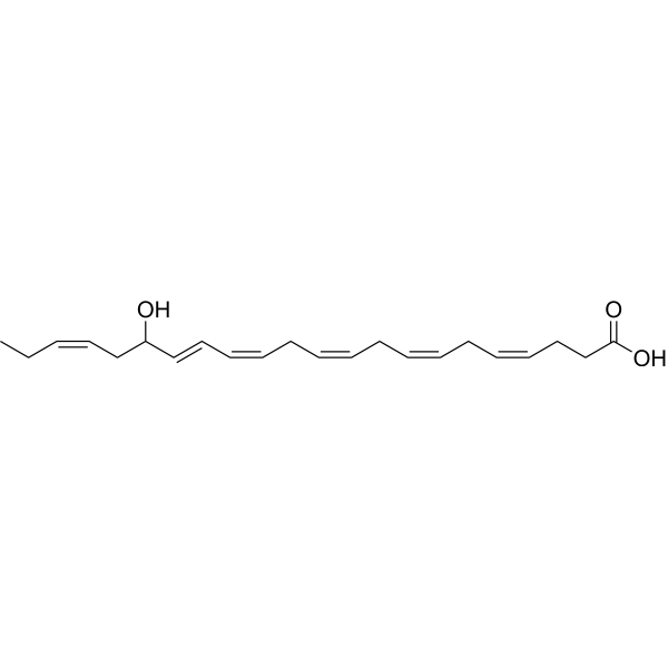
17-HDHA is a DHA-derived specialized pro-resolving mediator (SPM). 17-HDHA inhibits NF-κB activation by activating PPARγ and upregulating IκBα, targets FPR2 and Bcl-6, and also acts as a precursor of RvD1 and PD1. 17-HDHA inhibits pulmonary artery smooth muscle cell proliferation, regulates the expression of adiponectin, GLUT-4 and inflammatory cytokines, suppresses IgE production by B cells and promotes antibody secretion. 17-HDHA reduces adipose tissue inflammation, improves insulin sensitivity and glucose tolerance, alleviates pathological processes associated with pulmonary hypertension, and enhances antibody responses and virus neutralizing activity against influenza HA. 17-HDHA can be used in research related to obesity-associated inflammation, insulin resistance, glucose intolerance, pulmonary hypertension and influenza virus infection.
商品は「研究用試薬」です。人や動物の医療用・臨床診断用・食品用の製品ではありません。
研究用途以外に使用した場合、当社は一切の責任を負いかねます。
- 純度 : 98.86%
- CAS 番号: 90780-52-2
- 分子式: C22H32O3
- 分子量:344.49
-
保管条件:
Solution, -20°C, 2 years

To place orders, for customer services and technical support, please contact: MedChemExpress USA
Tel: 609-228-6898 E-mail: [email protected] [email protected]


-
 生物活性
生物活性
-
化学情報
- プロトコル
- 純度とドキュメンテーション
- 参考文献
-
 Help & FAQs


-
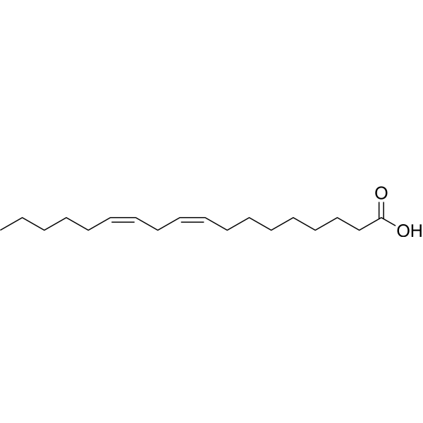
HY-N0729
Linoleic acidHuman Endogenous Metabolite -
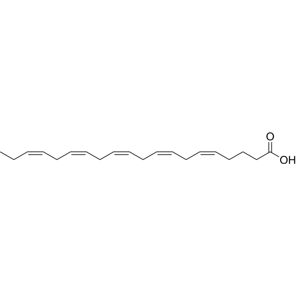
HY-B0660
Eicosapentaenoic AcidLC-PUFA -
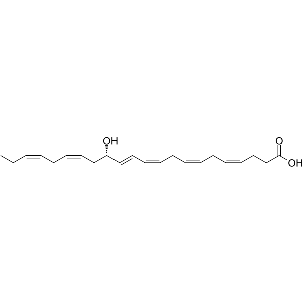
HY-130239
14(S)-HDHADHA Oxygenation Product -
HY-129692
WithanoneiGluR Inhibitor -

HY-141636
1-PalMitoyl-2-arachidoyllecithin -
HY-120891
trans-EKODE-(E)-IbPeroxide -

HY-129692R
Withanone (Standard)Standard of Withanone -
HY-130304
8-Isoprostaglandin E2Thromboxane A2 Receptor Ligand -
HY-120255
17(R)-HDHADecomposition-promoting Media -
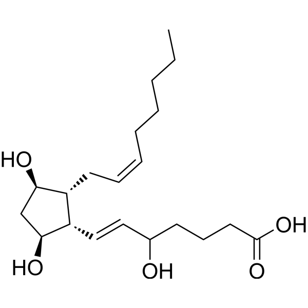
HY-137119
(±)5-iPF2α-VIRacemate -

HY-23201
A1-Phytoprostane-IMAPK Activator -
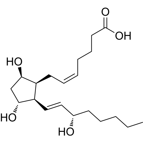
HY-138244
8-iso Prostaglandin F2βBiomarkers
Endogenous Metabolite アイソフォーム固有の製品をすべて表示
More

生物活性
製品説明
IC50 & Target
[1]|
PPARγ |
体外実験
Pretreatment with Dexamethasone (HY-14648) blocks the inhibitory effect of 17-HDHA (10-100 nM; 6 days) on IgE production in peripheral blood mononuclear cells from asthmatic patients not receiving oral corticosteroids[3].
17-HDHA (10-100 nM; 24-72 h) reduces IgE production, ε germline transcript levels and mature IgE mRNA levels in purified CD19+ B cells from healthy donors, and pretreatment with Dexamethasone blocks these inhibitory effects[3].
The ability of 17-HDHA (10-100 nM; 7 days) to reduce IgE production in purified CD19+ B cells and peripheral blood mononuclear cells from healthy donors is blocked by the 5-lipoxygenase inhibitory effect of zileuton[3].
17-HDHA (10-100 nM; 4 h) enhances the expression of Bcl-6 protein in purified CD19+ B cells derived from healthy donors, and pretreatment with Dexamethasone reduces Bcl-6 expression regardless of 17-HDHA treatment[3].
17-HDHA (10-100 nM; administered daily for 6 consecutive days, with 30 min pre-incubation prior to stimulation) upregulates the expression of CD80 and CD86 on purified naive CD19+ B cells from C57BL/6 mice activated by CpG combined with anti-IgM, without altering the expression of MHC class II[4].
17-HDHA (10-100 nM; administered daily for 6 consecutive days, with 30 min pre-incubation prior to stimulation) enhances the production of IgM and IgG in purified naive CD19+ B cells from C57BL/6 mice activated by CpG combined with anti-IgM, and upregulates the mRNA and protein levels of Blimp-1[4].
17-HDHA (300-1000 nM; 4-24 h) induces elevated levels of specific D-series resolvins and 10S,17S-dihydroxydocosahexaenoic acid (10S,17S-diHDHA) in human primary great saphenous vein endothelial cells (ECs) and vascular smooth muscle cells (VSMCs). Moreover, starting from treatment with 300 nM 17-HDHA, the production of RvD1 shows concentration- and time-dependent patterns[6].
17-HDHA (1 μM; 24 h) increases 5-LOX protein expression by 94% in primary human great saphenous vein endothelial cells (but not in vascular smooth muscle cells)[6].
17-HDHA (1 μM; 5 h) induces the translocation of 5-LOX from the nucleus to the cytoplasm in primary human great saphenous vein endothelial cells (ECs) and vascular smooth muscle cells (VSMCs), with the relative cytoplasmic 5-LOX expression levels increasing by 28% and 37% in ECs and VSMCs, respectively[6].
MedChemExpress (MCE) has not independently confirmed the accuracy of these methods. They are for reference only.
-
Cell Line:mouse pulmonary artery smooth muscle cells (PASMCs)
-
Concentration:10 nM
-
Incubation Time:24 h
-
Result:Promoted PPARγ nuclear translocation in PDGFbb-stimulated mouse PASMCs, reversing the PDGFbb-induced reduction in nuclear PPARγ.
Suppressed PDGFbb-induced Stat3 phosphorylation in these cells.
-
Cell Line:PBMCs from asthma patients; B cells from healthy donors
-
Concentration:10 and 100 nM
-
Incubation Time:6 days
-
Result:Significantly reduced spontaneous and induced IgE production; dexamethasone pretreatment blocked this effect.
-
Cell Line:B cells from healthy donors
-
Concentration:10 and 100 nM
-
Incubation Time:1 day (εGLT) or 3 days (mature IgE mRNA)
-
Result:Decreased εGLT and mature IgE mRNA levels; dexamethasone pretreatment blocked this effect.
-
Cell Line:B cells from healthy donors
-
Concentration:10 and 100 nM
-
Incubation Time:4 h
-
Result:Increased Bcl-6 protein expression in a concentration-dependent manner; dexamethasone pretreatment reduced basal Bcl-6 expression and blocked this effect.
-
Cell Line:Mouse B cells (CD19⁺)
-
Concentration:10 and 100 nM
-
Incubation Time:6 days
-
Result:Increased IgM and IgG production in a concentration-dependent manner, with a ~2-fold increase at 100 nM.
-
Cell Line:Endothelial cells and vascular smooth muscle cells
-
Concentration:1 μM
-
Incubation Time:5 h
-
Result:Induced translocation of 5-LOX from nucleus to cytoplasm, with a ~28% (ECs) and ~37% (VSMCs) increase in cytoplasmic/nuclear fluorescence ratio.
-
Cell Line:Human ECs and VSMCs
-
Concentration:300, 500 and 1000 nM
-
Incubation Time:4, 8 and 24 h
-
Result:Concentration- and time-dependently increased RvD1 production, with significant increases at 300 nM and peak at 8 h.
体内実験
17-HDHA (1 μg; intramuscular injection; administered concurrently with primary and booster immunizations) enhances the production of HA-specific IgG in mice immunized with HA combined with CpG ODN, with significantly elevated antibody titers detected at multiple time points post-immunization[4].
17-HDHA (1 μg; intramuscular injection; administered concurrently with each immunization during weeks 0-4) enhances the production of HA-specific antibodies and the differentiation of plasma cells in the bone marrow of mice immunized with HA alone, significantly increasing IgG titers and the frequency of antibody-secreting cells[4].
MedChemExpress (MCE) has not independently confirmed the accuracy of these methods. They are for reference only.
-
Animal Model:C57BL/6J (male, wild-type, diet-induced obesity via 60% kcal high-fat diet for 17 weeks)[1]
-
Dosage:50 ng/g body weight
-
Administration:i.p.; every 12 h; 8 days; osmotic pump; continuous; 15 days
-
Result:Reduced mRNA expression of inflammatory genes (MCP-1, TNF-α, IL-6, OPN, NF-κB) in gonadal adipose tissue.
Increased IκBα protein level in gonadal adipose tissue.
Reduced CD11c+/CD206+ adipose tissue macrophage ratio.
Enhanced mRNA expression of PPARγ, PPARα, GLUT-4, and adiponectin in gonadal adipose tissue.
Moderately improved glucose tolerance with reduced plasma insulin at 45 minutes post-glucose challenge.
Achieved 30% reduction in fasting insulin concentration and 40% lower HOMA-IR (non-significant trends).
Showed trend toward improved insulin tolerance at 60, 90, and 120 minutes post-insulin injection.
Significantly decreased fasting insulin concentration and HOMA-IR.
Showed trend toward reduced blood glucose at 30 minutes post-insulin injection.
Significantly lowered blood glucose at 15 minutes post-glucose challenge.
Reduced plasma insulin levels at baseline and 45 minutes post-glucose challenge.
-
Animal Model:C57BL/6J (male, 8-10 wk old) were immunized via intraperitoneal injection with OVA protein (10 μg/mouse) emulsified in complete Freund's adjuvant (CFA). Immediately after the primary immunization, the compound was administered via intraperitoneal injection at the same site. Blood samples were collected at 2 and 6 weeks after the primary immunization to measure OVA-specific IgM and IgG antibody levels. At week 10, mice received a booster immunization via intraperitoneal injection with OVA (10 μg/mouse) in PBS. Blood samples were collected again at 2 weeks after the booster immunization for antibody detection
[4] -
Dosage:1 μg
-
Administration:i.p.; single concurrent injection
-
Result:Showed significantly higher OVA-specific IgM titers at weeks 6 and 12 compared with vehicle controls.
Showed significantly higher OVA-specific IgG titers at week 6 compared with vehicle controls.
Showed a decreasing trend in OVA-specific IgE levels compared with vehicle controls.
-
Animal Model:C57BL/6J (male, 8-10 wk old) were primed via intramuscular injection at week 0 with recombinant HA protein (H1N1 A/Brisbane/59/2007 (HY-P74094), 5 μg/mouse), CpG ODN 1826 (HY-146245) (10 μg/mouse), and the compound (1 μg/mouse). Booster immunizations were given at weeks 2 and 4 in the same manner. [4]
-
Dosage:1 μg
-
Administration:i.m.; concurrent with primary and booster immunizations
-
Result:Showed significantly higher HA-specific IgG titers at weeks 3, 6, and 11 compared with vehicle controls.
Showed no significant differences in HA-specific IgM titers at any time point compared with vehicle controls.
-
Animal Model:C57BL/6J (male, 8-10 wk old) were immunized via intramuscular injection at weeks 0, 2, and 4 with recombinant HA protein (H1N1 A/California/04/2009, 2 μg/mouse) and the compound (1 μg/mouse)[4]
-
Dosage:1 μg
-
Administration:i.m.; concurrent with each immunization at weeks 0, 2, and 4
-
Result:Showed a 2-fold increase in HA-specific IgM titers at week 2 compared with mock and vehicle controls.
Showed a 9-fold increase in HA-specific IgG titers at week 2 compared with mock and vehicle controls.
Showed a 3-fold increase in HA-specific IgG titers at week 4 compared with vehicle controls.
Showed a 9-fold increase in HA-specific IgG titers at week 6 compared with vehicle controls.
Showed a 2-fold increase in the percentage of CD19+ CD138+ plasma cells in the bone marrow compared with mock and vehicle controls.
Showed a 2-fold increase in HA-specific IgG-secreting bone marrow cells compared with mock and vehicle controls.
Showed no differences in splenic CD19+ CD138+ plasma cells compared with controls.
-
Animal Model:C57BL/6J (male, 8-10 wk old) were immunized via intramuscular injection at weeks 0, 2, and 4 with recombinant HA protein (H1N1 A/California/04/2009, 2 μg/mouse) and 17-HDHA (1 μg/mouse) or vehicle control (3 immunizations in total). Blood was collected at 14 days after the last immunization (week 6), and serum neutralizing antibody titers were measured by GFP-based microneutralization assay. At 28 days after the last immunization (week 8), mice were challenged intranasally with 300 PFU/mouse of mouse-adapted live influenza virus H1N1 A/California/04/E3/2009[4]
-
Dosage:1 μg
-
Administration:i.m.; concurrent with each immunization at weeks 0, 2, and 4
-
Result:Showed 44% of treated mice with detectable neutralizing antibody titers (≥40) at week 6, compared with 0% of mock and vehicle controls.
Showed minimal weight loss following viral infection, while mock and vehicle-treated mice experienced dramatic weight loss.
Showed a 100% survival rate following viral infection, compared with 30% for mock controls and 80% for vehicle controls.
- RNA extraction experimental
- Research Protocol for Infectious Diseases
- BrdU Incorporation Assay
- Somatic Cell Culture
- CFSE Dye Dilution Proliferation Assay
- Dye-dilution cell tracking and proliferation staining
- CCK-8/WST-8 Cell Proliferation Assay
- Protocol for Cell Cycle
- Research Protocol for Cardiovascular Diseases
- Research Protocol for Inflammation-related Diseases
- Cell Counting-Based Growth Curve Assay
- Colony Formation (Clonogenic) Assay
- Research Protocol for Metabolic Diseases
- Cell Viability Determination by MTT Colorimetric Assay
- EdU Incorporation Assay (Click Chemistry-Based DNA Synthesis Measurement)
- Ki-67 Immunostaining Proliferation Assay
- PCNA Immunodetection Proliferation Assay
- LPS-Induced Endotoxemia/Systemic Inflammation
- Protocol for Cell Counting and Cell Density Analysis
- Large-size fat particle sorting
- Research Protocol for Endocrine Diseases
- MTT Cell Proliferation Assay


化学情報
-
CAS 番号 90780-52-2
-
性状 Liquid
-
分子量 344.49
-
分子式 C22H32O3
-
Color Colorless to light yellow
-
SMILES
CC/C=C\CC(O)/C=C/C=C\C/C=C\C/C=C\C/C=C\CCC(O)=O
-
輸送条件
Room temperature in continental US; may vary elsewhere.
-
保管条件
Solution, -20°C, 2 years


純度とドキュメンテーション
-
データシート (291 KB)
-
SDS (393 KB)
- English - EN (393 KB)
- Français - FR (393 KB)
- Deutsch - DE (393 KB)
- Norwegian - NO (393 KB)
- Español - ES (393 KB)
- Swedish - SV (393 KB)
- Italian - IT (393 KB)
- Korean - KR (393 KB)
- Portuguese - PT (393 KB)
-
取扱説明書 (2659 KB)
参考文献
[1]. Neuhofer A, et al. Impaired local production of proresolving lipid mediators in obesity and 17-HDHA as a potential treatment for obesity-associated inflammation. Diabetes. 2013 Jun;62(6):1945-56. [Content Brief]
[2]. Shu T, et al. Eosinophils protect against pulmonary hypertension through 14-HDHA and 17-HDHA. The European respiratory journal. 2023 Mar;61(3):2200582. [Content Brief]
[3]. Kim N, et al. Corticosteroids inhibit anti-IgE activities of specialized proresolving mediators on B cells from asthma patients. JCI insight. 2017 Feb 09;2(3):e88588. [Content Brief]
[4]. Ramon S, et al. The specialized proresolving mediator 17-HDHA enhances the antibody-mediated immune response against influenza virus: a new class of adjuvant?. Journal of immunology (Baltimore, Md. : 1950). 2014 Dec 15;193(12):6031-40. [Content Brief]
[5]. Tran M, et al. Investigating the catalytic efficiency of C22-Fatty acids with LOX human isozymes and the platelet response of the C22-oxylipin products. Archives of biochemistry and biophysics. 2023 Oct 01;747:109742. [Content Brief]
[6]. Chatterjee A, et al. Biosynthesis of proresolving lipid mediators by vascular cells and tissues. FASEB journal : official publication of the Federation of American Societies for Experimental Biology. 2017 Aug;31(8):3393-3402. [Content Brief]

Calculators
濃度 (開始) × 体積 (開始) = 濃度 (終了) × 体積 (終了)


17-HDHA 関連分類
HY-113512 関連分類
 Powered by Bioz