



Desipramine
Based on 18 publication(s) in Google Scholar
Desipramine is a first-generation tricyclic antidepressant. Desipramine selectively binds to norepinephrine transporter and blocks neuronal norepinephrine reuptake. Desipramine activates MAPK signaling via ERK1/2, JNK, and p38, represses NF-κB and AP-1 activity, and induces apoptosis via ROS elevation, mitochondrial membrane potential reduction, and intracellular calcium increase. Desipramine also shows anyi-inflammatory activity, inhibiting TNF-α production. Desipramine can be used for the research of hepatocellular cancer, inflammation, and neurological diseases.
Nur für Forschungszwecke. Wir verkaufen nicht an Patienten.
- CAS. Nr.: 50-47-5
- Formel: C18H22N2
- Molecular Weight:266.38
-
Speicherung:Powder -20°C, 3 years ; In solvent -80°C, 6 months , -20°C, 1 month

To place orders, for customer services and technical support, please contact: MedChemExpress USA
Tel: 609-228-6898 E-mail: [email protected] [email protected]


-
 Biologische Aktivität
-
Chemical Information
- Protokoll
- Reinheit & Dokumentation
- Verweise
-
 Help & FAQs


Publications Citing Use of MedChemExpress (MCE) Desipramine
More- Cell. 2025 Nov 26;188(24):6861-6872.e14. [Abstract]
- Acta Pharm Sin B. 2026 Jan 27.
- Neural Regen Res. 2021 Aug;16(8):1660-1670. [Abstract]
- Br J Pharmacol. 2024 Dec;181(23):4874-4889. [Abstract]
- J Med Chem. 2026 Feb 10. [Abstract]
- Pharmaceutics. 2022 Jul 22;14(8):1523. [Abstract]
- J Parkinson Dis. 2020;10(2):523-542. [Abstract]
- Front Endocrinol. 2026 May 5:17:1825379. [Abstract]
- Glia. 2022 Nov;70(11):2093-2107. [Abstract]
- Inflammation. 2021 Aug;44(4):1592-1606. [Abstract]
- Biochim Biophys Acta Mol Basis Dis. 2023 Mar 28;1869(5):166700. [Abstract]
- Neuropharmacology. 2026 Nov 1:298:111009. [Abstract]
- Neurotoxicology. 2022 Dec:93:173-185. [Abstract]
- Drug Metab Dispos. 2026 Jul 14;54(8):100365.
- Neuroscience. 2024 Mar 26:542:11-20. [Abstract]
- Parkinsons Dis. 2022 Nov 14:2022:1428817. [Abstract]
- Sci Total Environ. 2024 May 1:923:171405. [Abstract]
- Research Square Print. 2023 Feb 23.
 Customer Validation & Images

-
Cell Proliferation/Viability Assay
-
WB
-
Cell Imaging/Staining
-
Bio/Physico-chemical Assay
-
Bio/Physico-chemical Assay
Alle Adrenergic Receptor Isoform-spezifische Produkte anzeigen
MoreAlle AP-1 Isoform-spezifische Produkte anzeigen
MoreAlle TNF Receptor Isoform-spezifische Produkte anzeigen
More

Biologische Aktivität
Beschreibung
IC50 & Target
[1]|
ERK1 |
ERK2 |
Cellular Effect
|
Cell Line

|
Type | Value | Description | References |
|---|---|---|---|---|
| DG-75 | IC50 |
39.81 μM
Compound: Desipramine
|
Reduction in cell viability of human DG75 cells after 24 hrs by Alamar blue viability assay
Reduction in cell viability of human DG75 cells after 24 hrs by Alamar blue viability assay
|
[PMID: 23385211] |
| HEK293 | IC50 |
0.0524 μM
Compound: desipramine
|
Inhibition of [3H]serotonin uptake in human SERT K490T mutant transfected HEK293 cells
Inhibition of [3H]serotonin uptake in human SERT K490T mutant transfected HEK293 cells
|
[PMID: 17690258] |
| HEK293 | IC50 |
0.108 μM
Compound: desipramine
|
Inhibition of [3H]serotonin uptake in human wild type SERT transfected HEK293 cells
Inhibition of [3H]serotonin uptake in human wild type SERT transfected HEK293 cells
|
[PMID: 17690258] |
| HEK293 | IC50 |
17.7 μM
Compound: desipramine
|
Inhibition of [3H]dopamine uptake in human DAT I390V mutant transfected HEK293 cells
Inhibition of [3H]dopamine uptake in human DAT I390V mutant transfected HEK293 cells
|
[PMID: 17690258] |
| HEK293 | IC50 |
47.9 μM
Compound: desipramine
|
Inhibition of [3H]dopamine uptake in human wild type DAT transfected HEK293 cells
Inhibition of [3H]dopamine uptake in human wild type DAT transfected HEK293 cells
|
[PMID: 17690258] |
| HEK293 | IC50 |
50.5 μM
Compound: desipramine
|
Inhibition of [3H]dopamine uptake in human DAT P387A mutant transfected HEK293 cells
Inhibition of [3H]dopamine uptake in human DAT P387A mutant transfected HEK293 cells
|
[PMID: 17690258] |
| HEK293 | IC50 |
9.9 μM
Compound: desipramine
|
Inhibition of [3H]dopamine uptake in human DAT F472L mutant transfected HEK293 cells
Inhibition of [3H]dopamine uptake in human DAT F472L mutant transfected HEK293 cells
|
[PMID: 17690258] |
| HEK293 | IC50 |
0.0042 μM
Compound: Desipramine
|
Inhibition of [3H]norepinephrine uptake at human NET expressed in HEK293 cells
Inhibition of [3H]norepinephrine uptake at human NET expressed in HEK293 cells
|
[PMID: 17846138] |
| HEK293 | IC50 |
0.064 μM
Compound: Desipramine
|
Inhibition of [3H]5HT uptake at human SERT expressed in HEK293 cells
Inhibition of [3H]5HT uptake at human SERT expressed in HEK293 cells
|
[PMID: 17846138] |
| HEK293 | IC50 |
82 μM
Compound: Desipramine
|
Inhibition of [3H]dopamine uptake at human DAT expressed in HEK293 cells
Inhibition of [3H]dopamine uptake at human DAT expressed in HEK293 cells
|
[PMID: 17846138] |
| HEK293 | IC50 |
56.8 μM
Compound: desipramine
|
Inhibition of 4-(4-(dimethylamino)styryl)-N-methylpyridinium uptake at human OCT1 expressed in HEK293 cells by confocal microscopy
Inhibition of 4-(4-(dimethylamino)styryl)-N-methylpyridinium uptake at human OCT1 expressed in HEK293 cells by confocal microscopy
|
[PMID: 18788725] |
| HEK293 | IC50 |
1520 nM
Compound: Desipramine
|
Inhibition of sodium current measured using whole-cell patch clamp experiments in HEK-293 cells stably transfected with hNaV1.5 cDNA
Inhibition of sodium current measured using whole-cell patch clamp experiments in HEK-293 cells stably transfected with hNaV1.5 cDNA
|
[PMID: 21300721] |
| HEK293 | IC50 |
9.18 μM
Compound: Desipramine
|
Inhibition of human OCT1 expressed in HEK293 cells assessed as decrease in uptake of ASP+ after 2 mins by fluorescence assay
Inhibition of human OCT1 expressed in HEK293 cells assessed as decrease in uptake of ASP+ after 2 mins by fluorescence assay
|
[PMID: 28230985] |
| HeLa | IC50 |
17.78 μM
Compound: Desipramine
|
Reduction in cell viability of human HeLa cells after 24 hrs by Alamar blue viability assay
Reduction in cell viability of human HeLa cells after 24 hrs by Alamar blue viability assay
|
[PMID: 23385211] |
| HRPE | IC50 |
0.08 mM
Compound: Desipramine
|
TP_TRANSPORTER: inhibition of TEA uptake in Octn1-HRPE cells
TP_TRANSPORTER: inhibition of TEA uptake in Octn1-HRPE cells
|
[PMID: 10825452] |
| HRPE | IC50 |
68 μM
Compound: Desipramine
|
TP_TRANSPORTER: inhibition of MPP+ uptake (MPP+: 1 uM) in OCT3-expressing HRPE cells
TP_TRANSPORTER: inhibition of MPP+ uptake (MPP+: 1 uM) in OCT3-expressing HRPE cells
|
[PMID: 9830022] |
| MDCK | IC50 |
0.93 nM
Compound: desipramine
|
Displacement of [125I]RTI-55 from human NET expressed in MDCK cell membrane
Displacement of [125I]RTI-55 from human NET expressed in MDCK cell membrane
|
[PMID: 16989524] |
| MDCK | IC50 |
3.9 nM
Compound: desipramine
|
Inhibition of norepinephrine uptake at human NET expressed in MDCK cells
Inhibition of norepinephrine uptake at human NET expressed in MDCK cells
|
[PMID: 18771916] |
| MDCK | IC50 |
0.93 nM
Compound: desipramine
|
Displacement of [125I]RTI55 from human recombinant norepinephrine transporter expressed in MDCK cells after 3 hrs
Displacement of [125I]RTI55 from human recombinant norepinephrine transporter expressed in MDCK cells after 3 hrs
|
[PMID: 23403082] |
| MDCK | IC50 |
1.5 nM
Compound: Desipramine
|
Displacement of [3H]Nisoxetine from human recombinant NET over-expressed in dog MDCK cells
Displacement of [3H]Nisoxetine from human recombinant NET over-expressed in dog MDCK cells
|
[PMID: 24012181] |
| Splenocyte | IC50 |
22.9 μM
Compound: Desipramine
|
Cytotoxicity against C57BL/6J mouse splenocytes after 72 hrs by alamar blue assay
Cytotoxicity against C57BL/6J mouse splenocytes after 72 hrs by alamar blue assay
|
[PMID: 17846138] |
| Ventricular myocyte | IC50 |
1709 nM
Compound: Desipramine
|
Inhibition of calcium current (ICaL) measured using whole-cell patch clamp experiments in isolated guinea pig ventricular myocytes
Inhibition of calcium current (ICaL) measured using whole-cell patch clamp experiments in isolated guinea pig ventricular myocytes
|
[PMID: 21300721] |
| Ventricular myocyte | IC50 |
1.709 μM
Compound: Desipramine
|
Inhibition of L-type calcium channel measured using whole-cell patch clamp in guinea pig ventricular myocytes
Inhibition of L-type calcium channel measured using whole-cell patch clamp in guinea pig ventricular myocytes
|
[PMID: 22761000] |
| Ventricular myocyte | IC50 |
11.7 μM
Compound: Desipramine
|
Inhibition of L-type calcium channel measured using whole-cell patch clamp in rat ventricular myocytes
Inhibition of L-type calcium channel measured using whole-cell patch clamp in rat ventricular myocytes
|
[PMID: 22761000] |
In Vitro
Desipramine (3-500 µM; 24 h) reduces the viability of human hepatoma Hep3B cells in a dose-dependent manner[1].
Desipramine (10-500 µM; 24 h) induces cytotoxicity in human hepatoma Hep3B cells in a dose-dependent manner, as measured by increased LDH release[1].
Desipramine (3-100 µM; 24 h) increases intracellular ROS production in human hepatoma Hep3B cells in a dose-dependent manner[1].
Desipramine (3-100 µM µM; 24 h) has its induced cell death in human hepatoma Hep3B cells abolished by the ROS scavenger NAC (HY-B0215) at concentrations up to 30 µM, and partially reduced at 100 µM after 24 h[1].
Desipramine (3-100 µM; 24 h) reduces mitochondrial membrane potential in human hepatoma Hep3B cells in vitro in a dose-dependent manner[1].
Desipramine (100 µM; 1-8 h) activates ERK1/2, JNK, and p38 MAPK signaling in human hepatoma Hep3B cells-[1].
Desipramine (3-100 µM; 24 h) has its induced cell death in human hepatoma Hep3B cells abolished by inhibition of ERK1/2, p38, or JNK MAPK signaling[1].
Desipramine (30-500 µM) increases intracellular calcium levels in human hepatoma Hep3B cells in a concentration-dependent manner, with increases occurring via both extracellular calcium influx and intracellular calcium release[1].
Desipramine potently binds to the rat norepinephrine transporter with a Ki of 6.2 nM, showing 25-fold selectivity over the rat serotonin transporter (Ki = 158 nM) [3].
Desipramine (1 h) binds to the rat α2D-adrenoceptor with a Ki of 3.4 μM[3].
Desipramine potently binds to the human norepinephrine transporter with a Ki of 0.63 nM, has lower affinity for the human serotonin transporter (Ki = 22 nM), and lacks affinity for the human dopamine transporter (Ki > 10,000 nM)[3].
Desipramine (0.01-10 μM; 20 h) dose-dependently inhibits LPS (HY-D1056)-induced TNF-α release from primary human monocytes[4].
Desipramine (10 μM) reduces TNF-α-induced RANTES release and RANTES mRNA accumulation in A549 human lung epithelial cells[4].
Desipramine (10 μM) represses TNF-α-induced NF-κB activity in A549 human lung epithelial cells[4].
Desipramine (10 μM) reduces TPA (HY-18739)-induced AP-1 activity in A549 human lung epithelial cells[4].
MedChemExpress (MCE) has not independently confirmed the accuracy of these methods. They are for reference only.
-
Cell Line:human hepatoma Hep3B cells
-
Concentration:1 µM, 3 µM, 10 µM, 30 µM, 100 µM, 300 µM, 500 µM
-
Incubation Time:24 h
-
Result:Did not affect cell viability at 1 µM.
Reduced cell viability to 90.2% of control at 3 µM.
Reduced cell viability to 87.4% of control at 10 µM.
Reduced cell viability to 69.1% of control at 30 µM.
Reduced cell viability to 45.9% of control at 100 µM.
Reduced cell viability to 36.1% of control at 300 µM.
Reduced cell viability to 27.8% of control at 500 µM.
Caused statistically significant viability reductions at concentrations ≥3 µM compared to control.
-
Cell Line:human hepatoma Hep3B cells
-
Concentration:10 µM, 30 µM, 100 µM, 300 µM, 500 µM
-
Incubation Time:24 h
-
Result:Increased LDH activity to 105.7% of control at 10 µM.
Increased LDH activity to 109.8% of control at 30 µM.
Increased LDH activity to 150.3% of control at 100 µM.
Increased LDH activity to 227.7% of control at 300 µM.
Increased LDH activity to 255.7% of control at 500 µM.
Caused statistically significant increases in LDH release at all tested concentrations compared to control.
-
Cell Line:human hepatoma Hep3B cells
-
Concentration:100 µM
-
Incubation Time:1 h, 2 h, 4 h, 8 h
-
Result:Increased phosphorylation of ERK1/2 to 185.2% of control at 1 h, 151.5% of control at 2 h, and 138.5% of control at 4 h.
Increased phosphorylation of p38 to 135.1% of control at 1 h and 116.4% of control at 2 h.
Increased phosphorylation of JNK to 118.9% of control at 1 h and 113.9% of control at 2 h.
Maintained unchanged total levels of ERK1/2, JNK, and p38 across all time points.
Caused statistically significant increases in MAPK phosphorylation at the specified time points compared to control.
In Vivo
Desipramine (10 mg/kg; i.p.; daily; 8 days, 30 minutes pre-Ovalbumin nebulization) reduces lung inflammation by decreasing macrophage and lymphocyte counts in Ovalbumin (HY-W250978)-sensitized rats but does not attenuate bronchial hyperresponsiveness[4].
MedChemExpress (MCE) has not independently confirmed the accuracy of these methods. They are for reference only.
-
Animal Model:LPS-induced BALB/c mice (5-week-old, 17-21 g)[4]
-
Dosage:5, 10, 20 mg/kg
-
Administration:i.p.; single dose (preventive, 30 minutes pre-LPS)
-
Result:Significantly inhibited LPS-induced serum TNF-α production.
Increased mouse survival rate to 70% with preventive 20 mg/kg treatment (vs 10% in saline controls).
-
Animal Model:Ovalbumin-sensitized Brown Norway rats (10-week-old)[4]
-
Dosage:10 mg/kg
-
Administration:i.p.; daily; 8 days (30 minutes pre-ovalbumin nebulization)
-
Result:Did not reduce bronchial hyperresponsiveness to metacholine (measured by enhanced pause, Penh).
Reduced total number of inflammatory cells in bronchoalveolar lavages.
Decreased macrophage count by 52% compared to saline-treated sensitized rats.
Decreased lymphocyte count by 21% compared to saline-treated sensitized rats.
Did not reduce neutrophil or eosinophil counts compared to saline-treated sensitized rats.
- Detection of Apoptosis
- TUNEL staining for apoptotic DNA fragmentation
- Mitochondrial membrane-potential fluorescent assay
- Annexin V plus membrane-impermeant dye apoptosis staining
- ROS/oxidative-stress fluorescent staining
- Fluorescent plasma-membrane potential dye assay
- Mitochondrial membrane-potential and mitochondrial mass staining
- Apoptosis Solutions
- Calcium Spark Assay
- Research Protocol for Inflammation-related Diseases
- Ca2+ Staining Technique


Chemical Information
-
CAS. Nr. 50-47-5
-
Appearance Solid
-
Molecular Weight 266.38
-
Formel C18H22N2
-
Color White to off-white
-
SMILES
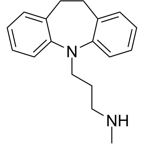
CNCCCN1C2=CC=CC=C2CCC3=CC=CC=C31
-
Versand
Room temperature in continental US; may vary elsewhere.
-
Speicherung
Powder -20°C 3 years In solvent -80°C 6 months -20°C 1 month


Publications (18)
-
Journal Impact Factor
-
Most Recent
-
Cell
2025 Nov 26;188(24):6861-6872.e14. PMID: 41138730 -
-
Neural Regen Res
Glucagon-like peptide-1/glucose-dependent insulinotropic polypeptide dual receptor agonist DA-CH5 is superior to exendin-4 in protecting neurons in the 6-hydroxydopamine rat Parkinson model. [Abstract]2021 Aug;16(8):1660-1670. PMID: 33433498 -
Br J Pharmacol
Serotonergic transmission plays differentiated roles in the rapid and sustained antidepressant-like effects of ketamine. [Abstract]2024 Dec;181(23):4874-4889. PMID: 39238235 -
J Med Chem
Bioisostere-Driven Discovery of SePP: A Selenium-Containing Polypharmacological Agent Relevant to Fragile X Syndrome. [Abstract]2026 Feb 10. PMID: 41666325 -
Pharmaceutics
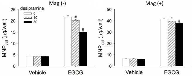
Laminin Receptor-Mediated Nanoparticle Uptake by Tumor Cells: Interplay of Epigallocatechin Gallate and Magnetic Force at Nano-Bio Interface. [Abstract]2022 Jul 22;14(8):1523. PMID: 35893779
Desipramine purchased from MedChemExpress. Usage Cited in: Pharmaceutics. 2022 Jul 22;14(8):1523. [Abstract]
Figure shows that 10 and 30 μM of Desipramine hydrochloride attenuated EGCG-induced enhancement of MNPcell by 8.9% and 39.3% in the Mag− group, respectively. In the Mag+ group, MNPcell was enhanced up to 6.6-fold by EGCG compared with the control group, which was minorly attenuated by 5.7% and 11.2% by Desipramine hydrochloride at 10 and 30 μM, respectively.
-
J Parkinson Dis
The Novel Dual GLP-1/GIP Receptor Agonist DA-CH5 Is Superior to Single GLP-1 Receptor Agonists in the MPTP Model of Parkinson's Disease. [Abstract]2020;10(2):523-542. PMID: 31958096 -
Front Endocrinol
The novel GLP-1/GIP dual receptor agonist DA5-CH is superior to tirzepatide and exendin-4 in the 6-OHDA Parkinson rat model. [Abstract]2026 May 5:17:1825379. PMID: 42165019 -
Glia
Potassium channel Kir 4.1 regulates oligodendrocyte differentiation via intracellular pH regulation. [Abstract]2022 Nov;70(11):2093-2107. PMID: 35775976 -
Inflammation
Acid Sphingomyelinase and Acid β-Glucosidase 1 Exert Opposite Effects on Interleukin-1β-Induced Interleukin 6 Production in Rheumatoid Arthritis Fibroblast-Like Synoviocytes. [Abstract]2021 Aug;44(4):1592-1606. PMID: 33665756 -
Biochim Biophys Acta Mol Basis Dis
Involvement of Kir4.1 in pain insensitivity of the BTBR mouse model of autism spectrum disorder. [Abstract]2023 Mar 28;1869(5):166700. PMID: 36990129 -
Neuropharmacology
Antidepressant-like activity of JZ-1201 in male rodents: a novel selective 5-HT/NE reuptake inhibitor and 5-HT1A receptor partial agonist. [Abstract]2026 Nov 1:298:111009. PMID: 42107524 -
Neurotoxicology
Ropivacaine-induced seizures evoked pain sensitization in rats: Participation of 5-HT/5-HT3R. [Abstract]2022 Dec:93:173-185. PMID: 36209936 -
-
Neuroscience
Acute Administration of Lactate Exerts Antidepressant-like Effect Through cAMP-dependent Protein Synthesis. [Abstract]2024 Mar 26:542:11-20. PMID: 38336096 -
Parkinsons Dis
DA5-CH and Semaglutide Protect against Neurodegeneration and Reduce α-Synuclein Levels in the 6-OHDA Parkinson's Disease Rat Model. [Abstract]2022 Nov 14:2022:1428817. PMID: 36419409 -
Sci Total Environ
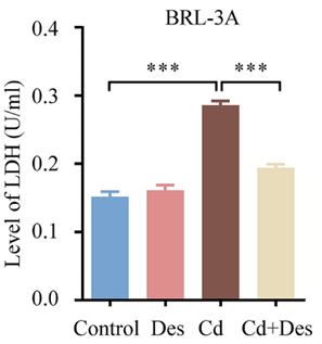
Cadmium exacerbates liver injury by remodeling ceramide metabolism: Multiomics and laboratory evidence. [Abstract]2024 May 1:923:171405. PMID: 38432385
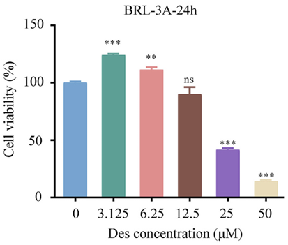
Desipramine purchased from MedChemExpress. Usage Cited in: Sci Total Environ. 2024 May 1:923:171405. [Abstract]
Desipramine hydrochloride (Des; 0-50 μM; 24 h). Cell viability of BRL-3A cells after Des intervention was detected by CCK-8. Other groups were compared with 0 μM group.
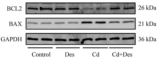
Desipramine purchased from MedChemExpress. Usage Cited in: Sci Total Environ. 2024 May 1:923:171405. [Abstract]
Desipramine hydrochloride (Des; 12.5 μM; 24 h). The expression levels of apoptosis-related proteins in BRL-3A cells were detected by western blotting.
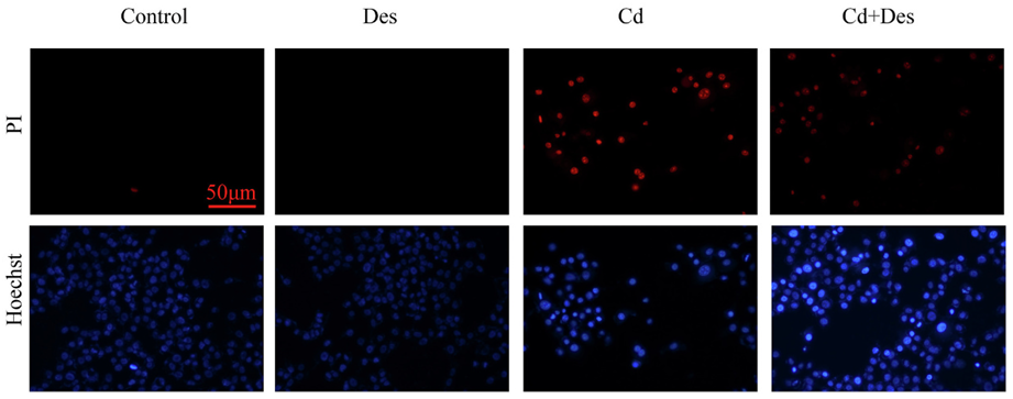
Desipramine purchased from MedChemExpress. Usage Cited in: Sci Total Environ. 2024 May 1:923:171405. [Abstract]
Desipramine hydrochloride (Des; 12.5 μM; 24 h). Hoechst/PI double staining: Hoechst/PI was used to double stain and observe them under fluorescence microscope (200×). Hoechst 33342 can stain the nucleus with blue fluorescence. PI can stain necrotic cells with red fluorescence.
Desipramine purchased from MedChemExpress. Usage Cited in: Sci Total Environ. 2024 May 1:923:171405. [Abstract]
Desipramine hydrochloride (Des; 12.5 μM; 24 h). The level of LDH in cell culture medium after 12.5 μM Des intervention and/or 20 μM Cadmium exposure for 24 h.
-


Reinheit & Dokumentation
-
Data Sheet (287 KB)
-
SDS (393 KB)
- English - EN (393 KB)
- Français - FR (393 KB)
- Deutsch - DE (393 KB)
- Norwegian - NO (393 KB)
- Español - ES (393 KB)
- Swedish - SV (393 KB)
- Italian - IT (393 KB)
- Korean - KR (393 KB)
- Portuguese - PT (393 KB)
-
Handling Instructions (2659 KB)
Verweise
[1]. Yang DK, et al. Desipramine induces apoptosis in hepatocellular carcinoma cells. Oncol Rep. 2017;38(2):1029-1034. [Content Brief]
[2]. Kishore-Kumar R, et al. Desipramine relieves postherpetic neuralgia. Clin Pharmacol Ther. 1990;47(3):305-312. [Content Brief]
[3]. Deupree JD, et al. Pharmacological properties of the active metabolites of the antidepressants desipramine and citalopram. Eur J Pharmacol. 2007;576(1-3):55-60. [Content Brief]
[4]. Roumestan C, et al. Anti-inflammatory properties of desipramine and fluoxetine. Respir Res. 2007;8(1):35. Published 2007 May 3. [Content Brief]

Calculators
Konzentration (Stammlösung) × Volumen (Stammlösung) = Konzentration (Ziellösung) × Volumen (Ziellösung)


Desipramine Related Classifications
HY-B1272A Related Classifications
 Powered by Bioz
Keywords