



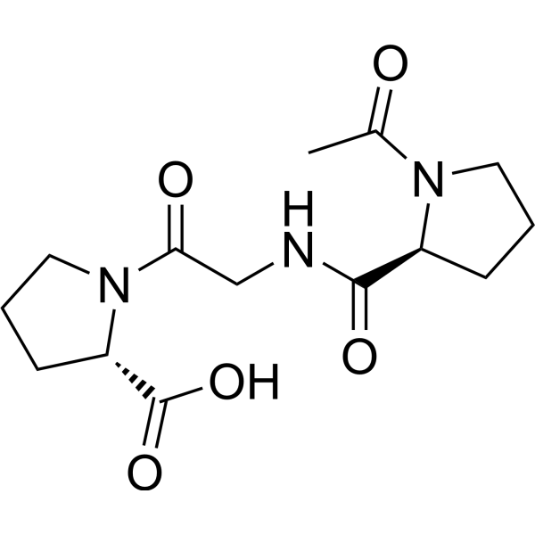
Ac-Pro-Gly-Pro-OH
Based on 2 publication(s) in Google Scholar
Ac-Pro-Gly-Pro-OH is an endogenous degradation product of extracellular collagen and acts as a CXCR2 agonist. Ac-Pro-Gly-Pro-OH exerts bactericidal activity by generating hydrogen peroxide, inhibits pulmonary inflammation, and reduces immune cell apoptosis (apoptosis). Ac-Pro-Gly-Pro-OH promotes the production of IFN-γ and inhibits the production of TNF-α and IL-6 in leukocytes. Ac-Pro-Gly-Pro-OH increases the survival rate of mice in sepsis models, enhances the bactericidal activity of neutrophils, acts as a neutrophil chemoattractant, induces neutrophil polarization, and regulates inflammatory and repair processes. Ac-Pro-Gly-Pro-OH induces chronic inflammation and tissue remodeling through sustained action. Ac-Pro-Gly-Pro-OH is released via alkaline hydrolysis of corneal proteins in alkali-injured eyes, thereby driving the early infiltration of neutrophils into the cornea. Ac-Pro-Gly-Pro-OH is applicable to research related to sepsis, chronic obstructive pulmonary disease, cystic fibrosis, bronchiolitis obliterans syndrome, severe asthma, idiopathic pulmonary fibrosis, and corneal ulcer.
商品は「研究用試薬」です。人や動物の医療用・臨床診断用・食品用の製品ではありません。
研究用途以外に使用した場合、当社は一切の責任を負いかねます。
- 純度 : 99.49%
- CAS 番号: 292171-04-1
- 分子式: C14H21N3O5
- 分子量:311.33
-
保管条件:
Sealed storage, away from moisture.
Powder -80°C, 2 years , -20°C, 1 year* In solvent : -80°C, 6 months; -20°C, 1 month (sealed storage, away from moisture)

To place orders, for customer services and technical support, please contact: MedChemExpress USA
Tel: 609-228-6898 E-mail: [email protected] [email protected]


-
 生物活性
生物活性
-
化学情報
-
溶剤 & 溶解度
- プロトコル
- 純度とドキュメンテーション
- 参考文献
-
 Help & FAQs


-
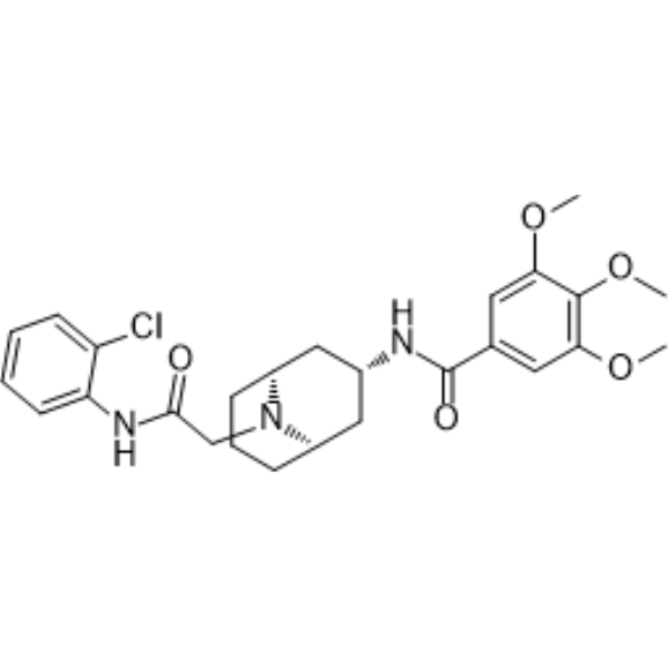
HY-122197
ML339CXCR6 Antagonist -
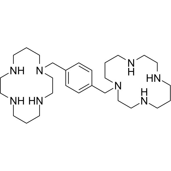
HY-10046
PlerixaforCXCR4 Antagonist -
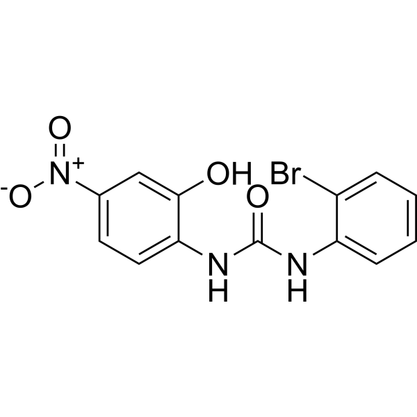
HY-16711
SB225002CXCR2 Antagonist -
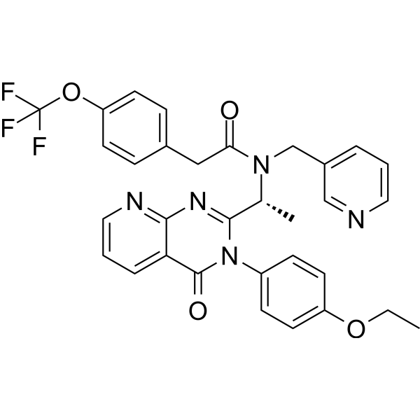
HY-15319
AMG 487CXCR3 Antagonist -
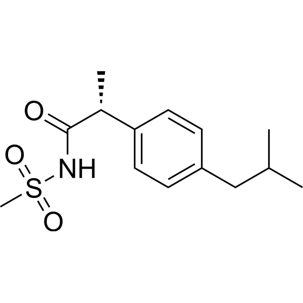
HY-15251
ReparixinCXCR1/2 Inhibitor -
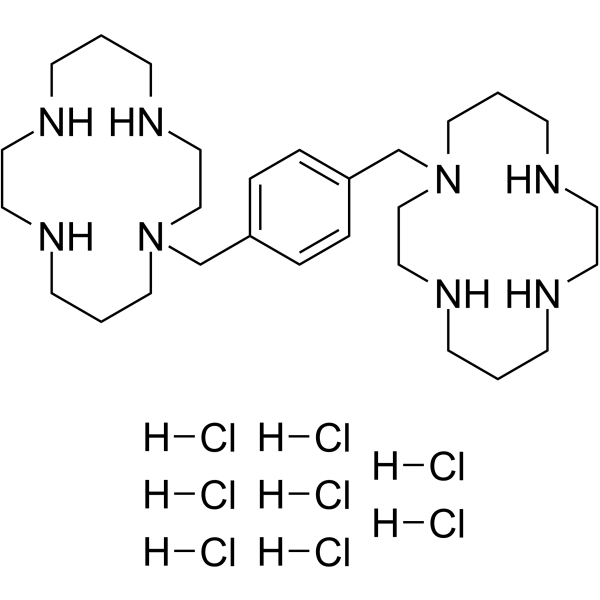
HY-50912
Plerixafor octahydrochlorideCXCR4 Antagonist -
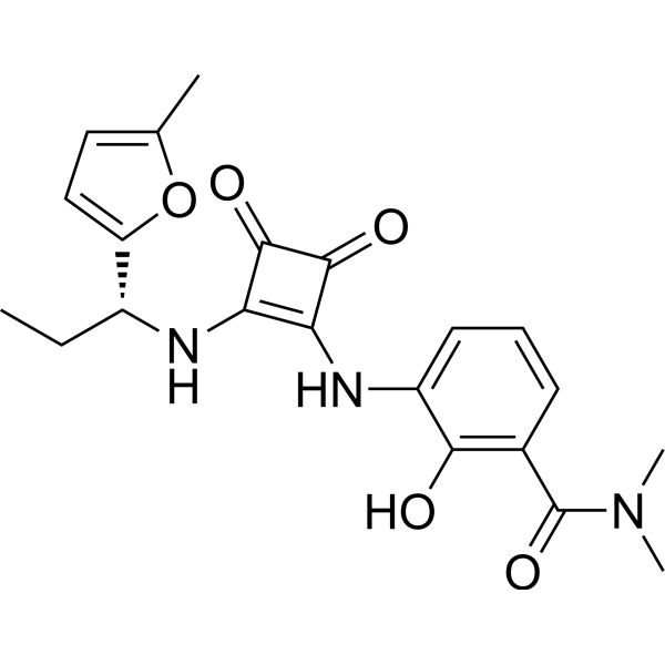
HY-10198
NavarixinCXCR1/2 Antagonist -
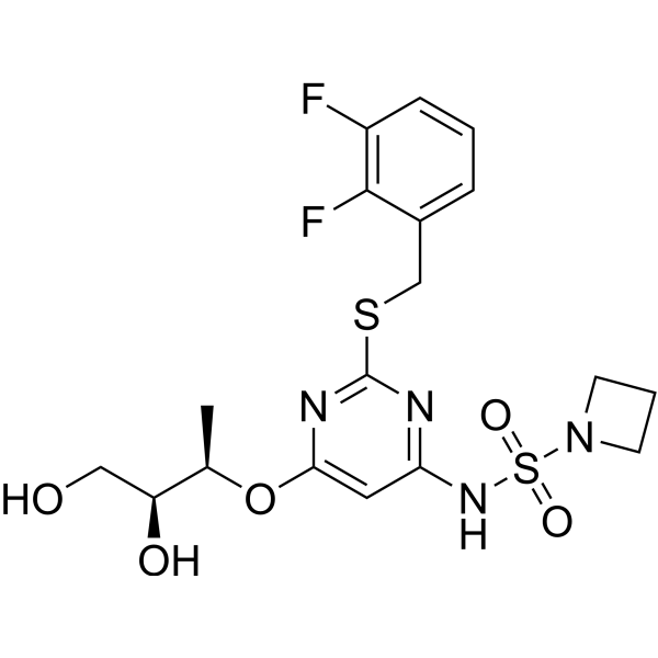
HY-19855
AZD-5069CXCR2 Antagonist -
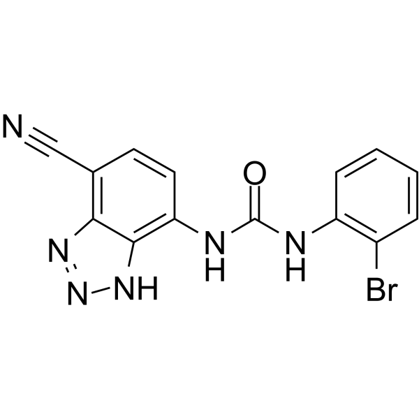
HY-50688
SB-265610CXCR2 Antagonist -
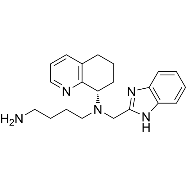
HY-50101
MavorixaforCXCR4 Antagonist -
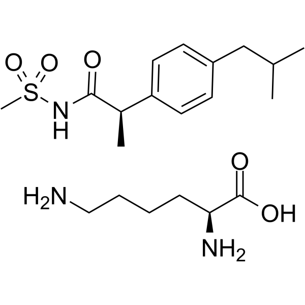
HY-15252
Reparixin L-lysine saltCXCR Inhibitor -
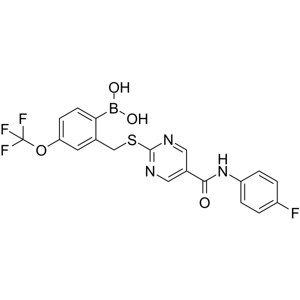
HY-119339
SX-682CXCR1/2 Inhibitor
MedChemExpress(MCE)の使用を引用している文献 Ac-Pro-Gly-Pro-OH
More

生物活性
製品説明
IC50 & Target
[1]|
CXCR2 |
体外実験
Ac-Pro-Gly-Pro-OH (0-50 μM; 10 min) induces concentration-dependent hydrogen peroxide (H2O2) production in isolated mouse neutrophils via CXCR2[1].
Ac-Pro-Gly-Pro-OH (0-50 μM; 1 h) enhances the concentration-dependent bactericidal activity of isolated murine neutrophils[1].
Ac-Pro-Gly-Pro-OH (1-20 μM; 30 min pre-incubation, 24 h LPS stimulation) modulates cytokine production in LPS-stimulated mouse splenocytes, inhibits the pro-inflammatory cytokines TNF-α and IL-6 in a concentration-dependent manner, while enhancing the type 1 cytokine IFN-γ, with the most potent effect observed at 20 μM[1].
Ac-Pro-Gly-Pro-OH (0.1-50 μM; 1 h) enhances the concentration-dependent bactericidal activity of isolated human neutrophils, reaching a maximum killing rate (approximately 95%) at 20 μM and 50 μM[1].
Ac-Pro-Gly-Pro-OH (0-20 μM; 30 min pre-incubation, 3 h LPS stimulation) regulates cytokine production in LPS (HY-D1056)-stimulated human neutrophils and PBMCs. It inhibits the pro-inflammatory cytokine TNF-α and enhances the type 1 cytokine IFN-γ in a concentration-dependent manner, with the strongest effect observed at 20 μM[1].
Ac-Pro-Gly-Pro-OH activates neutrophils, mediates superoxide anion production, MMP-9 release and CXCL8 release by binding to CXCR1/2, and induces G protein-dependent neutrophil chemotaxis[2].
Ac-Pro-Gly-Pro-OH promotes CXCR2-dependent paracellular permeability in endothelial cells and the release of endothelin-1 in aortic endothelial cells[2].
Ac-Pro-Gly-Pro-OH induces CXCR2-dependent migration, proliferation and tube formation of endothelial progenitor cells[2].
Ac-Pro-Gly-Pro-OH resists degradation mediated by LTA4H, but is susceptible to degradation by ACE[2].
Ac-Pro-Gly-Pro-OH induces polarization in isolated human neutrophils with an EC50 of 0.50 mM[3].
MedChemExpress (MCE) has not independently confirmed the accuracy of these methods. They are for reference only.
-
Cell Line:mouse splenocytes
-
Concentration:1, 5, 10, and 20 μM
-
Incubation Time:30 min pre-incubation, 3 h LPS stimulation
-
Result:Modulated cytokine production in LPS-stimulated mouse splenocytes, inhibited the pro-inflammatory cytokines TNF-α and IL-6 in a concentration-dependent manner, while enhancing the type 1 cytokine IFN-γ, with the most potent effect observed at 20 μM
体内実験
Ac-Pro-Gly-Pro-OH drives neutrophil recruitment and corneal ulcer formation in an alkali-injured rabbit corneal model[2].
Ac-Pro-Gly-Pro-OH induces dose-dependent airway neutrophilic inflammation in mice via the CXCR2 signaling pathway, triggers emphysema in *Mus musculus*, abrogates bleomycin hydrochloride (HY-17565A)-induced pulmonary fibrosis in mice, and promotes cutaneous neovascularization and wound healing in mice[2].
MedChemExpress (MCE) has not independently confirmed the accuracy of these methods. They are for reference only.
-
Animal Model:ICR (male, wild-type albino)[1]
-
Dosage:7.5 mg/kg; 12.5 mg/kg; 17.5 mg/kg (survival, organ inflammation, bactericidal activity, cytokine modulation); 17.5 mg/kg (5 doses, survival); 17.5 mg/kg (delayed 4 doses, survival); 17.5 mg/kg (2 doses, organ inflammation)
-
Administration:s.c.; 4 doses at 2,14,26,38 hours post-CLP; 5 doses at 2,14,26,38,50 hours post-CLP; 4 doses at 10,22,34,48 hours post-CLP; 2 doses at 2,14 hours post-CLP/4 doses at 2,14,26,38 hours post-E. coli injection/4 doses at 2,14,26,38 hours post-LPS injection
-
Result:Increased survival in a dose-dependent manner.
Further improved survival when 17.5 mg/kg was given as 5 doses vs fewer doses.
Showed therapeutic effect on survival with delayed 17.5 mg/kg (4 doses starting 10 hours post-CLP).
Reduced CLP-induced acute lung injury score, thrombi and hyaline membrane score, and lung wet-to-dry weight ratio.
Inhibited CLP-induced liver inflammation, splenic and thymic tissue damage, and reduced TUNEL-positive cells in spleen and thymus by ~50-60% compared with vehicle controls.
Reduced intraperitoneal bacterial colony counts by 82.1% at 24 hours after CLP.
Blunted CLP-induced increases in peritoneal fluid TNF-α, IL-1β, and IL-6 levels, while enhancing production of type 1 cytokines IFN-γ, IL-12, and IL-2 in peritoneal fluid.
Reduced the incidence of fatal sepsis compared with vehicle controls.
- Phagocytosis Functional Assay
- Fibrosis/Collagen Morphometry
- Collagen: Sirius Red Staining
- Detection of Apoptosis
- TUNEL staining for apoptotic DNA fragmentation
- LPS-Induced Endotoxemia/Systemic Inflammation
- Annexin V plus membrane-impermeant dye apoptosis staining
- Cotton Pellet Granuloma
- Connective Tissue: Masson's Trichrome/Collagen Trichrome Staining
- Apoptosis Solutions
- Research Protocol for Inflammation-related Diseases
- Bacterial live/dead nucleic-acid viability staining


化学情報
-
CAS 番号 292171-04-1
-
性状 Solid
-
分子量 311.33
-
分子式 C14H21N3O5
-
Color White to off-white
-
配列
Ac-Pro-Gly-Pro-OH
-
シーケンスの短縮
Ac-PGP-OH
-
輸送条件
Room temperature in continental US; may vary elsewhere.
-
保管条件
Sealed storage, away from moisture
Powder -80°C 2 years -20°C 1 year * In solvent : -80°C, 6 months; -20°C, 1 month (sealed storage, away from moisture)


Publications (2)
-
Journal Impact Factor
-
Most Recent
-
Adv Sci (Weinh)
Diabetes Mellitus Facilitates Gallstone Formation Through CXCR2-NETs-Mediated Liver-Bile Barrier Damage. [Abstract]2026 Apr;13(24):e19500. PMID: 41698066 -
CNS Neurosci Ther
Network Pharmacology-Based and Experimental Validation Elucidate the Target Mechanism of Vinorine in Ameliorating Secondary Brain Injury After Intracerebral Hemorrhage. [Abstract]2025 Sep;31(9):e70609. PMID: 40994248


溶剤 & 溶解度
体外:
H2O : ≥ 50 mg/mL (160.60 mM)
* "≥" means soluble, but saturation unknown.
Please refer to the solubility information to select the appropriate solvent. Once prepared, please aliquot and store the solution to prevent product inactivation from repeated freeze-thaw cycles.
Storage method and period of stock solution: -80°C, 6 months; -20°C, 1 month (sealed storage, away from moisture). When stored at -80°C, please use it within 6 months. When stored at -20°C, please use it within 1 month.
* Note: If you choose water as the stock solution, please dilute it to the working solution, then filter and sterilize it with a 0.22 μm filter before use.
Please refer to the solubility information to select the appropriate solvent. Once prepared, please aliquot and store the solution to prevent product inactivation from repeated freeze-thaw cycles.
Storage method and period of stock solution: -80°C, 6 months; -20°C, 1 month (sealed storage, away from moisture). When stored at -80°C, please use it within 6 months. When stored at -20°C, please use it within 1 month.
* Note: If you choose water as the stock solution, please dilute it to the working solution, then filter and sterilize it with a 0.22 μm filter before use.
濃度 (開始) × 体積 (開始) = 濃度 (終了) × 体積 (終了)


純度とドキュメンテーション
-
データシート (286 KB)
-
SDS (393 KB)
- English - EN (393 KB)
- Français - FR (393 KB)
- Deutsch - DE (393 KB)
- Norwegian - NO (393 KB)
- Español - ES (393 KB)
- Swedish - SV (393 KB)
- Italian - IT (393 KB)
- Korean - KR (393 KB)
- Portuguese - PT (393 KB)
-
取扱説明書 (2659 KB)
参考文献
[1]. Kim SD, et al. Activation of CXCR2 by extracellular matrix degradation product acetylated Pro-Gly-Pro has therapeutic effects against sepsis. Am J Respir Crit Care Med. 2011;184(2):243-251. [Content Brief]
[2]. Patel DF, et al. The multifaceted roles of the matrikine Pro-Gly-Pro in pulmonary health and disease. Eur Respir Rev. 2018;27(148):180017. Published 2018 Jun 27. [Content Brief]
[3]. Lee YC, et al. NMR conformational analysis of cis and trans proline isomers in the neutrophil chemoattractant, N-acetyl-proline-glycine-proline. Biopolymers. 2001;58(6):548-561. [Content Brief]
[4]. Wu JW, et al. Network Pharmacology-Based and Experimental Validation Elucidate the Target Mechanism of Vinorine in Ameliorating Secondary Brain Injury After Intracerebral Hemorrhage. CNS Neurosci Ther. 2025;31(9):e70609. [Content Brief]


Complete Stock Solution Preparation Table
Please refer to the solubility information to select the appropriate solvent. Once prepared, please aliquot and store the solution to prevent product inactivation from repeated freeze-thaw cycles.
Storage method and period of stock solution: -80°C, 6 months; -20°C, 1 month (sealed storage, away from moisture). When stored at -80°C, please use it within 6 months. When stored at -20°C, please use it within 1 month.
| Optional Solvent | Concentration Solvent Mass | 1 mg | 5 mg | 10 mg | 25 mg |
|---|---|---|---|---|---|
| H2O | 1 mM | 3.2120 mL | 16.0601 mL | 32.1203 mL | 80.3006 mL |
| 5 mM | 0.6424 mL | 3.2120 mL | 6.4241 mL | 16.0601 mL | |
| 10 mM | 0.3212 mL | 1.6060 mL | 3.2120 mL | 8.0301 mL | |
| 15 mM | 0.2141 mL | 1.0707 mL | 2.1414 mL | 5.3534 mL | |
| 20 mM | 0.1606 mL | 0.8030 mL | 1.6060 mL | 4.0150 mL | |
| 25 mM | 0.1285 mL | 0.6424 mL | 1.2848 mL | 3.2120 mL | |
| 30 mM | 0.1071 mL | 0.5353 mL | 1.0707 mL | 2.6767 mL | |
| 40 mM | 0.0803 mL | 0.4015 mL | 0.8030 mL | 2.0075 mL | |
| 50 mM | 0.0642 mL | 0.3212 mL | 0.6424 mL | 1.6060 mL | |
| 60 mM | 0.0535 mL | 0.2677 mL | 0.5353 mL | 1.3383 mL | |
| 80 mM | 0.0402 mL | 0.2008 mL | 0.4015 mL | 1.0038 mL | |
| 100 mM | 0.0321 mL | 0.1606 mL | 0.3212 mL | 0.8030 mL |
* Note: If you choose water as the stock solution, please dilute it to the working solution, then filter and sterilize it with a 0.22 μm filter before use.


Ac-Pro-Gly-Pro-OH 関連分類
HY-P4846 関連分類
 Powered by Bioz