



TriDAP
Based on 1 publication(s) in Google Scholar
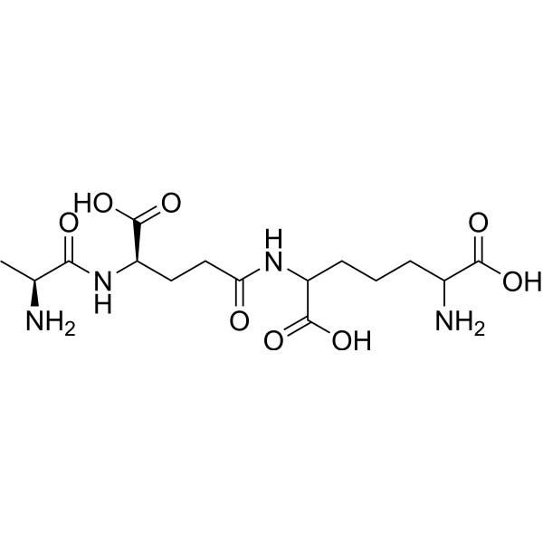
TriDAP (L-Ala-γ-D-Glu-meso-diaminopimelic acid) is a NOD1 agonist with a Kd value of 34.5 μM. TriDAP enhances the binding of NOD1-RICK, promotes RICK phosphorylation, and activates the NF-κB, TAK1, MEK/ERK, p38 and interferon response pathways. TriDAP downregulates Runx2 via increasing ubiquitination and reduces trabecular bone parameters. TriDAP decreases IκBα levels and increases p65 levels. TriDAP induces the secretion of proinflammatory mediators IL-8 and prostaglandins, triggers tissue inflammation and innate immune activation, and inhibits SARS-CoV-2 replication in lung epithelial cells. TriDAP increases the RANKL/OPG ratio in mice, reduces bone mass and enhances osteoclast activity, and inhibits new bone formation by decreasing the mineralization deposition rate in mice. TriDAP can be used in research related to pulpitis, chronic ulcerative colitis, Crohn's disease and SARS-CoV-2 infection.
For research use only. We do not sell to patients.
- CAS No.: 877462-71-0
- Formula: C15H26N4O8
- Molecular Weight:390.39
-
Storage:
Please store the product under the recommended conditions in the Certificate of Analysis.

To place orders, for customer services and technical support, please contact: MedChemExpress USA
Tel: 609-228-6898 E-mail: [email protected] [email protected]


-
 Biological Activity
-
Chemical Information
- Protocols
- Purity & Documentation
- References
-
 Help & FAQs



Publications Citing Use of MedChemExpress (MCE) TriDAP
MoreAll MAP3K Isoforms
MoreAll MEK Isoforms
More

Biological Activity
Description
IC50 & Target
[4]|
hNOD1 34.5 μM (Kd) |
IL-8 |
In Vitro
TriDAP (0-10 μg/mL; 6 days) dose-dependently inhibits osteogenic differentiation of primary mouse calvarial osteoblast precursors and enhances osteoclast differentiation in the co-culture system of mouse BMMs and primary calvarial osteoblast precursors[1].
TriDAP (0-10 μg/mL; 2 days) downregulates the expression of osteogenic marker genes including Alp, Bsp and Runx2 in MC3T3-E1 mouse preosteoblasts in a dose-dependent manner[1].
TriDAP (0-10 μg/mL; 18 h) dose-dependently inhibits the transcriptional activity of Runx2 in MC3T3-E1 mouse preosteoblasts[1].
TriDAP (1 μg/mL; 2-24 h) reduces the stability of Runx2 protein in transiently transfected HEK293 cells. After 8 hours of co-incubation with Cycloheximide (HY-12320), the relative level of Runx2 decreases by approximately 50%, which enhances Smurf1-mediated Runx2 degradation in cells[1].
TriDAP (1-10 μg/mL; 24 h) increases the ubiquitination level of Runx2 in transiently transfected HEK293 cells in a dose-dependent manner[1].
TriDAP (0.1-10 μg/mL; 2 days) dose-dependently increases the RANKL/OPG ratio in primary mouse calvarial osteoprogenitor cells, through a mechanism that decreases OPG secretion and increases RANKL secretion[1].
TriDAP (1 μg/mL; 15-90 min) activates the NF-κB pathway in MC3T3-E1 mouse osteoprogenitor cells by reducing IκBα levels and increasing p65 levels, and induces nuclear translocation of NF-κB p65[1].
TriDAP (0.1-10 μg/mL; 24 h) dose-dependently enhances the transcriptional activity of NF-κB in MC3T3-E1 mouse preosteoblasts[1].
TriDAP (10 μg/mL; 10 days) inhibits osteogenic mineralization of wild-type primary mouse calvarial preosteoblasts, but this effect is abolished in NOD1-deficient preosteoblasts, indicating that this action is dependent on NOD1[1].
TriDAP (10 μg/mL; 16 h) preferentially activates the transcriptional activity of NF-κB in HEK293 cells transfected with NOD1, whereas the activity is extremely low in cells transfected with NOD2[1].
TriDAP (10-100 μM) binds directly to purified full-length NOD1 protein, with a measured Kd value of 34.5 μM via SPR[4].
Binding of TriDAP (0.1 mM) to purified full-length NOD1 protein increases the binding affinity between purified RICK protein and NOD1, with SPR assays showing that its Kd value decreases from 4.13 μM to 3.26 μM[4].
TriDAP (0-5 mM; 1-4 h) induces the expression of NOD1 and secretion of IL-8 in Caco2-BBE intestinal epithelial cells, and this pro-inflammatory response depends on the expression of NOD1[4].
TriDAP activates the NOD1 pathway in HEK-Blue hNOD1 cells, with an EC50 of 700 ng/mL[5].
TriDAP (0.4-50 µM; 6-24 h) increases the proportion of IL-8+ human lung epithelial A549 cells by 3.29-fold, elevates the proportion of IL-8+ human lung epithelial A549-Dual cells, does not reduce cell viability, and activates the NF-κB and ISRE pathways in human lung epithelial A549-Dual cells[5].
TriDAP (50 µM; 8 h) upregulates the mRNA expression levels of IL-8, CXCL10 and ISG15 in the human lung epithelial cell line A549-Dual[5].
TriDAP (2-50 µM; 3-hour pretreatment; 48 hours post-infection) dose-dependently inhibits SARS-CoV-2 replication in human lung epithelial A549-Dual cells, with an EC50 of 7.75 µM, and reduces the number of infected cells by 49% at a concentration of 50 µM[5].
MedChemExpress (MCE) has not independently confirmed the accuracy of these methods. They are for reference only.
-
Cell Line:primary murine calvarial osteoblast precursors
-
Concentration:0.1, 1, 10 μg/mL
-
Incubation Time:6 days
-
Result:Inhibited osteoblast differentiation in a dose-dependent manner, as evidenced by reduced ALP staining intensity with increasing TriDAP concentrations.
-
Cell Line:MC3T3-E1 murine osteoblast precursor cells
-
Concentration:1, 10 μg/mL
-
Incubation Time:2 days
-
Result:Decreased the expression of osteogenic marker genes Alp, Bsp, and Runx2 in a dose-dependent manner compared to untreated cells.
-
Cell Line:transiently transfected HEK293 cells
-
Concentration:1 μg/mL
-
Incubation Time:2, 4, 8 h
-
Result:Decreased the half-life of Runx2 protein; reduced relative Runx2 levels significantly compared to untreated cells at 4 and 8 h.
-
Cell Line:primary murine calvarial osteoblast precursors
-
Concentration:0.1, 1, 10 μg/mL
-
Incubation Time:2 days
-
Result:Decreased OPG levels and increased RANKL levels in a dose-dependent manner, resulting in a dose-dependent increase in the RANKL/OPG ratio; caused statistically significant changes at all tested concentrations compared to untreated cells.
-
Cell Line:MC3T3-E1 murine osteoblast precursor cells
-
Concentration:1 μg/mL
-
Incubation Time:15 min
-
Result:Induced translocation of p65 from the cytoplasm to the nucleus within 15 minutes of treatment.
-
Cell Line:human lung epithelial A549-Dual cells
-
Concentration:50 µM
-
Incubation Time:8 h
-
Result:Induced a 1.68-fold increase in IL-8 mRNA compared to untreated control.
Significantly upregulated CXCL10 mRNA compared to untreated control.
Significantly upregulated ISG15 mRNA compared to untreated control.
In Vivo
MedChemExpress (MCE) has not independently confirmed the accuracy of these methods. They are for reference only.
-
Animal Model:C57BL/6 (6-week-old male)[1]
-
Dosage:1.25 mg/kg
-
Administration:i.p.; on days 0 and 4/ on days 1 and 5
-
Result:Caused significant reductions in trabecular bone volume (BV/TV), trabecular thickness (Tb.Th), and trabecular number (Tb.N) compared to PBS controls.
Left trabecular separation (Tb.Sp) unchanged.
Confirmed decreased trabecular bone in distal femurs via H&E staining.
Increased TRAP-positive surface area on bone surfaces, indicating enhanced osteoclast activity.
Significantly increased the RANKL/OPG ratio in bone marrow extracellular fluids relative to PBS controls.
Resulted in a significant reduction in the calcein-labeled mineralized surface of distal femurs compared to PBS controls.
Caused a significant decrease in mineral apposition rate compared to PBS controls.
- Kinase activity and phosphorylation assays
- Western Blot
- Research Protocol for Infectious Diseases
- Mesenchymal stromal/stem cell osteogenic differentiation
- DSS-Induced Colitis
- TNBS-Induced Colitis
- Protocol for Kinase activity and phosphorylation assays
- Research Protocol for Inflammation-related Diseases
- Osteoclast differentiation from monocyte/macrophage precursors


Chemical Information
-
CAS No. 877462-71-0
-
Molecular Weight 390.39
-
Formula C15H26N4O8
-
Synonyms
L-Ala-γ-D-Glu-meso-diaminopimelic acid
-
Sequence
Ala-{d-γGlu}-{meso-Diaminopimelic acid}
-
Sequence Shortening
A-{d-γGlu}-{meso-Diaminopimelic acid}
-
Shipping
Room temperature in continental US; may vary elsewhere.
-
Storage
Please store the product under the recommended conditions in the Certificate of Analysis.


Publications (1)
-
Journal Impact Factor
-
Most Recent
-
Exploration
A Bioorthogonal and Programmable Bacterial Delivery System for Spatiotemporally Targeted Therapy of Solid Tumors. [Abstract]2025 Dec 18;5(6):20240396. PMID: 41476656


Purity & Documentation
References
[1]. Enoksson M, et al. Human cord blood-derived mast cells are activated by the Nod1 agonist M-TriDAP to release pro-inflammatory cytokines and chemokines. J Innate Immun. 2011;3(2):142-149. [Content Brief]
[2]. Laroui H, et al. L-Ala-γ-D-Glu-meso-diaminopimelic acid (DAP) interacts directly with leucine-rich region domain of nucleotide-binding oligomerization domain 1, increasing phosphorylation activity of receptor-interacting serine/threonine-protein kinase 2 and its interaction with nucleotide-binding oligomerization domain 1. J Biol Chem. 2011;286(35):31003-31013. [Content Brief]
[3]. Garcia-Vidal E, et al. Nucleotide-Binding Oligomerization Domain 1 (NOD1) Agonists Prevent SARS-CoV-2 Infection in Human Lung Epithelial Cells through Harnessing the Innate Immune Response. Int J Mol Sci. 2024;25(10):5318. Published 2024 May 13. [Content Brief]
[4]. Chang MC, et al. Inducing phospholipase A2 and cyclooxygenase-2 expression and prostaglandins' production of human dental pulp cells by activation of NOD receptor and its downstream signaling. Int J Biol Macromol. 2025;292:139193. [Content Brief]
[5]. Park OJ, et al. The dual roles of peptidoglycans: NOD1 and NOD2 inversely regulate bone metabolism. Exp Mol Med. 2025;57(8):1837-1846. [Content Brief]

Calculators
Concentration (start) × Volume (start) = Concentration (final) × Volume (final)


TriDAP Related Classifications
HY-P5522 Related Classifications
 Powered by Bioz