



β-Eudesmol
Based on 2 publication(s) in Google Scholar
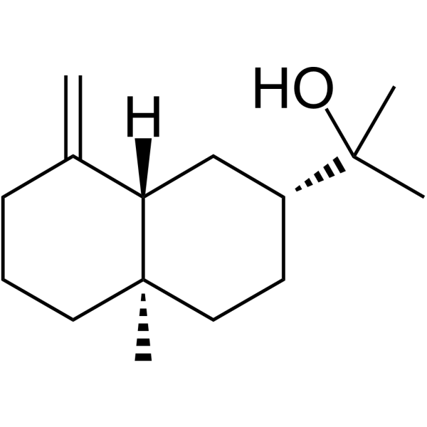
β-Eudesmol has anticancer and anti-inflammatory activities. Beta-Eudesmol can induce apoptosis. β-Eudesmol is a neostigmine antagonist. β-Eudesmol can antagonize neostigmine-induced neuromuscular failure. β-Eudesmoll can be used in the study of sepsis diseases. β-Eudesmol is a sesquiterpene-like compound that can be extracted from the rhizome of Atractylodes lancea.
For research use only. We do not sell to patients.
- Purity : 99.49%
- CAS No.: 473-15-4
- Formula: C15H26O
- Molecular Weight:222.37
-
Storage:
-20°C, stored under nitrogen
* In solvent : -80°C, 6 months; -20°C, 1 month (stored under nitrogen)

To place orders, for customer services and technical support, please contact: MedChemExpress USA
Tel: 609-228-6898 E-mail: [email protected] [email protected]


-
 Biological Activity
-
Chemical Information
-
Solvent & Solubility
- Protocols
- Purity & Documentation
- References
-
 Help & FAQs


-
HY-107324
β-ElemeneApoptosis Activator -
HY-17568
NonivamideTRP Channel Agonist -
HY-N0134
Tanshinone IPhospholipase Inhibitor -
HY-15064
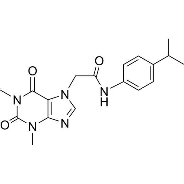
HC-030031TRP Channel Inhibitor -
HY-W012815
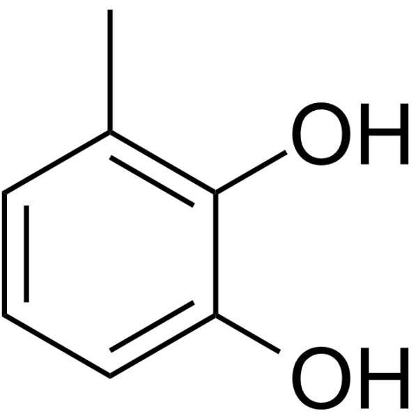
3-MethylcatecholMolecular Building Block -
HY-N0451
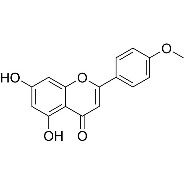
AcacetinAutophagy Inducer -

HY-17568R
Nonivamide (Standard)TRP Channel Agonist -
HY-101795
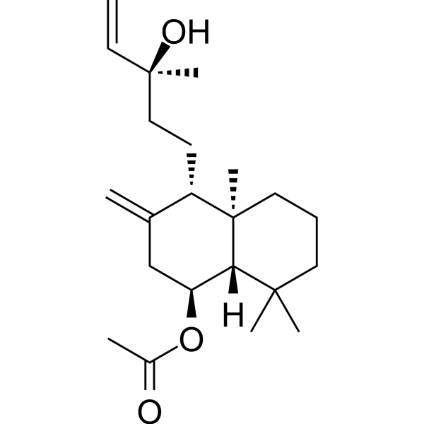
Larixyl acetateTRPC6 Inhibitor -
HY-19608
GSK1016790ATRPV4 Channel Agonist -
HY-N1109
UvaolTriterpene -

HY-N0134R
Tanshinone I (Standard)Phospholipase Inhibitor -
HY-15699A
SAR7334 hydrochlorideTRPC6 Inhibitor
Publications Citing Use of MedChemExpress (MCE) β-Eudesmol
MoreAll Caspase Isoforms
More

Biological Activity
Description
In Vitro
β-Eudesmol (30-100 μM; 6 days) can inhibit the cell proliferation induced by vascular endothelial growth factor (VEGF) (30 ng/ml) and basic fibroblast growth factor (bFGF) (30 ng/ml) in HUVEC cells, thereby inhibiting angiogenesis[1].
β-Eudesmol (10-100 μM; 72 h) inhibits cell proliferation in a time- and dose-dependent manner in tumor in tumer cells HeLa, SGC-7901 and BEL-7402[1].
β-Eudesmol (10-120 μM; 4 h) can reduce cell activity in HL-60 cells[4].
β-Eudesmol (40-80 μM; 4 h) can activate caspase-3 by inducing the release of cytochrome c, thereby inducing cell apoptosis in HL-60 cells[4].
MedChemExpress (MCE) has not independently confirmed the accuracy of these methods. They are for reference only.
-
Cell Line:HL-60
-
Concentration:40 μM, 80 μM
-
Incubation Time:4 h
-
Result:Increased the expression of caspase-3 protein at 80 µM.
-
Cell Line:HL-60
-
Concentration:10 μM, 20 μM, 40 μM, 60 μM, 80 μM, 120 μM
-
Incubation Time:4 h
-
Result:Didn’t affect cell activity at a dose of 40 μM, but decreased cell activity at a dose of 80 μM, and cell viability decreased by 51.6%.
-
Cell Line:HL-60
-
Concentration:40 μM, 80 μM
-
Incubation Time:4 h
-
Result:Had no effect on apoptosis at the dose of 40 μM, but induced apoptosis at the dose of 80 μM, and the apoptosis rate was 31.7%.
In Vivo
β-Eudesmol (50-100 mg/kg; Intraperitoneal injection; Single dose) can inhibit the NF-κb signaling pathway and has anti-inflammatory and antioxidant effects in C57BL/6 mice[2].
MedChemExpress (MCE) has not independently confirmed the accuracy of these methods. They are for reference only.
-
Animal Model:Male KM mice model[1]
-
Dosage:1.25 mg/kg, 2.5mg/kg, 5 mg/kg
-
Administration:Intraperitoneal injection (i.p.); Once daily for 7 days
-
Result:Inhibited the growth of H22 tumor cells with inhibition rates of 39.9% (2.5 mg/kg) and 49.3%(5 mg/kg), respectively.
Inhibited the growth of S108 tumor cells with inhibition rates of 22.4% (2.5 mg/kg) and 42.2%(5 mg/kg), respectively.
Significantly decreased vascular index with inhibition rates of 34.4% (2.5 mg/kg) and 44.3% (5 mg/kg).
-
Animal Model:Male C57BL/6 mice model[2]
-
Dosage:50 mg/kg, 100 mg/kg
-
Administration:Intraperitoneal injection (i.p.); Single dose. 2 h before the cecal ligation and puncture (CLP) operation.
-
Result:Increased the expression of TNF-α, IL-1β and IL-6 in a dose-dependent manner.
Increased the levels of superoxide dismutase (SOD) and glutathione (GSH), and decreased the levels of malondialdehyde (MDA) and myeloperoxidase (MPO).
Inhibit the phosphorylation of p65.


Chemical Information
-
CAS No. 473-15-4
-
Appearance Solid
-
Molecular Weight 222.37
-
Formula C15H26O
-
Color White to off-white
-
SMILES
C[C@]12[C@@](C(CCC2)=C)([H])C[C@H](C(C)(O)C)CC1
-
Structure Classification
-
Initial Source
-
Shipping
Room temperature in continental US; may vary elsewhere.
-
Storage
-20°C, stored under nitrogen
* In solvent : -80°C, 6 months; -20°C, 1 month (stored under nitrogen)


Publications (2)
-
Journal Impact Factor
-
Most Recent
-
ACS Omega
2025 Aug 21;10(34):39192-39202. PMID: 40918390 -


Solvent & Solubility
In Vitro:
DMSO : 50 mg/mL (224.85 mM; Need ultrasonic; Hygroscopic DMSO has a significant impact on the solubility of product, please use newly opened DMSO)
H2O : < 0.1 mg/mL (insoluble)
Please refer to the solubility information to select the appropriate solvent. Once prepared, please aliquot and store the solution to prevent product inactivation from repeated freeze-thaw cycles.
Storage method and period of stock solution: -80°C, 6 months; -20°C, 1 month (stored under nitrogen). When stored at -80°C, please use it within 6 months. When stored at -20°C, please use it within 1 month.
Please refer to the solubility information to select the appropriate solvent. Once prepared, please aliquot and store the solution to prevent product inactivation from repeated freeze-thaw cycles.
Storage method and period of stock solution: -80°C, 6 months; -20°C, 1 month (stored under nitrogen). When stored at -80°C, please use it within 6 months. When stored at -20°C, please use it within 1 month.
Concentration (start) × Volume (start) = Concentration (final) × Volume (final)
In Vivo:
Select the appropriate dissolution method based on your experimental animal and administration route.
- For the following dissolution methods, please ensure to first prepare a clear stock solution using an In Vitro approach and then sequentially add co-solvents:
- To ensure reliable experimental results, the clarified stock solution can be appropriately stored based on storage conditions. As for the working solution for In Vivo experiments, it is recommended to prepare freshly and use it on the same day.
- The percentages shown for the solvents indicate their volumetric ratio in the final prepared solution. If precipitation or phase separation occurs during preparation, heat and/or sonication can be used to aid dissolution.
Add each solvent one by one: 10% DMSO 40% PEG300 5% Tween-80 45% Saline
Solubility: 2.5 mg/mL (11.24 mM); Suspended solution; Need ultrasonic
This protocol yields a suspended solution of 2.5 mg/mL. Suspended solution can be used for oral and intraperitoneal injection.
Taking 1 mL working solution as an example, add 100 μL DMSO stock solution (25.0 mg/mL) to 400 μL PEG300, and mix evenly; then add 50 μL Tween-80 and mix evenly; then add 450 μL Saline to adjust the volume to 1 mL.
Preparation of Saline: Dissolve 0.9 g sodium chloride in ddH₂O and dilute to 100 mL to obtain a clear Saline solution.
Add each solvent one by one: 10% DMSO 90% (20% SBE-β-CD in Saline)
Solubility: ≥ 2.5 mg/mL (11.24 mM); Clear solution
This protocol yields a clear solution of ≥ 2.5 mg/mL (saturation unknown).
Taking 1 mL working solution as an example, add 100 μL DMSO stock solution (25.0 mg/mL) to 900 μL 20% SBE-β-CD in Saline, and mix evenly.
Preparation of 20% SBE-β-CD in Saline (4°C, storage for one week): 2 g SBE-β-CD powder is dissolved in 10 mL Saline, completely dissolve until clear.
In Vivo Dissolution Calculator
Please enter the basic information of animal experiments:
-
-
-
-
Recommended: Prepare an additional quantity of animals to account for potential losses during experiments.
Please enter your animal formula composition:
-
%DMSO +
Recommended: Keep the proportion of DMSO in working solution below 2% if your animal is weak.
-
%+
-
+%Tween-80 + +
-
%Saline +
The co-solvents required include: DMSO, . All of co-solvents are available by MedChemExpress (MCE). , Tween 80. All of co-solvents are available by MedChemExpress (MCE).
Working solution concentration: 0.22 mg/mL
Method for preparing stock solution: mg drug dissolved in μL DMSO. Stock solution concentration: mg/mL. * In solvent : -80°C, 6 months; -20°C, 1 month (stored under nitrogen)
1. Take μL DMSO stock solution;
2. Add μL .
μL , mix evenly;
3. Then add μL Tween 80, mix evenly;
4. Then add μL
Please ensure that the stock solution in the first step is dissolved to a clear state, and add co-solvents in sequence. You can use ultrasonic heating (ultrasonic cleaner, recommended frequency 20-40 kHz), vortexing, etc. to assist dissolution.


Purity & Documentation
-
Data Sheet (283 KB)
-
SDS (393 KB)
- English - EN (393 KB)
- Français - FR (393 KB)
- Deutsch - DE (393 KB)
- Norwegian - NO (393 KB)
- Español - ES (393 KB)
- Swedish - SV (393 KB)
- Italian - IT (393 KB)
- Korean - KR (393 KB)
- Portuguese - PT (393 KB)
-
Handling Instructions (2659 KB)
References
[1]. Ma EL,et al. Beta-eudesmol suppresses tumour growth through inhibition of tumour neovascularisation and tumour cell proliferation. J Asian Nat Prod Res. 2008 Jan-Feb;10(1-2):159-67. [Content Brief]
[3]. Chiou LC, et al. Antagonism by beta-eudesmol of neostigmine-induced neuromuscular failure in mouse diaphragms. Eur J Pharmacol. 1992 Jun 5;216(2):199-206. [Content Brief]


Complete Stock Solution Preparation Table
Please refer to the solubility information to select the appropriate solvent. Once prepared, please aliquot and store the solution to prevent product inactivation from repeated freeze-thaw cycles.
Storage method and period of stock solution: -80°C, 6 months; -20°C, 1 month (stored under nitrogen). When stored at -80°C, please use it within 6 months. When stored at -20°C, please use it within 1 month.
| Optional Solvent | Concentration Solvent Mass | 1 mg | 5 mg | 10 mg | 25 mg |
|---|---|---|---|---|---|
| DMSO | 1 mM | 4.4970 mL | 22.4850 mL | 44.9701 mL | 112.4252 mL |
| 5 mM | 0.8994 mL | 4.4970 mL | 8.9940 mL | 22.4850 mL | |
| 10 mM | 0.4497 mL | 2.2485 mL | 4.4970 mL | 11.2425 mL | |
| 15 mM | 0.2998 mL | 1.4990 mL | 2.9980 mL | 7.4950 mL | |
| 20 mM | 0.2249 mL | 1.1243 mL | 2.2485 mL | 5.6213 mL | |
| 25 mM | 0.1799 mL | 0.8994 mL | 1.7988 mL | 4.4970 mL | |
| 30 mM | 0.1499 mL | 0.7495 mL | 1.4990 mL | 3.7475 mL | |
| 40 mM | 0.1124 mL | 0.5621 mL | 1.1243 mL | 2.8106 mL | |
| 50 mM | 0.0899 mL | 0.4497 mL | 0.8994 mL | 2.2485 mL | |
| 60 mM | 0.0750 mL | 0.3748 mL | 0.7495 mL | 1.8738 mL | |
| 80 mM | 0.0562 mL | 0.2811 mL | 0.5621 mL | 1.4053 mL | |
| 100 mM | 0.0450 mL | 0.2249 mL | 0.4497 mL | 1.1243 mL |


β-Eudesmol Related Classifications
HY-N6018 Related Classifications
 Powered by Bioz