



Ceftiofur hydrochloride
Based on 6 publication(s) in Google Scholar
Ceftiofur hydrochloride is a cell wall synthesis inhibitor that targets bacterial penicillin-binding proteins (PBPs) and has anti-inflammatory effects in endotoxemia. Ceftiofur hydrochloride exerts bactericidal effects by inhibiting the synthesis of bacterial cell wall peptidoglycan, leading to bacterial cell lysis. Ceftiofur hydrochloride also inhibits the activation of NF-κB and MAPKs, thereby reducing the secretion of pro-inflammatory cytokines such as TNF-α, IL-1β, and IL-6.
For research use only. We do not sell to patients.
- Purity : 98.97%
- CAS No.: 103980-44-5
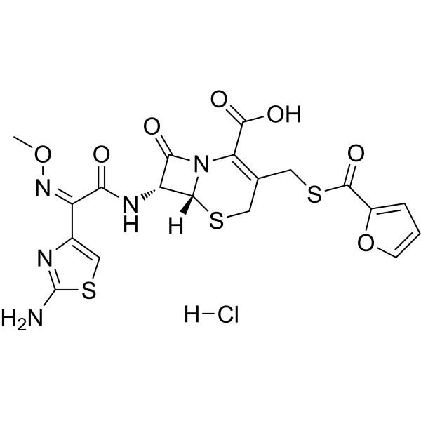
- Formula: C19H18ClN5O7S3
- Molecular Weight:560.02
-
Storage:
-20°C, sealed storage, away from moisture
* In solvent : -80°C, 6 months; -20°C, 1 month (sealed storage, away from moisture)

To place orders, for customer services and technical support, please contact: MedChemExpress USA
Tel: 609-228-6898 E-mail: [email protected] [email protected]


-
 Biological Activity
-
Chemical Information
-
Solvent & Solubility
- Protocols
- Purity & Documentation
- References
-
 Help & FAQs


-

HY-B0026R
Ceftiofur hydrochloride (Standard)Bacterial Inhibitor -
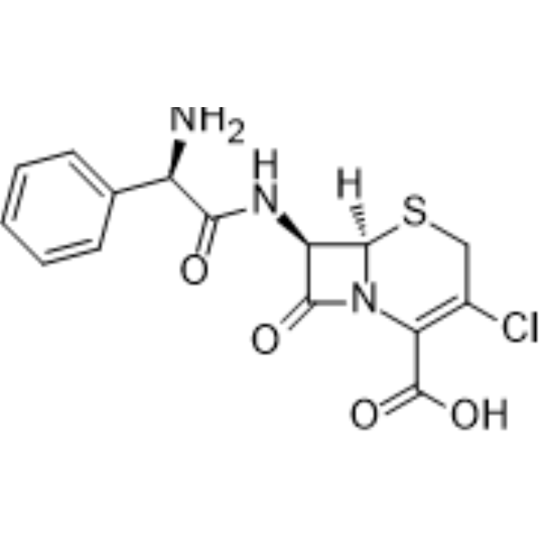
HY-B0198
CefaclorAntibiotic -
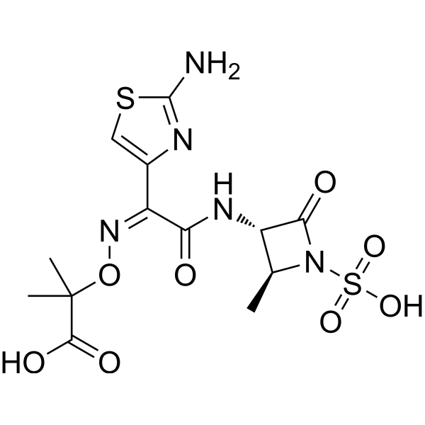
HY-B0129
AztreonamBacterial Inhibitor -
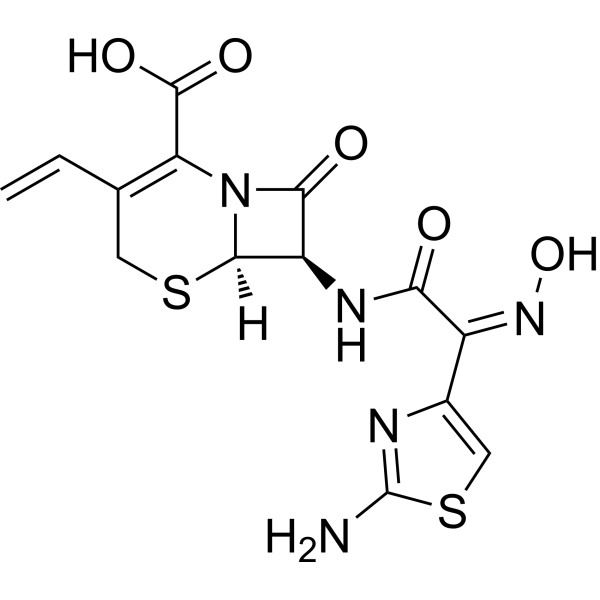
HY-B0136
CefdinirBacterial Inhibitor -
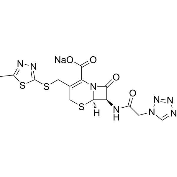
HY-B1078
Cefazolin sodiumCephalosporin Antibiotic -
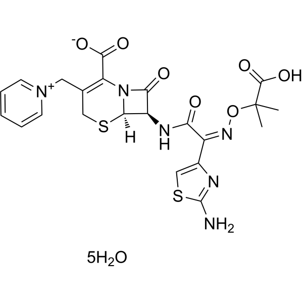
HY-B0593A
Ceftazidime pentahydrateAntibotic -
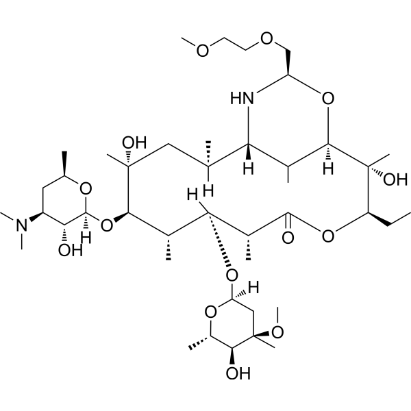
HY-B0643
DirithromycinAntibiotic -
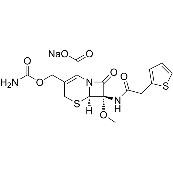
HY-B1117
Cefoxitin sodiumAntibiotic -
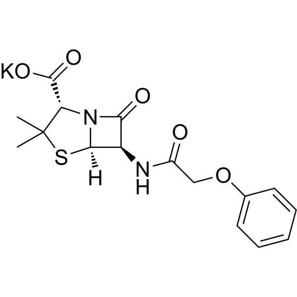
HY-B0975
Penicillin V PotassiumAntibiotic -
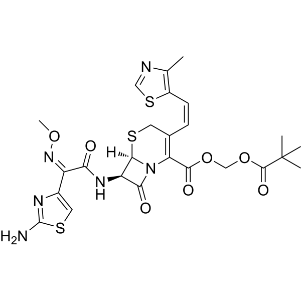
HY-17452A
Cefditoren PivoxilAntibacterial Agent -

HY-B0129R
Aztreonam (Standard)Bacterial Inhibitor -

HY-B0593AR
Ceftazidime pentahydrate (Standard)Antibotic
Publications Citing Use of MedChemExpress (MCE) Ceftiofur hydrochloride
MoreAll Antibiotic Isoforms
More

Biological Activity
Description
IC50 & Target
bacterial[1]
In Vitro
Ceftiofur (1, 5, 10 mg/L; pretreatment for 1 h+LPS stimulation for 12 h) hydrochloride can significantly inhibit the secretion of TNF-α, IL-1β, and IL-6 in RAW 264.7 cells, but has no significant effect on the secretion of IL-10[2].
Ceftiofur hydrochloride (1, 5, 10 mg/L; pretreatment for 1 h+LPS stimulation for 30 min) can significantly inhibit the phosphorylation of ERK, JNK, and p38 and the nuclear translocation of NF-κB p65 in RAW 264.7 cells[2].
MedChemExpress (MCE) has not independently confirmed the accuracy of these methods. They are for reference only. Further protocols information, click here.
-
Cell Line:RAW 264.7 cells
-
Concentration:1, 5, 10 mg/L
-
Incubation Time:1 h pretreatment with Ceftiofur followed by 30 min LPS stimulation
-
Result:Dramatically decreased the phosphorylation levels of ERK, JNK, and p38 in Ceftiofur-treated cells compared to the LPS-treated control cells, while the total protein levels of MAPKs showed no change.
-
Cell Line:RAW 264.7 cells
-
Concentration:1, 5, 10 mg/L
-
Incubation Time:1 h pretreatment with Ceftiofur followed by 30 min LPS stimulation
-
Result:Nuclear translocation of NF-κB p65 induced by LPS was strongly inhibited in a dose-dependent manner in Ceftiofur-pretreated cells.
In Vivo
MedChemExpress (MCE) has not independently confirmed the accuracy of these methods. They are for reference only.
-
Animal Model:Female C57BL/6 mice (18-20 g, 6-8 weeks old), LPS-induced endotoxemia model[3]
-
Dosage:20 mg/kg Ceftiofur (dissolved in saline)
-
Administration:Subcutaneous injection, single dose, administered 1 h before or 0, 1, 4, 12 h after intraperitoneal injection of 30 mg/kg LPS.
-
Result:Pretreatment with 20 mg/kg Ceftiofur 1 h before LPS challenge significantly increased the survival rate of mice (70% survival at 144 h vs. 0% in LPS control group).
When administered 0 or 1 h after LPS injection, Ceftiofur still provided significant protection, but protection decreased when delayed to 4 h and was absent at 12 h.
Plasma levels of TNF-α, IL-1β, and IL-6 in Ceftiofur-pretreated mice were significantly lower than those in the LPS control group at specific time points (1 h for TNF-α, 3 h for IL-6, 12 h for IL-1β), while IL-10 levels were significantly higher at 1 h after LPS challenge.


Chemical Information
-
CAS No. 103980-44-5
-
Appearance Solid
-
Molecular Weight 560.02
-
Formula C19H18ClN5O7S3
-
Color White to light yellow
-
SMILES
[H]Cl.O=C(C(N12)=C(CSC(C3=CC=CO3)=O)CS[C@]2([H])[C@H](NC(/C(C4=CSC(N)=N4)=N\OC)=O)C1=O)O
-
Shipping
Room temperature in continental US; may vary elsewhere.
-
Storage
-20°C, sealed storage, away from moisture
* In solvent : -80°C, 6 months; -20°C, 1 month (sealed storage, away from moisture)


Publications (6)
-
Journal Impact Factor
-
Most Recent
-
Nat Commun
Selective inhibition of cancer cell self-renewal through a Quisinostat-histone H1.0 axis. [Abstract]2020 Apr 14;11(1):1792. PMID: 32286289 -
Mol Syst Biol
2022 Sep;18(9):e11081. PMID: 36065847 -
-
Microorganisms
Isolation, Antimicrobial Susceptibility, and Genotypes of Three Pasteurellaeae Species Prevalent on Pig Farms in China Between 2021 and 2023. [Abstract]2025 Apr 18;13(4):938. PMID: 40284774 -
Vet Microbiol
Discovery of the tigecycline resistance gene cluster tmexCD3-toprJ1 in Pasteurella multocida strains isolated from pigs in China. [Abstract]2024 May:292:110046. PMID: 38471428 -


Solvent & Solubility
In Vitro:
DMSO : 116.67 mg/mL (208.33 mM; Need ultrasonic; Hygroscopic DMSO has a significant impact on the solubility of product, please use newly opened DMSO)
Please refer to the solubility information to select the appropriate solvent. Once prepared, please aliquot and store the solution to prevent product inactivation from repeated freeze-thaw cycles.
Storage method and period of stock solution: -80°C, 6 months; -20°C, 1 month (sealed storage, away from moisture). When stored at -80°C, please use it within 6 months. When stored at -20°C, please use it within 1 month.
Please refer to the solubility information to select the appropriate solvent. Once prepared, please aliquot and store the solution to prevent product inactivation from repeated freeze-thaw cycles.
Storage method and period of stock solution: -80°C, 6 months; -20°C, 1 month (sealed storage, away from moisture). When stored at -80°C, please use it within 6 months. When stored at -20°C, please use it within 1 month.
Concentration (start) × Volume (start) = Concentration (final) × Volume (final)

Protocols
-
LPS-Induced Endotoxemia/Systemic Inflammation
Lipopolysaccharide (LPS)-induced endotoxemia is a widely used in vivo model of acute systemic inflammation in which LPS, a Gram-negative bacterial endotoxin, activates innate immune signaling primarily through TLR4, leading to rapid and transient induction of pro-inflammatory cytokines such as TNF-α, IL-6, and IL-1β in circulation and tissues. This cytokine surge is commonly used as a measurable readout of systemic inflammatory activation and immune dysregulation, and is typically assessed within hours after intraperitoneal LPS administration in mouse models of endotoxemia. The model captures key features of systemic inflammatory response syndrome, including cytokine release, immune cell activation, and downstream tissue responses, and has been used to evaluate anti-inflammatory interventions such as cytokine modulation, lipid mediators, and immune cell-targeting therapies.
-
Protein Extraction
Protein extraction uses physical, chemical or biological methods, such as ultrasonic disruption, salting out, cell lysis, electrophoresis, etc., to destroy the cell membrane structure and to separate the proteins from different components according to their characteristics.
-
Research Protocol for Inflammation-related Diseases
The NLRP3 inflammasome is a cytosolic innate immune signaling platform that integrates priming signals and danger-signal activation to promote caspase-1 activation, maturation of IL-1β and IL-18, and gasdermin D-mediated pyroptotic cell death. The core experimental logic is to determine whether inflammatory disease phenotypes are driven by increased NLRP3 expression, ASC-containing inflammasome assembly, caspase-1 cleavage, GSDMD cleavage, and extracellular release of IL-1β/IL-18 rather than by nonspecific cell injury alone. The pathway is strongly linked to inflammation-related disease phenotypes because monosodium urate crystals activate NALP3/NLRP3 inflammasome signaling in gout-like crystal inflammation, cholesterol crystals activate NLRP3 inflammasomes in atherogenesis models, and DSS-induced intestinal inflammation has been reported to involve NLRP3 inflammasome activity. However, experimental colitis studies also show context-dependent protective effects of NLRP3 inflammasome co
-
Pyroptosis Solutions
Pyroptosis is a lytic inflammatory cell-death pathway executed by gasdermin pores, most classically through inflammasome-mediated activation of caspase-1, cleavage of gasdermin D, membrane pore formation, LDH release, and secretion of IL-1β and IL-18. The canonical pathway is commonly modeled by priming cells with an inflammatory signal such as LPS to induce pro-IL-1β and inflammasome components, followed by an activation signal such as ATP or nigericin to activate NLRP3, ASC speck formation, caspase-1 cleavage, GSDMD cleavage, cytokine release, and pyroptotic membrane rupture. The non-canonical pathway is triggered when cytosolic LPS activates mouse caspase-11 or human caspase-4/5, leading to GSDMD cleavage and pyroptosis, and this can secondarily activate NLRP3-dependent IL-1β release. Pyroptosis is linked to inflammatory injury, infection, cancer, liver disease, ocular disease, placental inflammation, and other disease phenotypes, but unresolved questions include which gasdermin fam


Purity & Documentation
-
Data Sheet (282 KB)
-
SDS (394 KB)
- English - EN (394 KB)
- Français - FR (394 KB)
- Deutsch - DE (394 KB)
- Norwegian - NO (394 KB)
- Español - ES (394 KB)
- Swedish - SV (394 KB)
- Italian - IT (394 KB)
- Korean - KR (394 KB)
- Portuguese - PT (394 KB)
-
Handling Instructions (2659 KB)
References
[1]. Salmon SA, et al. In vitro activity of Ceftiofur and its primary metabolite, desfuroylCeftiofur, against organisms of veterinary importance. J Vet Diagn Invest. 1996 Jul;8(3):332-6. [Content Brief]
[2]. Ci X, et al. Ceftiofur impairs pro-inflammatory cytokine secretion through the inhibition of the activation of NF-kappaB and MAPK. Biochem Biophys Res Commun. 2008 Jul 18;372(1):73-7. [Content Brief]
[3]. Ci X, et al. Ceftiofur regulates LPS-induced production of cytokines and improves LPS-induced survival rate in mice. Inflammation. 2008 Dec;31(6):422-7. [Content Brief]


Complete Stock Solution Preparation Table
Please refer to the solubility information to select the appropriate solvent. Once prepared, please aliquot and store the solution to prevent product inactivation from repeated freeze-thaw cycles.
Storage method and period of stock solution: -80°C, 6 months; -20°C, 1 month (sealed storage, away from moisture). When stored at -80°C, please use it within 6 months. When stored at -20°C, please use it within 1 month.
| Optional Solvent | Concentration Solvent Mass | 1 mg | 5 mg | 10 mg | 25 mg |
|---|---|---|---|---|---|
| DMSO | 1 mM | 1.7857 mL | 8.9283 mL | 17.8565 mL | 44.6413 mL |
| 5 mM | 0.3571 mL | 1.7857 mL | 3.5713 mL | 8.9283 mL | |
| 10 mM | 0.1786 mL | 0.8928 mL | 1.7857 mL | 4.4641 mL | |
| 15 mM | 0.1190 mL | 0.5952 mL | 1.1904 mL | 2.9761 mL | |
| 20 mM | 0.0893 mL | 0.4464 mL | 0.8928 mL | 2.2321 mL | |
| 25 mM | 0.0714 mL | 0.3571 mL | 0.7143 mL | 1.7857 mL | |
| 30 mM | 0.0595 mL | 0.2976 mL | 0.5952 mL | 1.4880 mL | |
| 40 mM | 0.0446 mL | 0.2232 mL | 0.4464 mL | 1.1160 mL | |
| 50 mM | 0.0357 mL | 0.1786 mL | 0.3571 mL | 0.8928 mL | |
| 60 mM | 0.0298 mL | 0.1488 mL | 0.2976 mL | 0.7440 mL | |
| 80 mM | 0.0223 mL | 0.1116 mL | 0.2232 mL | 0.5580 mL | |
| 100 mM | 0.0179 mL | 0.0893 mL | 0.1786 mL | 0.4464 mL |


Ceftiofur hydrochloride Related Classifications
HY-B0026 Related Classifications
 Powered by Bioz