



Cepharanthine
Based on 15 publication(s) in Google Scholar
Cepharanthine is a natural product that can be isolated from the plant Stephania cephalantha Hayata. Cepharanthine has anti-severe acute respiratory syndrome coronavirus 2 (anti-SARS-CoV-2) activities. Cepharanthine has good effective in suppressing viral proliferation (half maximal (50%) inhibitory concentration (IC50) and 90% inhibitory concentration (IC90) values of 1.90 and 4.46 μM. Cepharanthine can also effectively reverses P-gp-mediated multidrug resistance in K562 cells and increase enhances the sensitivity of anticancer agents in xenograft mice model. Cepharanthine shows inhibitory effects of human liver cytochrome P450 enzymes CYP3A4, CYP2E1 and CYP2C9. Cepharanthine has antitumor, anti-inflammatory and antinociceptive effects.
For research use only. We do not sell to patients.
- Purity : 99.87%
- CAS No.: 481-49-2
- Formula: C37H38N2O6
- Molecular Weight:606.71
-
Storage:Powder -20°C, 3 years , 4°C, 2 years ; In solvent -80°C, 6 months , -20°C, 1 month

To place orders, for customer services and technical support, please contact: MedChemExpress USA
Tel: 609-228-6898 E-mail: [email protected] [email protected]


-
 Biological Activity
-
Chemical Information
-
Solvent & Solubility
- Protocols
- Purity & Documentation
- References
-
 Help & FAQs


-
HY-N1372A
FangchinolineAutophagy Inducer -
HY-N0453
HypericinPKC Inhibitor -
HY-13764
TetrandrineCalcium Channel Inhibitor -
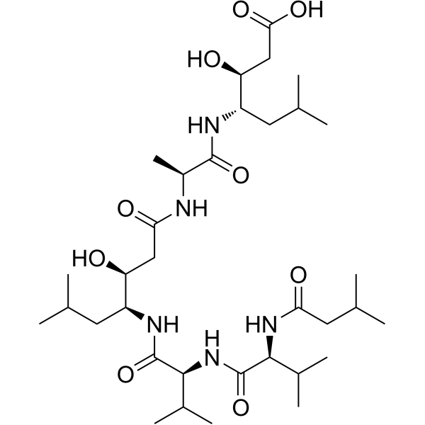
HY-P0018
PepstatinAspartic Protease Inhibitor -
HY-Y0873
PEG300Neutral Polymer -
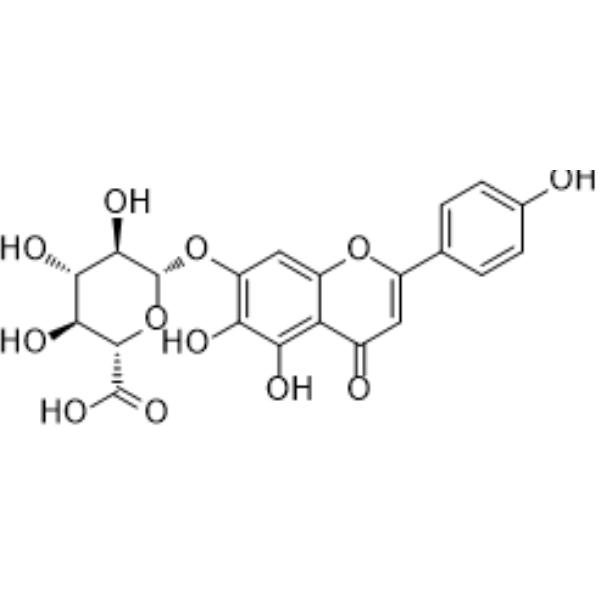
HY-N0751
ScutellarinSTAT Inhibitor -

HY-100579
Ferrostatin-1Ferroptosis Inhibitor -
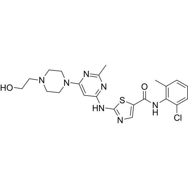
HY-10181
DasatinibSrc/Bcr-Abl Inhibitor -
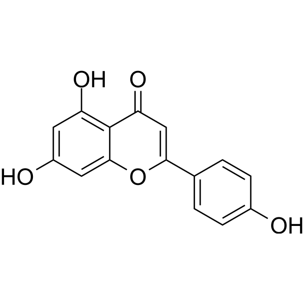
HY-N1201
ApigeninCytochrome P450 Inhibitor -
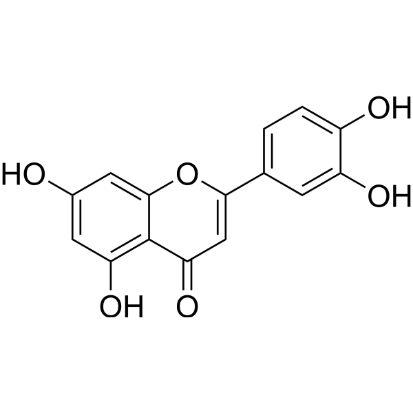
HY-N0162
LuteolinNrf2 Inhibitor -

HY-14648
DexamethasoneGR Agonist -

HY-Y0703
Sodium carboxymethyl cellulose (Viscosity:800-1200 mPa.s)Biochemical Assay Reagent
Publications Citing Use of MedChemExpress (MCE) Cepharanthine
More- J Transl Med. 2025 Jan 22;23(1):103. [Abstract]
- Am J Chin Med. 2026;54(1):329-348. [Abstract]
- Int Immunopharmacol. 2026 Sep 1:184:116935. [Abstract]
- Virol J. 2025 Jun 4;22(1):181. [Abstract]
- J Med Virol. 2026 Jul;98(7):e71062.
- Vet Microbiol. 2026 May:316:110992. [Abstract]
- Vet Microbiol. 2025 Sep:308:110647. [Abstract]
- Vet Microbiol. 2025 Jun:305:110519. [Abstract]
- Virology. 2025 Mar:604:110422. [Abstract]
- Arch Dermatol Res. 2025 Apr 24;317(1):734. [Abstract]
- Am J Transl Res. 2024 May 15;16(5):1602-1619. [Abstract]
- SSRN. 2026 Mar 15.
- SSRN. 2023 Sep 21.
- Oxid Med Cell Longev. 2022 Feb 9;2022:4295208. [Abstract]
- bioRxiv. 2020 Jun.
 Customer Validation & Images

-
Cell Proliferation/Viability Assay
-
Bio/Physico-chemical Assay
-
Flow Cytometry
-
WB
-
IF
All Parasite Isoforms
More

Biological Activity
Description
IC50 & Target
[4]|
CYP3A4 16.29 μM (IC50) |
CYP2E1 25.62 μM (IC50) |
CYP2C9 24.57 μM (IC50) |
Cellular Effect
|
Cell Line

|
Type | Value | Description | References |
|---|---|---|---|---|
| A-431 | ED50 |
2.1 μg/mL
Compound: 11
|
Cytotoxicity against human A431 cells after 3 days by sulforhodamine B assay
Cytotoxicity against human A431 cells after 3 days by sulforhodamine B assay
|
[PMID: 8450319] |
| A549 | IC50 |
5 μM
Compound: 7
|
Cytotoxicity against human A549 cells after 48 hrs by MTS assay
Cytotoxicity against human A549 cells after 48 hrs by MTS assay
|
[PMID: 23621840] |
| A673 | GI50 |
4.5 μM
Compound: Cepheranthine
|
Antiproliferative activity against human A673 assessed as cell growth inhibition after 48 hrs by SRB assay
Antiproliferative activity against human A673 assessed as cell growth inhibition after 48 hrs by SRB assay
|
[PMID: 33226219] |
| ECa-109 cell line | IC50 |
>10 μM
Compound: 7
|
Cytotoxicity against human ECA109 cells after 48 hrs by MTT assay
Cytotoxicity against human ECA109 cells after 48 hrs by MTT assay
|
[PMID: 23621840] |
| ECa-109 cell line | IC50 |
436.7 nM
Compound: CEP
|
Reversal of VCR -resistance in human Eca-109 cells assessed as cell viability at 5 uM incubated for 48 hrs by CCK-8 method (Rvb = 6830.0+/-537.0 nM)
Reversal of VCR -resistance in human Eca-109 cells assessed as cell viability at 5 uM incubated for 48 hrs by CCK-8 method (Rvb = 6830.0+/-537.0 nM)
|
[PMID: 36892076] |
| HCC1806 | GI50 |
7.2 μM
Compound: Cepheranthine
|
Antiproliferative activity against human HCC1806 assessed as cell growth inhibition after 48 hrs by SRB assay
Antiproliferative activity against human HCC1806 assessed as cell growth inhibition after 48 hrs by SRB assay
|
[PMID: 33226219] |
| HCC1937 | GI50 |
6 μM
Compound: Cepheranthine
|
Antiproliferative activity against human HCC1937 assessed as cell growth inhibition after 48 hrs by SRB assay
Antiproliferative activity against human HCC1937 assessed as cell growth inhibition after 48 hrs by SRB assay
|
[PMID: 33226219] |
| HCC70 | GI50 |
5.8 μM
Compound: Cepheranthine
|
Antiproliferative activity against human HCC70 assessed as cell growth inhibition after 48 hrs by SRB assay
Antiproliferative activity against human HCC70 assessed as cell growth inhibition after 48 hrs by SRB assay
|
[PMID: 33226219] |
| HEK-293T | CC50 |
2.1 μM
Compound: 3; cep
|
Cytotoxicity against HEK293T cells assessed as reduction in cell viability by CellTiter-Glo assay
Cytotoxicity against HEK293T cells assessed as reduction in cell viability by CellTiter-Glo assay
|
[PMID: 37043739] |
| HeLa | IC50 |
1.53 μM
Compound: Cepharanthine
|
Inhibition of Ebolavirus glycoprotein/matrix protein VP40 entry in human HeLa cells after 4.5 hrs beta-lactamase reporter assay
Inhibition of Ebolavirus glycoprotein/matrix protein VP40 entry in human HeLa cells after 4.5 hrs beta-lactamase reporter assay
|
[PMID: 29624387] |
| HL-60 | IC50 |
9.2 μM
Compound: 7
|
Cytotoxicity against human HL60 cells after 48 hrs by MTS assay
Cytotoxicity against human HL60 cells after 48 hrs by MTS assay
|
[PMID: 23621840] |
| HT-1080 | ED50 |
6.1 μg/mL
Compound: 11
|
Cytotoxicity against human HT1080 cells after 3 days by sulforhodamine B assay
Cytotoxicity against human HT1080 cells after 3 days by sulforhodamine B assay
|
[PMID: 8450319] |
| KB | ED50 |
5.9 μg/mL
Compound: 11
|
Cytotoxicity against human KB cells after 3 days by sulforhodamine B assay
Cytotoxicity against human KB cells after 3 days by sulforhodamine B assay
|
[PMID: 8450319] |
| KB | ED50 |
9700 nM
Compound: 12
|
Cytotoxicity against human KB cells after 72 hrs by SRB assay
Cytotoxicity against human KB cells after 72 hrs by SRB assay
|
[PMID: 9917283] |
| KB-V1 | ED50 |
0.9 μg/mL
Compound: 11
|
Cytotoxicity against human KBV1 cells after 3 days by sulforhodamine B assay
Cytotoxicity against human KBV1 cells after 3 days by sulforhodamine B assay
|
[PMID: 8450319] |
| LNCaP | ED50 |
5.6 μg/mL
Compound: 11
|
Cytotoxicity against human LNCAP cells after 3 days by sulforhodamine B assay
Cytotoxicity against human LNCAP cells after 3 days by sulforhodamine B assay
|
[PMID: 8450319] |
| MCF7 | IC50 |
2.9 μM
Compound: 7
|
Cytotoxicity against human MCF7 cells after 48 hrs by MTS assay
Cytotoxicity against human MCF7 cells after 48 hrs by MTS assay
|
[PMID: 23621840] |
| MDA-MB-231 | GI50 |
5.3 μM
Compound: Cepheranthine
|
Antiproliferative activity against human MDA-MB-231 assessed as cell growth inhibition after 48 hrs by SRB assay
Antiproliferative activity against human MDA-MB-231 assessed as cell growth inhibition after 48 hrs by SRB assay
|
[PMID: 33226219] |
| MDA-MB-453 | GI50 |
5.5 μM
Compound: Cepheranthine
|
Antiproliferative activity against human MDA-MB-453 assessed as cell growth inhibition after 48 hrs by SRB assay
Antiproliferative activity against human MDA-MB-453 assessed as cell growth inhibition after 48 hrs by SRB assay
|
[PMID: 33226219] |
| MOLT-4 | CC50 |
10 μg/mL
Compound: CEP, Cepharanthine
|
Cytotoxicity against human MOLT4 cells assessed as cell growth inhibition after 24 hrs by MTT assay
Cytotoxicity against human MOLT4 cells assessed as cell growth inhibition after 24 hrs by MTT assay
|
[PMID: 24704028] |
| P388 | ED50 |
0.3 μg/mL
Compound: 11
|
Cytotoxicity against mouse P388 cells after 2 days by sulforhodamine B assay
Cytotoxicity against mouse P388 cells after 2 days by sulforhodamine B assay
|
[PMID: 8450319] |
| SJRH30 | GI50 |
3.8 μM
Compound: Cepheranthine
|
Antiproliferative activity against human SJRH30 assessed as cell growth inhibition after 48 hrs by SRB assay
Antiproliferative activity against human SJRH30 assessed as cell growth inhibition after 48 hrs by SRB assay
|
[PMID: 33226219] |
| SK-MEL-2 | ED50 |
14.5 μg/mL
Compound: 11
|
Cytotoxicity against human SK-MEL-2 cells after 3 days by sulforhodamine B assay
Cytotoxicity against human SK-MEL-2 cells after 3 days by sulforhodamine B assay
|
[PMID: 8450319] |
| SMMC-7721 | IC50 |
9.9 μM
Compound: 7
|
Cytotoxicity against human SMMC7721 cells after 48 hrs by MTS assay
Cytotoxicity against human SMMC7721 cells after 48 hrs by MTS assay
|
[PMID: 23621840] |
| SW480 | IC50 |
4.7 μM
Compound: 7
|
Cytotoxicity against human SW480 cells after 48 hrs by MTS assay
Cytotoxicity against human SW480 cells after 48 hrs by MTS assay
|
[PMID: 23621840] |
| U-937 | IC50 |
>50 μM
Compound: 8, NSC 623442
|
Cytotoxicity against human U937 cells after 24 hrs by alamar blue assay
Cytotoxicity against human U937 cells after 24 hrs by alamar blue assay
|
[PMID: 22766217] |
| Vero | CC50 |
>50 μM
Compound: Cepharanthine
|
Cell viability measured by CellTiter-Glo assay in Vero cells at MOI 0.05 after 72hr
Cell viability measured by CellTiter-Glo assay in Vero cells at MOI 0.05 after 72hr
|
10.1101/2020.03.20.999730 |
| Vero | IC50 |
4.47 μM
Compound: Cepharanthine
|
Antiviral activity against SARS-CoV-2 (viral titer) measured by plaque assay in Vero cells at MOI 0.0125 after 24 hr
Antiviral activity against SARS-CoV-2 (viral titer) measured by plaque assay in Vero cells at MOI 0.0125 after 24 hr
|
10.1101/2020.03.20.999730 |
| Vero C1008 | EC50 |
0.98 μM
Compound: Cepharanthine
|
Antiviral activity against SARS-Co-V-2 infected in Vero E6 cells assessed as reduction in viral replication
Antiviral activity against SARS-Co-V-2 infected in Vero E6 cells assessed as reduction in viral replication
|
[PMID: 37229831] |
| ZR-75-1 | ED50 |
1.2 μg/mL
Compound: 11
|
Cytotoxicity against human ZR-75-1 cells after 3 days by sulforhodamine B assay
Cytotoxicity against human ZR-75-1 cells after 3 days by sulforhodamine B assay
|
[PMID: 8450319] |
In Vitro
Cepharanthine (CEP) (2 μM, 48 h) inhibits cell viability and colony formation and induces apoptosis via the mitochondrial pathway in human TNBC cells[2].
Cepharanthine (2 μM, 48 h) Combinates with Epirubicin (HY-13624) impairs mitochondrial function and causes mitochondrial fission and apoptosis in MDA-MB-231 cells[2].
Cepharanthine (5 μM, 24 h) potently enhances the sensitivity of anticancer agents Doxorubicin (HY-15142A) and Vincristine (HY-N0488) and enhanced apoptosis induced by anticancer agents in K562 cells[3].
Cepharanthine (10-50 μM, 0.5-1 h) changes the distribution of Doxorubicin (HY-15142A) from cytoplasmic vesicles to nucleoplasm in K562 cells by inhibiting the acidification of cytoplasmic organellesin[3].
Cepharanthine (0-50 μM, 30 min) shows inhibitory effects of human liver cytochrome P450 enzymes CYP3A4,CYP2E1 and CYP2C9in vitro[4].
Cepharanthine(0-4 μM, 48 hours) blocks P. falciparum development in ring stage with IC50s of 3.059, 0.927, 2.276, and 1.803 μM for FCM29, W2, 3D7 and K1, respectively[5].
MedChemExpress (MCE) has not independently confirmed the accuracy of these methods. They are for reference only. Further protocols information, click here.
-
Cell Line:MDAMB-231 and BT549 cells
-
Concentration:2 μM
-
Incubation Time:48 h
-
Result:Cepharanthine alone minimally increased apoptosis (~5% to ~10%), whereas combinated with Epirubicin (HY-13624) markedly increased apoptosis (~50%).
-
Cell Line:MDAMB-231 cells
-
Concentration:2 μM
-
Incubation Time:48 h
-
Result:Combinated with Epirubicin (HY-13624) markedly resulted in oxidation of the actin-remodeling protein cofilin, which promoted formation of an intramolecular disulfide bridge between Cys39, Cys80 and Ser3 dephosphorylation, leading to mitochondria translocation of cofilin.
Combinated with Epirubicin (HY-13624) induced mitochondrial fission in MDA-MB-231 cells.
-
Cell Line:K562 cells or MIA-PaCa-2 cells
-
Concentration:10,20,25,50 μM
-
Incubation Time:0.5 h or 1 h
-
Result:Made the intracellular localization of Doxorubicin (HY-15142A) in cytoplasmic vesicles shifted to the nucleoplasm.
Decreased red AO (weakly basic fluorescence probe) fluorescence by dose-dependent mannar in K562 cells.
-
Cell Line:P. falciparum cultivated in type A+ human erythrocytes
-
Concentration:2 μM
-
Incubation Time:48 h
-
Result:Blocked P. falciparum development in ring stage with IC50s of 3.059, 0.927, 2.276, and 1.803 μM for FCM29, W2, 3D7 and K1, respectively.
In Vivo
Cepharanthine (10 mg/kg, i.p., single dose) prevents LPS-induced pulmonary vascular injury in rats by inhibiting leukocyte activation[6].
Cepharanthine (CE)(10 mg/kg, i.p., single dose) exerts anti-inflammatory effects via NF-kB inhibition in a LPS-induced rat model of systemic inflammation[7].
Cepharanthine (20-180 mg/kg, i.p.) results in a dose-dependent antinociceptive effect with an ED50 value of 24.5 mg/kg in mice pain models[8].
MedChemExpress (MCE) has not independently confirmed the accuracy of these methods. They are for reference only.
-
Animal Model:MDA-MB-231 cell xenografts in mice[1]
-
Dosage:12 mg/kg
-
Administration:Intraperitoneal injection (i.p.), once daily for 36 days
-
Result:Combinated with Epirubicin (HY-13624) greatly enhanced the therapeutic efficacy compared with administration of either drug alone.
-
Animal Model:LPS-induced pulmonary vascular injury in male Wistar rats[6]
-
Dosage:10 mg/kg
-
Administration:Intraperitoneal injection (i.p.), single dose
-
Result:Decreased LPS-induced pulmonary vascular injury.
Significantly inhibited the increases in plasma tumor necrosis factor-a (TNF-a) concentrations.
-
Animal Model:LPS-induced Wistar rat model of systemic inflammation[7]
-
Dosage:10 mg/kg
-
Administration:Intraperitoneal injection (i.p.), single dose
-
Result:Significantly inhibited the increase in LPS-induced IL-6, TNF-α and nitrate/nitrite levels.
Reduced interstitial edema and inflammatory cell compared with the control group.
Reduced pathologic abnormalities, such as vacuolization, dot necrosis, striped necrosis, and bridging necrosis appeared, and inflammatory cells compared with the control group.
group compared with the LPS group.
-
Animal Model:Mice pain models in Kunming (KM) strain male and female mice [8]
-
Dosage:10 mg/kg
-
Administration:Intraperitoneal injection (i.p.)
-
Result:Resulted in a dose-dependent antinociceptive effect with an ED50 value of 24.5 mg/kg in mice pain models.
Significantly decreased the intestinal propulsion with maximal inhibition at 33.6%.
Clinical Trial
| NCT Number | Sponsor | Condition | Start Date |
Phase
|
|---|---|---|---|---|
| NCT01329991 | Plexxikon| | 2011-05 | PHASE1 |


Chemical Information
-
CAS No. 481-49-2
-
Appearance Solid
-
Molecular Weight 606.71
-
Formula C37H38N2O6
-
Color White to yellow
-
SMILES
[H][C@]12N(C)CCC(C1=C3)=CC(OC)=C3OC4=C5C(OCO5)=CC6=C4[C@](N(C)CC6)([H])CC(C=C7)=CC=C7OC8=C(OC)C=CC(C2)=C8
-
Structure Classification
-
Shipping
Room temperature in continental US; may vary elsewhere.
-
Storage
Powder -20°C 3 years 4°C 2 years In solvent -80°C 6 months -20°C 1 month


Publications (15)
-
Journal Impact Factor
-
Most Recent
-
J Transl Med
Single-cell profiling of SLC family transporters: uncovering the role of SLC7A1 in osteosarcoma. [Abstract]2025 Jan 22;23(1):103. PMID: 39844299
Cepharanthine purchased from MedChemExpress. Usage Cited in: J Transl Med. 2025 Jan 22;23(1):103. [Abstract]
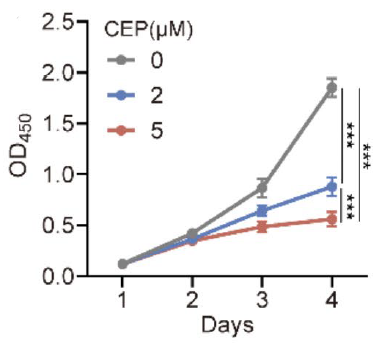
Cepharanthine (CPE) (2–5 μM) dose-dependently inhibited the proliferation of 143B cells.
Cepharanthine purchased from MedChemExpress. Usage Cited in: J Transl Med. 2025 Jan 22;23(1):103. [Abstract]
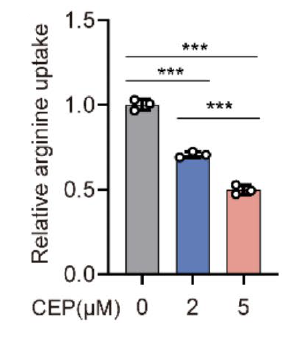
Cepharanthine (CPE) (2–5 μM) enhanced its inhibitory effects on arginine uptake in 143B cells with increasing concentration.
Cepharanthine purchased from MedChemExpress. Usage Cited in: J Transl Med. 2025 Jan 22;23(1):103. [Abstract]
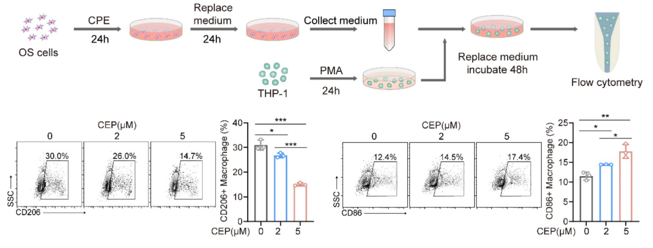
Cepharanthine (CPE) (2–5 μM; 48 h) pretreatment significantly downregulated CD206 expression and upregulated CD86 expression in THP1-derived macrophages in a dose-dependent manner.
-
Am J Chin Med
Cepharanthine Triggers Ferroptosis in Gastric Cancer by PINK1/FUNDC1-Mediated Mitophagy-Dependent GPX4 Degradation. [Abstract]2026;54(1):329-348. PMID: 41684093 -
Int Immunopharmacol
Cepharanthine inhibits NLRP3 inflammasome-induced pyroptosis through ARIH2-mediated degradation of NLRP3. [Abstract]2026 Sep 1:184:116935. PMID: 42217341 -
Virol J
Bisbenzylisoquinoline alkaloids inhibit influenza virus replication by disrupting endosomal acidification. [Abstract]2025 Jun 4;22(1):181. PMID: 40468427
Cepharanthine purchased from MedChemExpress. Usage Cited in: Virol J. 2025 Jun 4;22(1):181. [Abstract]
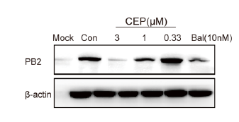
Cepharanthine (CPE) (0.33-3 nM; 24 h) dose-dependently suppressed protein levels of the viral PB2 in A549 cells.
Cepharanthine purchased from MedChemExpress. Usage Cited in: Virol J. 2025 Jun 4;22(1):181. [Abstract]
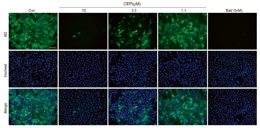
Cepharanthine (CPE) (0.33-3 nM; 24 h) significantly reduced the expression of the IAV M2 protein in MDCK cells.
-
-
Vet Microbiol
The Chinese medicine monomer Schisandrin C inhibits PRRSV infection by regulating the OGT-PI3K/AKT/mTOR signaling pathway. [Abstract]2026 May:316:110992. PMID: 41865607 -
Vet Microbiol
Antiviral activity of Stephania japonica extract against porcine epidemic diarrhea virus infection. [Abstract]2025 Sep:308:110647. PMID: 40706130 -
Vet Microbiol
Cepharanthine mitigates NIBV-induced pyroptosis via the MDA5/NF-κB/NLRP3 signaling pathway. [Abstract]2025 Jun:305:110519. PMID: 40273551 -
Virology
Cepharanthine: A promising natural compound against feline infectious peritonitis virus infection and associated inflammation. [Abstract]2025 Mar:604:110422. PMID: 39884162 -
Arch Dermatol Res
2025 Apr 24;317(1):734. PMID: 40274641 -
Am J Transl Res
Cepharanthine inhibits migration, invasion, and EMT of bladder cancer cells by activating the Rap1 signaling pathway in vitro. [Abstract]2024 May 15;16(5):1602-1619. PMID: 38883391 -
-
-
Oxid Med Cell Longev
Cepharanthine Attenuates Early Brain Injury after Subarachnoid Hemorrhage in Mice via Inhibiting 15-Lipoxygenase-1-Mediated Microglia and Endothelial Cell Ferroptosis. [Abstract]2022 Feb 9;2022:4295208. PMID: 35186185 -


Solvent & Solubility
In Vitro:
DMSO : 50 mg/mL (82.41 mM; Need ultrasonic; Hygroscopic DMSO has a significant impact on the solubility of product, please use newly opened DMSO)
Please refer to the solubility information to select the appropriate solvent. Once prepared, please aliquot and store the solution to prevent product inactivation from repeated freeze-thaw cycles.
Storage method and period of stock solution: -80°C, 6 months; -20°C, 1 month. When stored at -80°C, please use it within 6 months. When stored at -20°C, please use it within 1 month.
Please refer to the solubility information to select the appropriate solvent. Once prepared, please aliquot and store the solution to prevent product inactivation from repeated freeze-thaw cycles.
Storage method and period of stock solution: -80°C, 6 months; -20°C, 1 month. When stored at -80°C, please use it within 6 months. When stored at -20°C, please use it within 1 month.
Concentration (start) × Volume (start) = Concentration (final) × Volume (final)
In Vivo:
Select the appropriate dissolution method based on your experimental animal and administration route.
- For the following dissolution methods, please ensure to first prepare a clear stock solution using an In Vitro approach and then sequentially add co-solvents:
- To ensure reliable experimental results, the clarified stock solution can be appropriately stored based on storage conditions. As for the working solution for In Vivo experiments, it is recommended to prepare freshly and use it on the same day.
- The percentages shown for the solvents indicate their volumetric ratio in the final prepared solution. If precipitation or phase separation occurs during preparation, heat and/or sonication can be used to aid dissolution.
Add each solvent one by one: 10% DMSO 90% (20% SBE-β-CD in Saline)
Solubility: 2.5 mg/mL (4.12 mM); Suspended solution; Need ultrasonic
This protocol yields a suspended solution of 2.5 mg/mL. Suspended solution can be used for oral and intraperitoneal injection.
Taking 1 mL working solution as an example, add 100 μL DMSO stock solution (25.0 mg/mL) to 900 μL 20% SBE-β-CD in Saline, and mix evenly.
Preparation of 20% SBE-β-CD in Saline (4°C, storage for one week): 2 g SBE-β-CD powder is dissolved in 10 mL Saline, completely dissolve until clear.
Add each solvent one by one: 10% DMSO 40% PEG300 5% Tween-80 45% Saline
Solubility: ≥ 2.08 mg/mL (3.43 mM); Clear solution
This protocol yields a clear solution of ≥ 2.08 mg/mL (saturation unknown).
Taking 1 mL working solution as an example, add 100 μL DMSO stock solution (20.8 mg/mL) to 400 μL PEG300, and mix evenly; then add 50 μL Tween-80 and mix evenly; then add 450 μL Saline to adjust the volume to 1 mL.
Preparation of Saline: Dissolve 0.9 g sodium chloride in ddH₂O and dilute to 100 mL to obtain a clear Saline solution.
For the following dissolution methods, please prepare the working solution directly:
It is recommended to prepare fresh solutions and use them promptly within a short period of time.
The percentages shown for the solvents indicate their volumetric ratio in the final prepared solution. If precipitation or phase separation occurs during preparation, heat and/or sonication can be used to aid dissolution.
Add each solvent one by one: 50% PEG300 50% Saline
Solubility: 6.02 mg/mL (9.92 mM); Suspended solution; Need ultrasonic
Add each solvent one by one: 15% Cremophor EL 85% Saline
Solubility: 6.02 mg/mL (9.92 mM); Suspended solution; Need ultrasonic
In Vivo Dissolution Calculator
Please enter the basic information of animal experiments:
-
-
-
-
Recommended: Prepare an additional quantity of animals to account for potential losses during experiments.
Please enter your animal formula composition:
-
%DMSO +
Recommended: Keep the proportion of DMSO in working solution below 2% if your animal is weak.
-
%+
-
+%Tween-80 + +
-
%Saline +
The co-solvents required include: DMSO, . All of co-solvents are available by MedChemExpress (MCE). , Tween 80. All of co-solvents are available by MedChemExpress (MCE).
Working solution concentration: 0.22 mg/mL
Method for preparing stock solution: mg drug dissolved in μL DMSO. Stock solution concentration: mg/mL.
1. Take μL DMSO stock solution;
2. Add μL .
μL , mix evenly;
3. Then add μL Tween 80, mix evenly;
4. Then add μL
Please ensure that the stock solution in the first step is dissolved to a clear state, and add co-solvents in sequence. You can use ultrasonic heating (ultrasonic cleaner, recommended frequency 20-40 kHz), vortexing, etc. to assist dissolution.

Protocols
-
Cell Cytotoxicity Assay
Cytotoxicity assays are usually based on the assessment of cell membrane damage, which can also be indirectly detected by measuring cell viability. Detection methods include MTT assay, CKK-8 assay, LDH assay and ATP assay, etc.
-
Patient-Derived Xenograft (PDX)
Patient-derived xenograft (PDX) models are generated by engrafting primary human tumor tissue directly into immunodeficient mice, allowing in vivo propagation of patient tumor biology without initial in vitro adaptation. These models are used to preserve key histopathological and molecular characteristics of the original tumor and enable assessment of tumor growth dynamics and therapeutic response in a living organism. The biological readout is tumor engraftment and subsequent growth in the murine host, which reflects the ability of human tumor cells to survive, vascularize, and expand in an immunocompromised microenvironment.
-
Splenic/Portal-Vein Liver Metastasis Xenograft
Splenic and portal-vein liver metastasis xenograft models deliver tumor cells into the portal circulation so that cells reach the liver first and form hepatic metastatic lesions; splenic injection uses the spleen as an access route to the portal system, while direct portal-vein injection introduces cells into the portal vein without requiring splenectomy. The assay detects liver colonization, intrahepatic tumor growth, tumor distribution, treatment response, survival, and liver-metastasis microenvironment changes; readouts include bioluminescence or fluorescence imaging, gross liver nodule counts, liver weight or tumor burden, histology, and survival.
-
Research Protocol for Inflammation-related Diseases
The NLRP3 inflammasome is a cytosolic innate immune signaling platform that integrates priming signals and danger-signal activation to promote caspase-1 activation, maturation of IL-1β and IL-18, and gasdermin D-mediated pyroptotic cell death. The core experimental logic is to determine whether inflammatory disease phenotypes are driven by increased NLRP3 expression, ASC-containing inflammasome assembly, caspase-1 cleavage, GSDMD cleavage, and extracellular release of IL-1β/IL-18 rather than by nonspecific cell injury alone. The pathway is strongly linked to inflammation-related disease phenotypes because monosodium urate crystals activate NALP3/NLRP3 inflammasome signaling in gout-like crystal inflammation, cholesterol crystals activate NLRP3 inflammasomes in atherogenesis models, and DSS-induced intestinal inflammation has been reported to involve NLRP3 inflammasome activity. However, experimental colitis studies also show context-dependent protective effects of NLRP3 inflammasome co
-
Subcutaneous Cell-Line-Derived Xenograft
Subcutaneous cell-line-derived xenograft (CDX) models are established by implanting cultured human cancer cell lines into immunodeficient mice, where the injected cells form localized tumors that can be monitored in vivo as a measure of tumorigenic potential, growth kinetics, and treatment response. These models are widely used in oncology research because they allow reproducible tumor formation and enable comparative assessment of tumor growth between different cell lines or genetic manipulations in a controlled in vivo microenvironment. Subcutaneous implantation of cancer cells in immunodeficient mice is a standard approach for evaluating tumor growth behavior and therapeutic response across multiple cancer types, including prostate, esophageal, pancreatic, and colon cancer models.
-
Orthotopic Cell-Line Xenograft
Orthotopic cell-line xenograft models involve implantation of human cancer cell lines into the anatomically corresponding organ of immunodeficient mice to reproduce tumor growth within a native microenvironment, enabling more clinically relevant tumor behavior compared with subcutaneous models. These models are widely used because orthotopic placement better recapitulates tumor progression, including invasion and metastatic spread, which are often underrepresented in heterotopic implantation systems. Compared with conventional xenografts, orthotopic implantation is described as more technically complex but provides improved simulation of tumor-microenvironment interactions and metastatic behavior, making it particularly valuable for translational oncology research. Surgical orthotopic implantation approaches have been emphasized as enabling faithful reproduction of clinical cancer features, including metastasis and disease progression patterns that align with the tumor’s organ of origi
-
Cell Viability Determination by MTT Colorimetric Assay
The following protocol uses the MTT colorimetric assay as a classic literature-established method for assessing cell viability/metabolic activity in cultured mammalian cells. MTT[3-(4,5-dimethylthiazol-2-yl)-2,5-diphenyltetrazolium bromide] is reduced by metabolically active cells to a colored formazan product; the amount of formazan is quantified spectrophotometrically and provides an indirect measure of metabolically active viable cells. Importantly, MTT reduction reflects cellular oxidoreductase/metabolic activity rather than an absolute direct count of living cells, so changes in cellular metabolism can alter the signal independently of cell number.
-
Intraperitoneal/Peritoneal Dissemination Xenograft
Intraperitoneal (IP) or peritoneal dissemination xenograft models are based on the introduction of human cancer cells into the peritoneal cavity of immunodeficient mice, where they attach to peritoneal surfaces, form multicellular aggregates or spheroids, and progressively generate disseminated tumor nodules that mimic advanced peritoneal metastatic disease. These models are widely used to study ovarian cancer progression, tumor-microenvironment interactions, and intraperitoneal therapeutic responses, often incorporating bioluminescence or fluorescence imaging to longitudinally monitor tumor burden in vivo. The biological principle relies on the capacity of tumor cells such as SKOV3 or related ovarian carcinoma lines to survive in suspension, aggregate within ascites-like fluid, adhere to mesothelial surfaces, and invade peritoneal organs, thereby recapitulating human peritoneal carcinomatosis patterns observed in advanced disease.


Purity & Documentation
-
Data Sheet (297 KB)
-
SDS (393 KB)
- English - EN (393 KB)
- Français - FR (393 KB)
- Deutsch - DE (393 KB)
- Norwegian - NO (393 KB)
- Español - ES (393 KB)
- Swedish - SV (393 KB)
- Italian - IT (393 KB)
- Korean - KR (393 KB)
- Portuguese - PT (393 KB)
-
Handling Instructions (2659 KB)
References
[1]. Hijikata A, et al. Evaluating cepharanthine analogues as natural drugs against SARS-CoV-2. FEBS Open Bio. 2022;12(1):285-294 [Content Brief]
[2]. Shen LW, et.al. Cepharanthine sensitizes human triple negative breast cancer cells to chemotherapeutic agent epirubicin via inducing cofilin oxidation-mediated mitochondrial fission and apoptosis. Acta Pharmacol Sin. 2022 Jan;43(1):177-193. [Content Brief]
[3]. Ikeda R, et.al. Cepharanthine potently enhances the sensitivity of anticancer agents in K562 cells. Cancer Sci. 2005 Jun;96(6):372-6. [Content Brief]
[4]. Zhang X, et.al. In vitro inhibitory effects of cepharanthine on human liver cytochrome P450 enzymes. Pharm Biol. 2020 Dec;58(1):247-252. [Content Brief]
[5]. Hua P, et.al. Cepharanthine induces apoptosis through reactive oxygen species and mitochondrial dysfunction in human non-small-cell lung cancer cells. Biochem Biophys Res Commun. 2015 May 1;460(2):136-42. [Content Brief]
[6]. Desgrouas C, et.al. In vitro antiplasmodial activity of cepharanthine. Malar J. 2014 Aug 22;13:327. [Content Brief]
[7]. Murakami K, et.al. The prevention of lipopolysaccharide-induced pulmonary vascular injury by pretreatment with cepharanthine in rats. Am J Respir Crit Care Med. 2000 Jan;161(1):57-63. [Content Brief]
[8]. Wei XY, et,al. Antinociceptive activities and mechanism of action of Cepharanthine. Biochem Biophys Res Commun. 2022 Jul 23;614:219-224. [Content Brief]


Complete Stock Solution Preparation Table
Please refer to the solubility information to select the appropriate solvent. Once prepared, please aliquot and store the solution to prevent product inactivation from repeated freeze-thaw cycles.
Storage method and period of stock solution: -80°C, 6 months; -20°C, 1 month. When stored at -80°C, please use it within 6 months. When stored at -20°C, please use it within 1 month.
| Optional Solvent | Concentration Solvent Mass | 1 mg | 5 mg | 10 mg | 25 mg |
|---|---|---|---|---|---|
| DMSO | 1 mM | 1.6482 mL | 8.2412 mL | 16.4823 mL | 41.2058 mL |
| 5 mM | 0.3296 mL | 1.6482 mL | 3.2965 mL | 8.2412 mL | |
| 10 mM | 0.1648 mL | 0.8241 mL | 1.6482 mL | 4.1206 mL | |
| 15 mM | 0.1099 mL | 0.5494 mL | 1.0988 mL | 2.7471 mL | |
| 20 mM | 0.0824 mL | 0.4121 mL | 0.8241 mL | 2.0603 mL | |
| 25 mM | 0.0659 mL | 0.3296 mL | 0.6593 mL | 1.6482 mL | |
| 30 mM | 0.0549 mL | 0.2747 mL | 0.5494 mL | 1.3735 mL | |
| 40 mM | 0.0412 mL | 0.2060 mL | 0.4121 mL | 1.0301 mL | |
| 50 mM | 0.0330 mL | 0.1648 mL | 0.3296 mL | 0.8241 mL | |
| 60 mM | 0.0275 mL | 0.1374 mL | 0.2747 mL | 0.6868 mL | |
| 80 mM | 0.0206 mL | 0.1030 mL | 0.2060 mL | 0.5151 mL |


Cepharanthine Related Classifications
HY-N6972 Related Classifications
 Powered by Bioz