



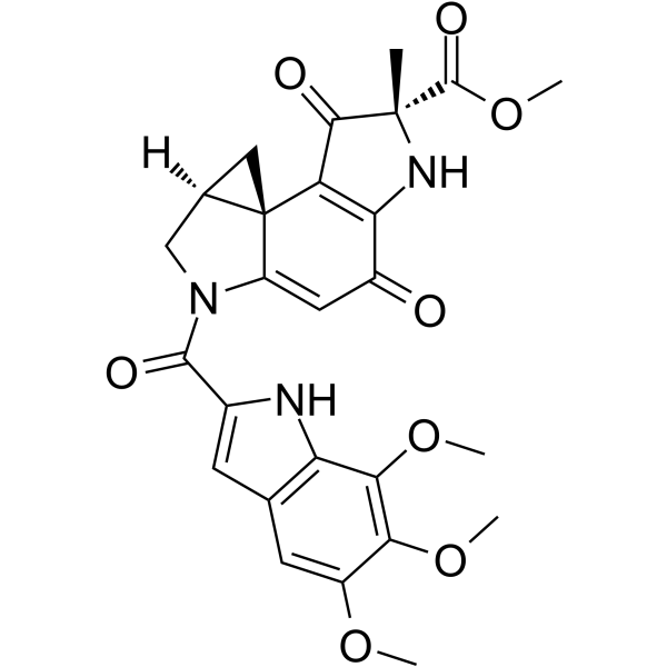
Duocarmycin A
Duocarmycin A is an antitumor antibiotic and DNA alkylating agent with broad-spectrum antibacterial activity, which can serve as a payload for synthesizing antibody-drug conjugates (ADCs). Duocarmycin A selectively binds to the AT-rich minor groove of DNA, forms covalent adducts by alkylating the adenine N3 residue, thereby disrupting DNA structure and inhibiting its replication and transcription. Duocarmycin A induces apoptosis, sub-G1 phase accumulation and chromatin condensation, reduces the levels of pro-caspase-3/9, and induces p53-independent p21 expression. Duocarmycin A is widely used in the research of various malignancies, including leukemia, sarcoma, glioblastoma, as well as multiple solid tumor models such as lung cancer, breast cancer, and colorectal cancer.
Nos produits utilisent uniquement pour la recherche. Nous ne vendons pas aux patients.
- CAS No.: 118292-34-5
- Formule: C26H25N3O8
- Masse moléculaire:507.49
-
Stockage:
Please store the product under the recommended conditions in the Certificate of Analysis.

To place orders, for customer services and technical support, please contact: MedChemExpress USA
Tel: 609-228-6898 E-mail: [email protected] [email protected]


-
 Activité biologique
-
Chemical Information
- Protocole
- Pureté et documentation
- Références
-
 Help & FAQs


-
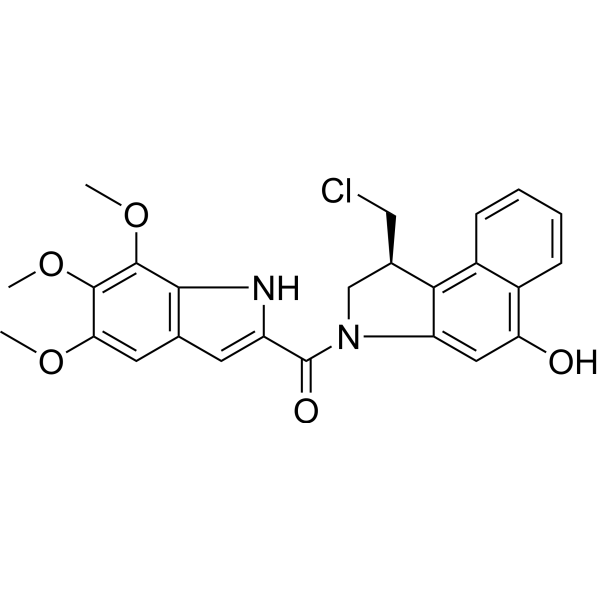
HY-107769
Duocarmycin TMDNA Alkylator -
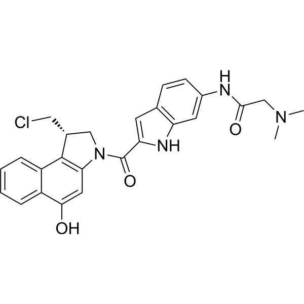
HY-128873
Duocarmycin GAADC Cytotoxin -
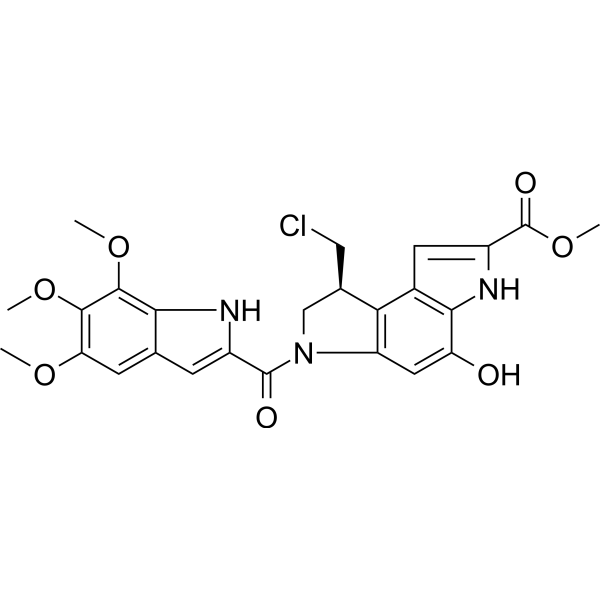
HY-129356A
(S)-Seco-Duocarmycin SAS-enantiomer -
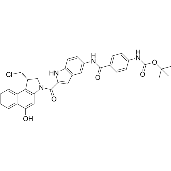
HY-129355
Duocarmycin analog-1DNA Alkylator/Crosslinker -

HY-107769R
Duocarmycin TM (Standard)Reference Standard -
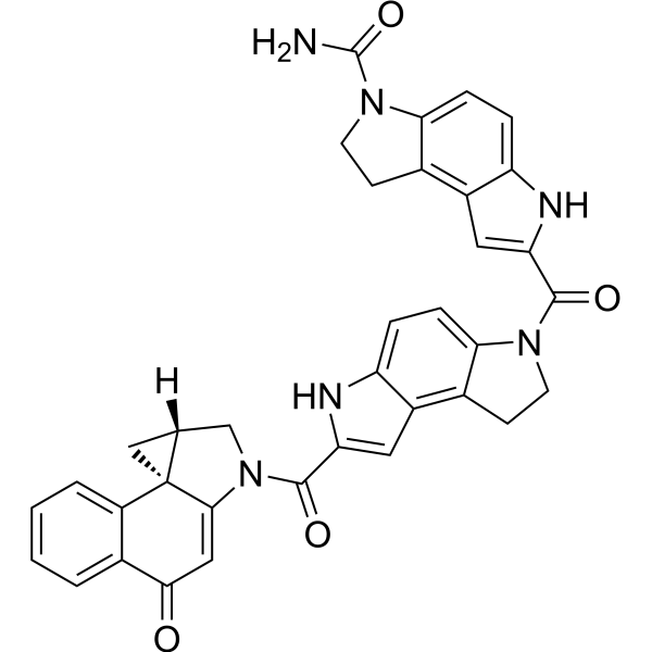
HY-128881
(+)-CBI-CDPI2DNA Alkylating Agent -
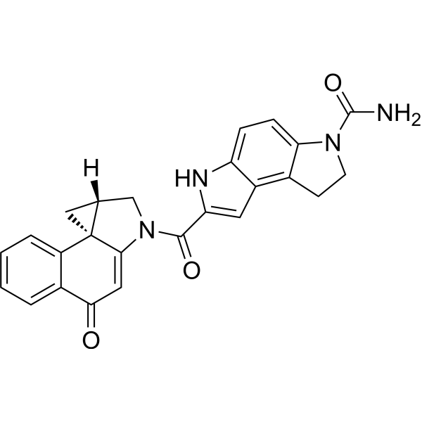
HY-128880
(+)-CBI-CDPI1DNA Alkylating Agent
Voir tous les produits spécifiques à Isoform ADC Payloads
MoreVoir tous les produits spécifiques à Isoform Antibiotic
MoreVoir tous les produits spécifiques à Isoform Caspase
More

Activité biologique
Description
IC50 & Target
|
Procaspase-3 |
Caspase-9 |
Duocarmycins |
In Vitro
Duocarmycin A (0.0016-0.0064 μg/ml; 0.032-42 μg/ml) potently inhibits the growth of Gram-positive bacterial strains (MIC 0.0016-0.0064 μg/ml) and exhibits variable inhibitory activity against Gram-negative bacterial and fungal strains (MIC 0.032-42 μg/ml)[1].
Duocarmycin A (0.003-0.03 μg/mL) induces intrinsic pathway apoptosis and G1 phase cell cycle arrest in HLC-2 cells in vitro in a dose-dependent manner, with altered expression of key apoptotic proteins (caspase-3, caspase-9, Bax) at 0.03 μg/mL[2].
Duocarmycin A (0.003-0.03 μg/ml; 72 h) induces concentration-dependent apoptosis in HLC-2 cells, with 27% apoptosis at 0.003 μg/ml and 51% apoptosis at 0.03 μg/ml after 72 h of treatment, accompanied by characteristic apoptotic nuclear morphology[3].
Duocarmycin A (0.003-0.03 μg/ml; 24-72 h) induces time- and concentration-dependent accumulation of apoptotic sub-G1 phase cells in HLC-2 cultures, with higher sub-G1 populations observed at 0.03 μg/ml compared to 0.003 μg/ml across 24, 48, and 72 h treatments[3].
MedChemExpress (MCE) has not independently confirmed the accuracy of these methods. They are for reference only.
-
Cell Line:human acute myeloid leukemia HL-60 (H2O2-sensitive) and HP100 (H2O2-resistant) cells
-
Concentration:10 nM
-
Incubation Time:30 min, 1 h, 2 h, 3 h, 4 h
-
Result:Induced DNA fragments in both cell lines at 30 min. Triggered DNA ladder formation at 2 h in HL-60 cells and at 3-4 h in HP100 cells.
Reached maximal caspase-3 activity at 3 h in HL-60 cells and 4 h in HP100 cells; did not induce detectable caspase-1 activity in either cell line.
-
Cell Line:human lung carcinoma (HLC-2) cells
-
Concentration:0.003 μg/ml; 0.03 μg/ml
-
Incubation Time:24 h; 48 h; 72 h
-
Result:Resulted in sub-G1 phase (apoptotic) cell populations of 3.94% at 24 h, 12.37% at 48 h, and 6.86% at 72 h at 0.003 μg/ml. Resulted in sub-G1 phase cell populations of 5.56% at 24 h, 15.26% at 48 h, and 20.72% at 72 h at 0.03 μg/ml.
-
Cell Line:human lung carcinoma (HLC-2) cells
-
Concentration:0.003 μg/ml; 0.03 μg/ml
-
Incubation Time:72 h (procaspase-3, procaspase-9, procaspase-8, Bax, Bcl-2); 24, 48, 72 h (p53, p21)
-
Result:Decreased levels of the 32-kDa procaspase-3 and 47-kDa procaspase-9 precursors, and increased levels of pro-apoptotic Bcl-2 family protein Bax after 72 h of treatment; showed no change in procaspase-8 or Bcl-2 levels. Induced relatively high expression of p21, with extremely low accumulation of p53 across 24, 48, and 72 h of treatment.
In Vivo
Duocarmycin-based B2M-ADC selectively targets senescent cancer cells and reduces their viability in in vivo models, with its activity exhibiting senescence phenotype specificity[2].
Galactose-modified Duocarmycin can selectively induce cell death in senescent cancer cells in mice by targeting SA-β-gal activity[2].
MedChemExpress (MCE) has not independently confirmed the accuracy of these methods. They are for reference only.
-
Animal Model:ddY mice with Murine sarcoma 180[1]
-
Dosage:0.04 mg/kg (i.p.);
0.03 mg/kg (i.v.);
0.015 mg/kg (i.v.);
0.0075 mg/kg (i.v.) -
Administration:i.p. or i.v.; single dose
-
Result:Achieved a T/C ratio of 0.35 at 0.04 mg/kg i.p. single dose.\nExhibited T/C ratios ranging from 0.26 to 0.42 at 0.03-0.0075 mg/kg i.v. single doses.
- Research Protocol for Infectious Diseases
- RNA extraction experimental
- Detection of Apoptosis
- TUNEL staining for apoptotic DNA fragmentation
- Annexin V plus membrane-impermeant dye apoptosis staining
- Nuclear DNA counterstaining and nuclear morphology staining
- Apoptosis Solutions
- Bacterial live/dead nucleic-acid viability staining
- Breast Cancer Modeling


Chemical Information
-
CAS No. 118292-34-5
-
Masse moléculaire 507.49
-
Formule C26H25N3O8
-
SMILES
O=C([C@@](N1)(C)C(C([C@@]23[C@@](C3)([H])CN(C(C(N4)=CC5=C4C(OC)=C(OC)C(OC)=C5)=O)C2=C6)=C1C6=O)=O)OC
-
Livraison
Room temperature in continental US; may vary elsewhere.
-
Stockage
Please store the product under the recommended conditions in the Certificate of Analysis.


Pureté et documentation
Références
[2]. Morcos A, et al. A Comprehensive Review of the Antitumor Properties and Mechanistic Insights of Duocarmycin Analogs. Cancers (Basel). 2024;16(19):3293. Published 2024 Sep 27. [Content Brief]
[3]. Hirota M, et al. Distamycin A enhances the cytotoxicity of duocarmycin A and suppresses duocarmycin A-induced apoptosis in human lung carcinoma cells. Int J Biochem Cell Biol. 2007;39(5):988-996. [Content Brief]

Calculators
Concentration (start) × Volume (start) = Concentration (final) × Volume (final)


Duocarmycin A Related Classifications
HY-12455 Related Classifications
 Powered by Bioz