



Eupalinolide B

Based on 1 Customer Validation
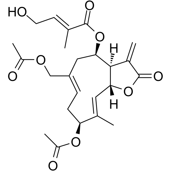
Eupalinolide B is a germ sesquiterpene. Eupalinolide B can be isolated from Eupatorium lindleyanum. Eupalinolide B induces Apoptosis, elevates ROS, promotes Autophagy. regulates GSK-3β/β-catenin, targets UBE2D3 and TAK1, activates ROS-ER-JNK, inhibits NF-κB and MAPKs. Eupalinolide B has anticancer activity against pancreatic cancer and liver cancer. Eupalinolide B relieves rheumatoid arthritis, acute lung injury, periodontitis, depression.
For research use only. We do not sell to patients.
- Purity : 99.27%
- CAS No.: 877822-41-8
- Formula: C24H30O9
- Molecular Weight:462.49
-
Storage:
-20°C, protect from light
* In solvent : -80°C, 6 months; -20°C, 1 month (protect from light)

To place orders, for customer services and technical support, please contact: MedChemExpress USA
Tel: 609-228-6898 E-mail: [email protected] [email protected]


-
 Biological Activity
-
Chemical Information
-
Solvent & Solubility
- Protocols
- Purity & Documentation
- References
-
 Help & FAQs


All MAP3K Isoforms
More

Biological Activity
Description
In Vitro
Eupalinolide B (0-10 μM; 24 h) inhibits the viability of pancreatic cancer cell lines MiaPaCa-2, PANC-1, and PL-45 more significantly than normal pancreatic cells HPNE[1].
Eupalinolide B (0-32 μM; 48 h) reduces the viability of RA-FLS in a concentration-dependent manner[2].
Eupalinolide B (0-10 μM; 1 h) inhibits the production of NO in LPS-stimulated RAW264.7 cells in a dose-dependent manner, with an IC50 value of 2.24 μM[3].
Eupalinolide B (6-24 μM; 24-72 h) significantly inhibits the growth of human hepatocarcinoma cell lines SMMC-7721 and HCCLM3[4].
EB (6-24 μM; 24 h) inhibits the migration of human hepatic carcinoma cells (SMMC-7721 and HCCLM3) via activating ROS-ER-JNK signaling pathway[4].
Eupalinolide B (0.125-1 μM; 48 h) promotes Corticosterone (HY-B1618)-induced PC12 cell proliferation and improve cell viability[6].
MedChemExpress (MCE) has not independently confirmed the accuracy of these methods. They are for reference only.
-
Cell Line:RA-FLS
-
Concentration:4, 8, 16 μM
-
Incubation Time:
-
Result:Increased the ratios of p-AMPK/t-AMPK and p-ULK-1/t-ULK-1 and decreased the p-mTOR/t-mTOR ratio.
-
Cell Line:RA-FLS
-
Concentration:4, 8, 16 μM
-
Incubation Time:48 h
-
Result:Elevated LC3B levels and decreased P62 expression.
In Vivo
Eupalinolide B (8-16 mg/kg; i.p.; daily; 2 weeks) alleviates rheumatoid arthritis in adjuvant-induced arthritis (AIA) rats[2].
Eupalinolide B (5-20 mg/kg; i.p.; administered 2 h before and 6 h, 18 h after LPS treatment) significantly alleviates LPS-induced acute lung injury in mice, manifested as a reduction in lung injury score, myeloperoxidase activity[3].
Eupalinolide B (25-50 mg/kg; i.p.; every 2 days; 3 weeks) significantly inhibits the growth of tumors formed by human hepatocarcinoma cell lines SMMC-7721 or HCCLM3 in nude mice, reducing tumor volume and weight[4].
Eupalinolide B (30 mg/kg; i.p.; daily; 14 days) can effectively improve periodontal inflammatory damage in a mouse periodontitis model and inhibit alveolar bone resorption[5].
Eupalinolide B (5-20 mg/kg; i.p.; once a day; 22 to 35 days after CUMS exposure) can improve the depressive-like behavior of CUMS rats, reduce hippocampal tissue pathological damage, and promote hippocampal neurogenesis[6].
MedChemExpress (MCE) has not independently confirmed the accuracy of these methods. They are for reference only.
-
Animal Model:Balb/c nude mice (male, 5-week-old); xenograft tumor model of pancreatic cancer[1]
-
Dosage:20 mg/kg or 40 mg/kg (dissolved in saline)
-
Administration:Intraperitoneal injection, daily for 4 weeks
-
Result:Significantly slowed tumor growth in the nude mice.
Reduced tumor volume and weigh.
Revealed a decrease in Ki-67 expression.
Inhibited cell proliferation.
-
Animal Model:Sprague-Dawley rats (male, 4-week-old); adjuvant-induced arthritis (AIA) model[2]
-
Dosage:8 mg/kg or 16 mg/kg
-
Administration:Intraperitoneal injection, daily for 2 weeks
-
Result:Reduced hind paw swelling and the arthritis index.
Down-regulated the serum levels of inflammatory cytokines TNF-α, IL-1β, and MCP-1.
Attenuated inflammatory cell infiltration, pannus formation, and synovial hyperplasia.
Reduced Ki67 expression, increased the number of apoptotic cells, and enhanced autophagy in the synovial tissues.
- Collagen-Induced Arthritis
- Detection of Apoptosis
- TUNEL staining for apoptotic DNA fragmentation
- Annexin V plus membrane-impermeant dye apoptosis staining
- ROS/oxidative-stress fluorescent staining
- Autophagy
- Apoptosis Solutions
- Lysosome and acidic-vesicle live-cell staining
- Research Protocol for Inflammation-related Diseases
- Liver Cancer Modeling
- Inhalation Toxicity Study
- Macroautophagy Solutions


Chemical Information
-
CAS No. 877822-41-8
-
Appearance Solid
-
Molecular Weight 462.49
-
Formula C24H30O9
-
Color White to off-white
-
SMILES
O=C(O[C@@H]1C/C(COC(C)=O)=C/C[C@H](OC(C)=O)/C(C)=C/[C@@]([C@]1([H])C2=C)([H])OC2=O)/C(C)=C/CO
-
Structure Classification
-
Shipping
Room temperature in continental US; may vary elsewhere.
-
Storage
-20°C, protect from light
* In solvent : -80°C, 6 months; -20°C, 1 month (protect from light)


Solvent & Solubility
In Vitro:
DMSO : 100 mg/mL (216.22 mM; Need ultrasonic; Hygroscopic DMSO has a significant impact on the solubility of product, please use newly opened DMSO)
Please refer to the solubility information to select the appropriate solvent. Once prepared, please aliquot and store the solution to prevent product inactivation from repeated freeze-thaw cycles.
Storage method and period of stock solution: -80°C, 6 months; -20°C, 1 month (protect from light). When stored at -80°C, please use it within 6 months. When stored at -20°C, please use it within 1 month.
Please refer to the solubility information to select the appropriate solvent. Once prepared, please aliquot and store the solution to prevent product inactivation from repeated freeze-thaw cycles.
Storage method and period of stock solution: -80°C, 6 months; -20°C, 1 month (protect from light). When stored at -80°C, please use it within 6 months. When stored at -20°C, please use it within 1 month.
Concentration (start) × Volume (start) = Concentration (final) × Volume (final)
In Vivo:
Select the appropriate dissolution method based on your experimental animal and administration route.
- For the following dissolution methods, please ensure to first prepare a clear stock solution using an In Vitro approach and then sequentially add co-solvents:
- To ensure reliable experimental results, the clarified stock solution can be appropriately stored based on storage conditions. As for the working solution for In Vivo experiments, it is recommended to prepare freshly and use it on the same day.
- The percentages shown for the solvents indicate their volumetric ratio in the final prepared solution. If precipitation or phase separation occurs during preparation, heat and/or sonication can be used to aid dissolution.
Add each solvent one by one: 10% DMSO 40% PEG300 5% Tween-80 45% Saline
Solubility: ≥ 2.5 mg/mL (5.41 mM); Clear solution
This protocol yields a clear solution of ≥ 2.5 mg/mL (saturation unknown).
Taking 1 mL working solution as an example, add 100 μL DMSO stock solution (25.0 mg/mL) to 400 μL PEG300, and mix evenly; then add 50 μL Tween-80 and mix evenly; then add 450 μL Saline to adjust the volume to 1 mL.
Preparation of Saline: Dissolve 0.9 g sodium chloride in ddH₂O and dilute to 100 mL to obtain a clear Saline solution.
Add each solvent one by one: 10% DMSO 90% (20% SBE-β-CD in Saline)
Solubility: ≥ 2.5 mg/mL (5.41 mM); Clear solution
This protocol yields a clear solution of ≥ 2.5 mg/mL (saturation unknown).
Taking 1 mL working solution as an example, add 100 μL DMSO stock solution (25.0 mg/mL) to 900 μL 20% SBE-β-CD in Saline, and mix evenly.
Preparation of 20% SBE-β-CD in Saline (4°C, storage for one week): 2 g SBE-β-CD powder is dissolved in 10 mL Saline, completely dissolve until clear.
In Vivo Dissolution Calculator
Please enter the basic information of animal experiments:
-
-
-
-
Recommended: Prepare an additional quantity of animals to account for potential losses during experiments.
Please enter your animal formula composition:
-
%DMSO +
Recommended: Keep the proportion of DMSO in working solution below 2% if your animal is weak.
-
%+
-
+%Tween-80 + +
-
%Saline +
The co-solvents required include: DMSO, . All of co-solvents are available by MedChemExpress (MCE). , Tween 80. All of co-solvents are available by MedChemExpress (MCE).
Working solution concentration: 0.22 mg/mL
Method for preparing stock solution: mg drug dissolved in μL DMSO. Stock solution concentration: mg/mL. * In solvent : -80°C, 6 months; -20°C, 1 month (protect from light)
1. Take μL DMSO stock solution;
2. Add μL .
μL , mix evenly;
3. Then add μL Tween 80, mix evenly;
4. Then add μL
Please ensure that the stock solution in the first step is dissolved to a clear state, and add co-solvents in sequence. You can use ultrasonic heating (ultrasonic cleaner, recommended frequency 20-40 kHz), vortexing, etc. to assist dissolution.


Purity & Documentation
-
Data Sheet (290 KB)
-
SDS (393 KB)
- English - EN (393 KB)
- Français - FR (393 KB)
- Deutsch - DE (393 KB)
- Norwegian - NO (393 KB)
- Español - ES (393 KB)
- Swedish - SV (393 KB)
- Italian - IT (393 KB)
- Korean - KR (393 KB)
- Portuguese - PT (393 KB)
-
Handling Instructions (2659 KB)
References
[1]. Huang Q, et al. Eupalinolide B suppresses pancreatic cancer by ROS generation and potential cuproptosis. iScience. 2024 Jul 14;27(8):110496. [Content Brief]
[2]. Gu SL, et al. Eupalinolide B alleviates rheumatoid arthritis through the promotion of apoptosis and autophagy via regulating the AMPK/mTOR/ULK-1 signaling axis. Int Immunopharmacol. 2025 Feb 20;148:114179. [Content Brief]
[3]. Yang L, et al. Eupalinolide B attenuates lipopolysaccharide-induced acute lung injury through inhibition of NF-κB and MAPKs signaling by targeting TAK1 protein. Int Immunopharmacol. 2022 Oct;111:109148. [Content Brief]
[4]. Zhang Y, et al. Eupalinolide B inhibits hepatic carcinoma by inducing ferroptosis and ROS-ER-JNK pathway. Acta Biochim Biophys Sin (Shanghai). 2022 Jul 25;54(7):974-986. [Content Brief]
[5]. Kuang W, et al. Eupalinolide B inhibits periodontitis development by targeting ubiquitin conjugating enzyme UBE2D3. MedComm (2020). 2025 Jan 14;6(1):e70034. [Content Brief]
[6]. Wang TT, et al. Eupalinolide B alleviates corticosterone-induced PC12 cell injury and improves depression-like behaviors in CUMS rats by regulating the GSK-3β/β-catenin pathway. Biochem Pharmacol. 2025 May;235:116831. [Content Brief]
[7]. Yang NY, et al. Cytotoxic sesquiterpene lactones from Eupatorium lindleyanum. J Asian Nat Prod Res. 2007 Apr-Aug;9(3-5):339-45. [Content Brief]


Complete Stock Solution Preparation Table
Please refer to the solubility information to select the appropriate solvent. Once prepared, please aliquot and store the solution to prevent product inactivation from repeated freeze-thaw cycles.
Storage method and period of stock solution: -80°C, 6 months; -20°C, 1 month (protect from light). When stored at -80°C, please use it within 6 months. When stored at -20°C, please use it within 1 month.
| Optional Solvent | Concentration Solvent Mass | 1 mg | 5 mg | 10 mg | 25 mg |
|---|---|---|---|---|---|
| DMSO | 1 mM | 2.1622 mL | 10.8110 mL | 21.6221 mL | 54.0552 mL |
| 5 mM | 0.4324 mL | 2.1622 mL | 4.3244 mL | 10.8110 mL | |
| 10 mM | 0.2162 mL | 1.0811 mL | 2.1622 mL | 5.4055 mL | |
| 15 mM | 0.1441 mL | 0.7207 mL | 1.4415 mL | 3.6037 mL | |
| 20 mM | 0.1081 mL | 0.5406 mL | 1.0811 mL | 2.7028 mL | |
| 25 mM | 0.0865 mL | 0.4324 mL | 0.8649 mL | 2.1622 mL | |
| 30 mM | 0.0721 mL | 0.3604 mL | 0.7207 mL | 1.8018 mL | |
| 40 mM | 0.0541 mL | 0.2703 mL | 0.5406 mL | 1.3514 mL | |
| 50 mM | 0.0432 mL | 0.2162 mL | 0.4324 mL | 1.0811 mL | |
| 60 mM | 0.0360 mL | 0.1802 mL | 0.3604 mL | 0.9009 mL | |
| 80 mM | 0.0270 mL | 0.1351 mL | 0.2703 mL | 0.6757 mL | |
| 100 mM | 0.0216 mL | 0.1081 mL | 0.2162 mL | 0.5406 mL |


Eupalinolide B Related Classifications
HY-N0753 Related Classifications
 Powered by Bioz