



KBU2046

Based on 1 Customer Validation
KBU2046 is an orally active transforming growth factor-β (TGF-β1) inhibitor. KBU2046 reduces integrin family protein expression, decreases Raf, RIPK1 and ERK phosphorylation to deactivate the ERK signaling pathway, and down-regulates genes linked to TGF-β1 maturation. KBU2046 suppresses tumor cell motility, impedes cancer invasion and metastasis, and inhibits human ESCC growth and metastasis in a murine model. KBU2046 can be used for the researches of triple-negative breast cancer and esophageal squamous cell carcinoma.
For research use only. We do not sell to patients.
- Purity : 98.85%
- CAS No.: 1143863-69-7
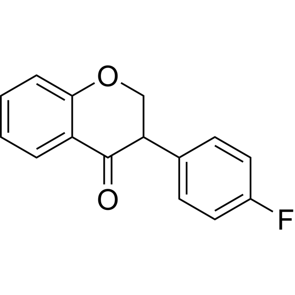
- Formula: C15H11FO2
- Molecular Weight:242.25
-
Storage:Powder -20°C, 3 years , 4°C, 2 years ; In solvent -80°C, 6 months , -20°C, 1 month

To place orders, for customer services and technical support, please contact: MedChemExpress USA
Tel: 609-228-6898 E-mail: [email protected] [email protected]


-
 Biological Activity
-
Chemical Information
-
Solvent & Solubility
- Protocols
- Purity & Documentation
- References
-
 Help & FAQs


-
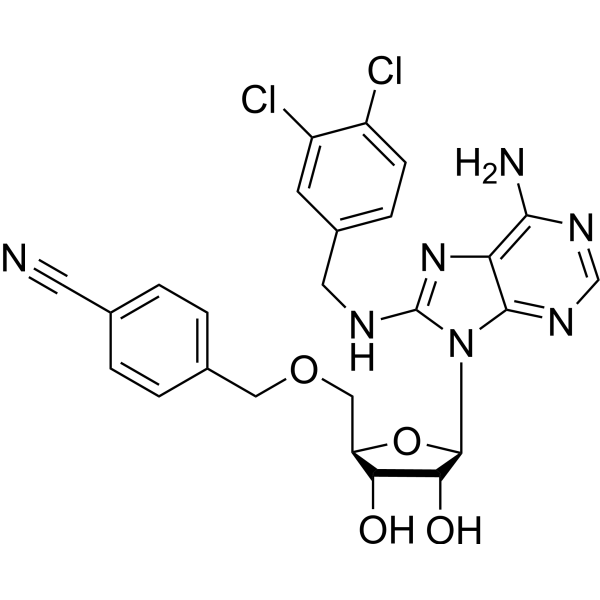
HY-10941
VER-155008Hsp70 Inhibitor -
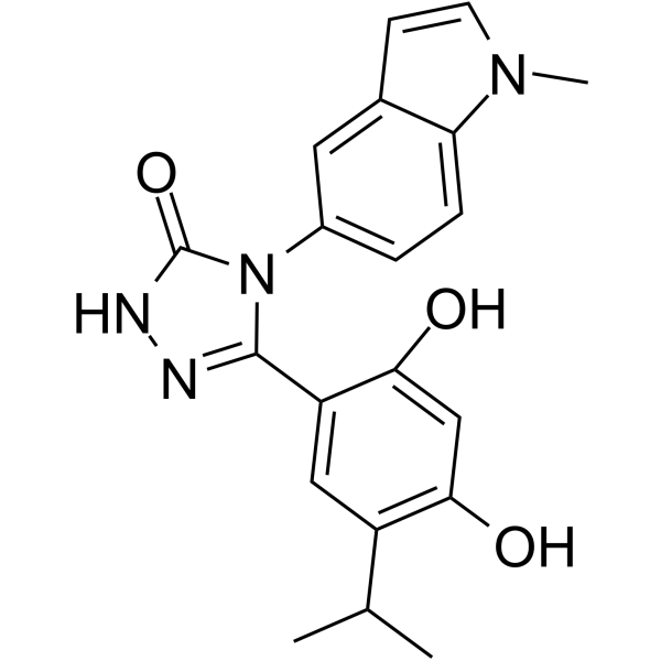
HY-15205
GanetespibHSP90 Inhibitor -
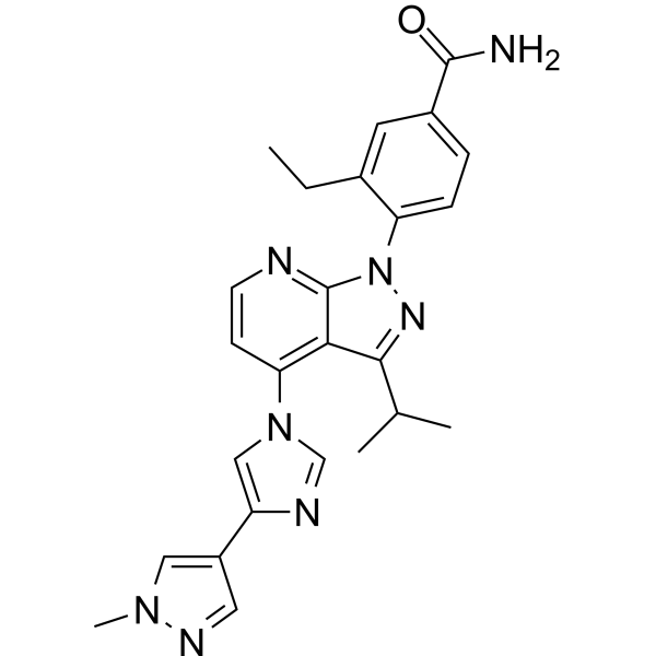
HY-15785
PimitespibHSP90α/HSP90β Inhibitor -
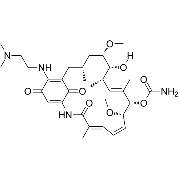
HY-10389
AlvespimycinHsp90 Inhibitor -
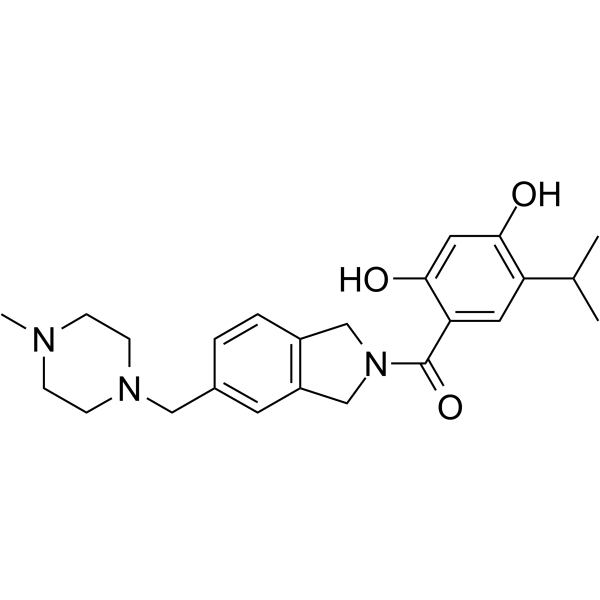
HY-14463
OnalespibHsp90 Inhibitor -
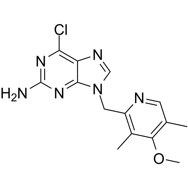
HY-10212
BIIB021HSP90 Inhibitor -
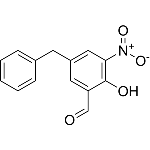
HY-124817
Col003Hsp47 Inhibitor -
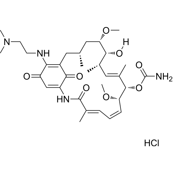
HY-12024
Alvespimycin hydrochloride -
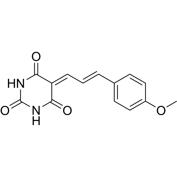
HY-18669
ML346Hsp70 Activator -
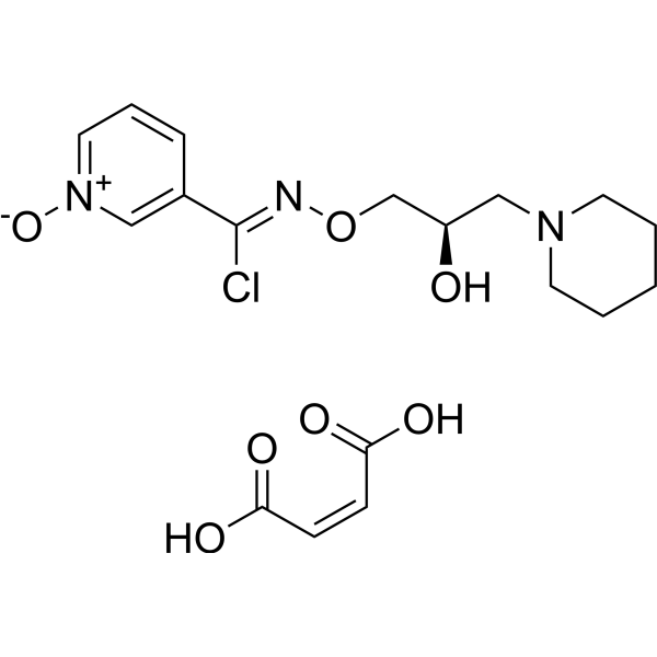
HY-106443A
Arimoclomol maleateHSP Co-Inducer -
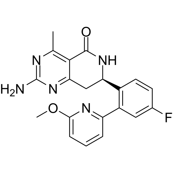
HY-15190
NVP-HSP990Apoptosis Inducer -
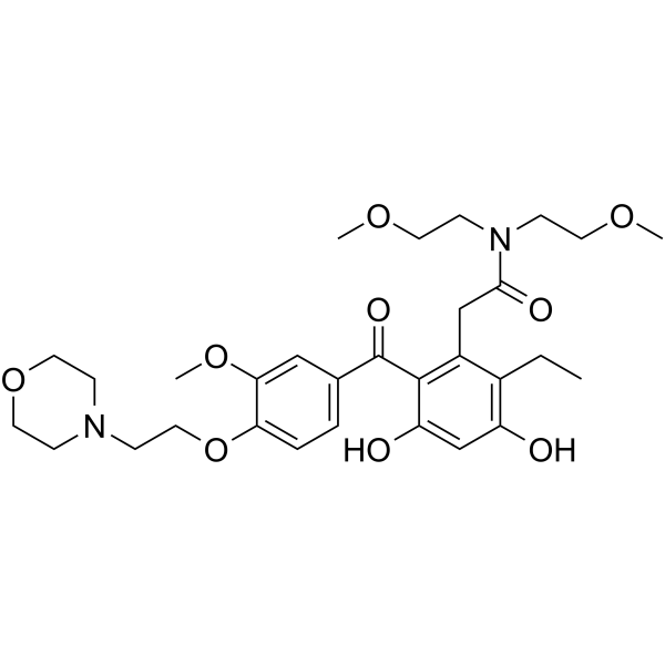
HY-13468
KW-2478


Biological Activity
Description
IC50 & Target
[1]|
RIPK1 |
ERK1 |
ERK2 |
In Vitro
KBU2046 (1-10 μM; 24-48 h) does not significantly alter the proliferation of MDA-MB-231 or BT-549 TNBC cells[1].
KBU2046 (1-10 μM; 24 h) dose-dependently inhibits TGF-β1-induced motility of MDA-MB-231 and BT-549 TNBC cells, with maximum inhibition at 10 μM, reducing motility to 50.77% and 36.09% of control levels, respectively[1].
KBU2046 (10 μM; 24 h) significantly down-regulates the mRNA expression of LRRC8E, LTBP3, DNAL1, and MAFF in MDA-MB-231 and BT-549 TNBC cells[1].
KBU2046 (1-10 μM; 48 h) dose-dependently suppresses integrin αv and integrin α6 protein expression in MDA-MB-231 and BT-549 TNBC cells, and reverses TGF-β1-mediated up-regulation of these integrins[1].
KBU2046 (10 μM; 15-60 min) time-dependently inhibits phosphorylation of Raf1 and ERK1/2 in MDA-MB-231 and BT-549 TNBC cells, with significant suppression evident within 15 min of treatment[1].
KBU2046 (1-10 μM; 24-48 h) inhibits collective migration of KYSE-30 and KYSE-410 ESCC cells in a time- and concentration-dependent manner[2].
KBU2046 (10 μM; 72 h) inhibits EGF and TGF-β-stimulated single-cell migration and invasion of KYSE-30 and KYSE-410 ESCC cells[2].
KBU2046 (1-10 μM; 72 h) does not inhibit proliferation of KYSE-30 or KYSE-410 ESCC cells after 72-hour incubation[2].
KBU2046 (10 μM; 30 min) inhibits EGF-stimulated phosphorylation of RIPK1 at Ser166 in KYSE-30 and KYSE-410 ESCC cells[2].
KBU2046 (1-10 μM) downregulates ITGA3, ITGA6, and ITGAV expression in KYSE-30 and KYSE-410 ESCC cells in a concentration-dependent manner[2].
KBU2046 (10 μM) selectively modulates expression of motility-related proteins in KYSE-30 ESCC cells, with significant downregulation of integrin family members and other ECM-adhesion proteins that are overexpressed in human ESCC[2].
MedChemExpress (MCE) has not independently confirmed the accuracy of these methods. They are for reference only.
-
Cell Line:MDA-MB-231, BT-549 cells
-
Concentration:1, 5, 10 μM
-
Incubation Time:24 h; 48 h;
-
Result:Exhibited no time- or concentration-dependent alterations in cell proliferation in either MDA-MB-231 or BT-549 cells.
-
Cell Line:MDA-MB-231, BT-549 cells
-
Concentration:1, 5, 10 μM
-
Incubation Time:24 h
-
Result:Reduced TGF-β1-induced cellular motility rates to 92.97% at 1 μM, 70.59% at 5 μM, and 50.77% at 10 μM in MDA-MB-231 cells compared to control.
Reduced TGF-β1-induced cellular motility rates to 78.43% at 1 μM, 43.55% at 5 μM, and 36.09% at 10 μM in BT-549 cells compared to control.
-
Cell Line:MDA-MB-231, BT-549 cells
-
Concentration:1, 5, 10 μM
-
Incubation Time:48 h (co-treatment with 10 ng/mL TGF-β1)
-
Result:Reduced integrin αv protein levels to 67.28% at 1 μM, 57.61% at 5 μM, and 42.29% at 10 μM, and integrin α6 levels to 87.15% at 1 μM, 76.54% at 5 μM, and 45.26% at 10 μM in MDA-MB-231 cells compared to control.
Reduced integrin αv levels to 96.13% at 1 μM, 58.68% at 5 μM, and 51.11% at 10 μM, and integrin α6 levels to 82.47% at 1 μM, 66.29% at 5 μM, and 61.89% at 10 μM in BT-549 cells compared to control.
Partially reversed TGF-β1-mediated up-regulation of integrin αv and α6 in both cell lines.
Exhibited no significant effects on integrin α3 or β8 protein levels.
-
Cell Line:MDA-MB-231, BT-549 cells
-
Concentration:10 μM
-
Incubation Time:15 min; 30 min; 60 min
-
Result:Reduced phosphorylation levels of Raf1 (Ser338) and ERK1/2 (Thr202/Tyr204) in a time-dependent manner in both MDA-MB-231 and BT-549 cells.
Showed statistically significant reductions at 15, 30, and 60 min compared to control levels.
-
Cell Line:KYSE-30, KYSE-410 esophageal squamous cell carcinoma (ESCC) cells
-
Concentration:1 μM, 10 μM
-
Incubation Time:24 h, 48 h
-
Result:Significantly inhibited wound gap closure in a time- and concentration-dependent manner.
Decreased wound closure by 46.7% in KYSE-30 cells and 47.5% in KYSE-410 cells at 48 h with 10 μM treatment compared to vehicle controls.
-
Cell Line:KYSE-30, KYSE-410 esophageal squamous cell carcinoma (ESCC) cells
-
Concentration:1 μM, 10 μM
-
Incubation Time:72 h
-
Result:Had no significant effect on cell viability of KYSE-30 or KYSE-410 cells after 72 h, indicating no inhibition of cell proliferation.
-
Cell Line:EGF-stimulated KYSE-30, KYSE-410 esophageal squamous cell carcinoma (ESCC) cells
-
Concentration:10 μM (in the presence of 5 ng/mL EGF)
-
Incubation Time:30 min
-
Result:Markedly inhibited EGF-stimulated phosphorylation of RIPK1 at Ser166 in both KYSE-30 and KYSE-410 cells.
In Vivo
KBU2046 (80 mg/kg; i.g.; every 2 days; 12 total doses) reduces mean ESCC tumor volume to 62% of control levels in male athymic nude mice, without causing significant systemic toxicity[2].
MedChemExpress (MCE) has not independently confirmed the accuracy of these methods. They are for reference only.
-
Animal Model:NOD-SCID (male, 4-6 weeks old, 16-18 g, tail vein injection of 1.5×10⁶ KYSE-30 ESCC cells)[2]
-
Dosage:80 mg/kg
-
Administration:p.o.; every other day; 12 total doses
-
Result:Completely inhibited pulmonary metastasis, with 0 of 4 treated mice showing pulmonary metastases compared to 3 of 4 control mice (one-sided t-test, P=0.03).
Showed no significant difference in body weight compared to control group.
Caused no histomorphological changes in heart, liver, or spleen.
-
Animal Model:athymic nude (male, 4-6 weeks old, SPF grade, subcutaneous implantation of 1×10⁶ KYSE-30 ESCC cells into the right flank)[2]
-
Dosage:80 mg/kg
-
Administration:p.o.; every 2 days; 12 total doses
-
Result:Reduced mean tumor volume to 62% of control levels, though the difference was not statistically significant.
Showed no significant difference in body weight compared to control group.
- RNA extraction experimental
- Western Blot
- Scratch/Wound-Healing Migration Assay
- Cell migration
- Breast Cancer Modeling
- Protocol For Protein Expression And Purification
- Kinase activity and phosphorylation assays
- Under-Agarose Cell Migration Assay
- Transwell/Boyden Chamber Migration Assay
- Cell invasion
- Protocol for Kinase activity and phosphorylation assays
- Cell-Exclusion Zone Migration Assay
- Orthotopic Cell-Line Xenograft


Chemical Information
-
CAS No. 1143863-69-7
-
Appearance Solid
-
Molecular Weight 242.25
-
Formula C15H11FO2
-
Color White to off-white
-
SMILES
O=C1C(C2=CC=C(F)C=C2)COC3=CC=CC=C13
-
Shipping
Room temperature in continental US; may vary elsewhere.
-
Storage
Powder -20°C 3 years 4°C 2 years In solvent -80°C 6 months -20°C 1 month


Solvent & Solubility
In Vitro:
DMSO : 50 mg/mL (206.40 mM; Need ultrasonic; Hygroscopic DMSO has a significant impact on the solubility of product, please use newly opened DMSO)
Please refer to the solubility information to select the appropriate solvent. Once prepared, please aliquot and store the solution to prevent product inactivation from repeated freeze-thaw cycles.
Storage method and period of stock solution: -80°C, 6 months; -20°C, 1 month. When stored at -80°C, please use it within 6 months. When stored at -20°C, please use it within 1 month.
Please refer to the solubility information to select the appropriate solvent. Once prepared, please aliquot and store the solution to prevent product inactivation from repeated freeze-thaw cycles.
Storage method and period of stock solution: -80°C, 6 months; -20°C, 1 month. When stored at -80°C, please use it within 6 months. When stored at -20°C, please use it within 1 month.
Concentration (start) × Volume (start) = Concentration (final) × Volume (final)
In Vivo:
Select the appropriate dissolution method based on your experimental animal and administration route.
- For the following dissolution methods, please ensure to first prepare a clear stock solution using an In Vitro approach and then sequentially add co-solvents:
- To ensure reliable experimental results, the clarified stock solution can be appropriately stored based on storage conditions. As for the working solution for In Vivo experiments, it is recommended to prepare freshly and use it on the same day.
- The percentages shown for the solvents indicate their volumetric ratio in the final prepared solution. If precipitation or phase separation occurs during preparation, heat and/or sonication can be used to aid dissolution.
Add each solvent one by one: 10% DMSO 90% (20% SBE-β-CD in Saline)
Solubility: ≥ 2.5 mg/mL (10.32 mM); Clear solution
This protocol yields a clear solution of ≥ 2.5 mg/mL (saturation unknown).
Taking 1 mL working solution as an example, add 100 μL DMSO stock solution (25.0 mg/mL) to 900 μL 20% SBE-β-CD in Saline, and mix evenly.
Preparation of 20% SBE-β-CD in Saline (4°C, storage for one week): 2 g SBE-β-CD powder is dissolved in 10 mL Saline, completely dissolve until clear.
In Vivo Dissolution Calculator
Please enter the basic information of animal experiments:
-
-
-
-
Recommended: Prepare an additional quantity of animals to account for potential losses during experiments.
Please enter your animal formula composition:
-
%DMSO +
Recommended: Keep the proportion of DMSO in working solution below 2% if your animal is weak.
-
%+
-
+%Tween-80 + +
-
%Saline +
The co-solvents required include: DMSO, . All of co-solvents are available by MedChemExpress (MCE). , Tween 80. All of co-solvents are available by MedChemExpress (MCE).
Working solution concentration: 0.22 mg/mL
Method for preparing stock solution: mg drug dissolved in μL DMSO. Stock solution concentration: mg/mL.
1. Take μL DMSO stock solution;
2. Add μL .
μL , mix evenly;
3. Then add μL Tween 80, mix evenly;
4. Then add μL
Please ensure that the stock solution in the first step is dissolved to a clear state, and add co-solvents in sequence. You can use ultrasonic heating (ultrasonic cleaner, recommended frequency 20-40 kHz), vortexing, etc. to assist dissolution.


Purity & Documentation
-
Data Sheet (285 KB)
-
SDS (394 KB)
- English - EN (394 KB)
- Français - FR (394 KB)
- Deutsch - DE (394 KB)
- Norwegian - NO (394 KB)
- Español - ES (394 KB)
- Swedish - SV (394 KB)
- Italian - IT (394 KB)
- Korean - KR (394 KB)
- Portuguese - PT (394 KB)
-
Handling Instructions (2659 KB)
References
[1]. Chen J, et al. Unveiling the therapeutic potential: KBU2046 halts triple-negative breast cancer cell migration by constricting TGF-β1 activation in vitro. Oncol Res. 2024;32(11):1791-1802. Published 2024 Oct 16. [Content Brief]
[2]. Li K, et al. KBU2046 exerts inhibition on chemokine gradient-mediated motility of esophageal squamous cell carcinoma through reducing integrin expression. Biochim Biophys Acta Mol Basis Dis. 2025;1871(5):167800. [Content Brief]


Complete Stock Solution Preparation Table
Please refer to the solubility information to select the appropriate solvent. Once prepared, please aliquot and store the solution to prevent product inactivation from repeated freeze-thaw cycles.
Storage method and period of stock solution: -80°C, 6 months; -20°C, 1 month. When stored at -80°C, please use it within 6 months. When stored at -20°C, please use it within 1 month.
| Optional Solvent | Concentration Solvent Mass | 1 mg | 5 mg | 10 mg | 25 mg |
|---|---|---|---|---|---|
| DMSO | 1 mM | 4.1280 mL | 20.6398 mL | 41.2797 mL | 103.1992 mL |
| 5 mM | 0.8256 mL | 4.1280 mL | 8.2559 mL | 20.6398 mL | |
| 10 mM | 0.4128 mL | 2.0640 mL | 4.1280 mL | 10.3199 mL | |
| 15 mM | 0.2752 mL | 1.3760 mL | 2.7520 mL | 6.8799 mL | |
| 20 mM | 0.2064 mL | 1.0320 mL | 2.0640 mL | 5.1600 mL | |
| 25 mM | 0.1651 mL | 0.8256 mL | 1.6512 mL | 4.1280 mL | |
| 30 mM | 0.1376 mL | 0.6880 mL | 1.3760 mL | 3.4400 mL | |
| 40 mM | 0.1032 mL | 0.5160 mL | 1.0320 mL | 2.5800 mL | |
| 50 mM | 0.0826 mL | 0.4128 mL | 0.8256 mL | 2.0640 mL | |
| 60 mM | 0.0688 mL | 0.3440 mL | 0.6880 mL | 1.7200 mL | |
| 80 mM | 0.0516 mL | 0.2580 mL | 0.5160 mL | 1.2900 mL | |
| 100 mM | 0.0413 mL | 0.2064 mL | 0.4128 mL | 1.0320 mL |


KBU2046 Related Classifications
HY-120548 Related Classifications
 Powered by Bioz