



Edaravone-d5

Based on 1 Customer Validation
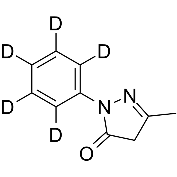
Edaravone-d5 is a deuterium labeled Edaravone. Edaravone is a strong novel free radical scavenger, and inhibits MMP-9-related brain hemorrhage in rats treated with tissue plasminogen activator.
For research use only. We do not sell to patients.
- Purity : 99.52%
- CAS No.: 1228765-67-0
- Formula: C10H5D5N2O
- Molecular Weight:179.23
-
Storage:Powder -20°C, 3 years , 4°C, 2 years ; In solvent -80°C, 6 months , -20°C, 1 month

To place orders, for customer services and technical support, please contact: MedChemExpress USA
Tel: 609-228-6898 E-mail: [email protected] [email protected]


-
 Biological Activity
-
Chemical Information
- Protocols
- Purity & Documentation
- References
-
 Help & FAQs




Biological Activity
Description
Application
1. This compound can be used as a tracer
2. This compound can be used as an internal standard for quantitative analysis by NMR, GC-MS, or LC-MS.


Chemical Information
-
CAS No. 1228765-67-0
-
Unlabeled CAS 89-25-8
-
Appearance Solid
-
Molecular Weight 179.23
-
Formula C10H5D5N2O
-
Color Off-white to light yellow
-
SMILES
O=C1N(C2=C([2H])C([2H])=C([2H])C([2H])=C2[2H])N=C(C)C1
-
Synonyms
MCI-186-d5
-
Shipping
Room temperature in continental US; may vary elsewhere.
-
Storage
Powder -20°C 3 years 4°C 2 years In solvent -80°C 6 months -20°C 1 month


Purity & Documentation
-
Data Sheet (271 KB)
-
SDS (393 KB)
- English - EN (393 KB)
- Français - FR (393 KB)
- Deutsch - DE (393 KB)
- Norwegian - NO (393 KB)
- Español - ES (393 KB)
- Swedish - SV (393 KB)
- Italian - IT (393 KB)
- Korean - KR (393 KB)
- Portuguese - PT (393 KB)
-
Handling Instructions (2659 KB)
References
[1]. Yoshida, H., et al. Neuroprotective effects of edaravone: a novel free radical scavenger in cerebrovascular injury. CNS Drug Rev, 2006. 12(1): p. 9-20. [Content Brief]
[2]. Okamura, K., et al. Edaravone, a free radical scavenger, attenuates cerebral infarction and hemorrhagic infarction in rats with hyperglycemia. Neurol Res, 2013. [Content Brief]
[3]. Garcia CA, et al. Edaravone reduces astrogliosis and apoptosis in young rats with kaolin-induced hydrocephalus. Childs Nerv Syst. 2016 Dec 17. [Epub ahead of print] [Content Brief]
[4]. Bai X, et al. Protective Effect of Edaravone on Glutamate-Induced Neurotoxicity in Spiral Ganglion Neurons. Neural Plast. 2016;2016:4034218. Epub 2016 Nov 10. [Content Brief]

Calculators
Concentration (start) × Volume (start) = Concentration (final) × Volume (final)


Edaravone-d5 Related Classifications
HY-B0099S Related Classifications
 Powered by Bioz