



Feprazone

Based on 1 Customer Validation
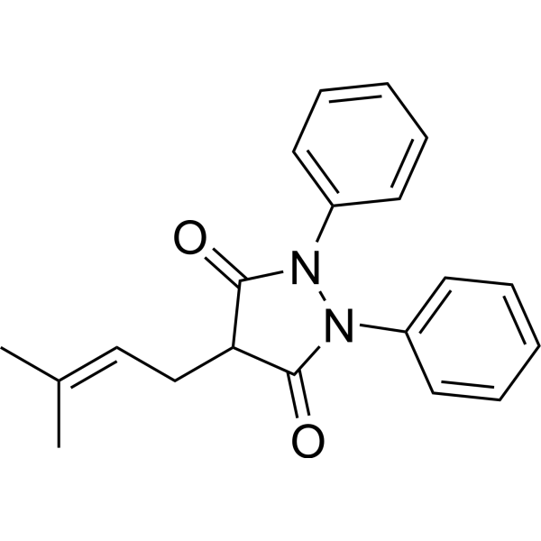
Feprazone (DA2370; Prenazone), an analogue of Phenylbutazone (HY-B0230), is a nonsteroidal anti-inflammatory agent with analgesic and antipyretic activities. Feprazone acts by inhibiting the activity of cyclooxygenase (COX)-2. Feprazone ameliorates free fatty acid (FFA)-induced oxidative stress by reducing the production of mitochondrial reactive oxygen species (ROS). Feprazone can decrease the expression of MMP-2 and MMP-9. Besides, Feprazone can suppress adipogenesis and increase lipolysis in differentiating 3 T3-L1 cells. Feprazone also can be used to research atherosclerosis and obesity.
For research use only. We do not sell to patients.
- Purity : 99.88%
- CAS No.: 30748-29-9
- Formula: C20H20N2O2
- Molecular Weight:320.39
-
Storage:
4°C, protect from light
* In solvent : -80°C, 6 months; -20°C, 1 month (protect from light)

To place orders, for customer services and technical support, please contact: MedChemExpress USA
Tel: 609-228-6898 E-mail: [email protected] [email protected]


-
 Biological Activity
-
Chemical Information
-
Solvent & Solubility
- Protocols
- Purity & Documentation
- References
-
 Help & FAQs


-

HY-114911R
Feprazone (Standard)Nonsteroidal Anti-inflammatory Agent -
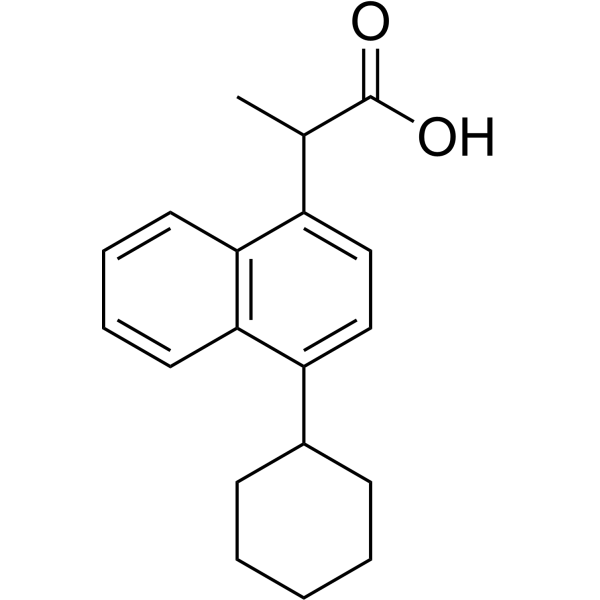
HY-118827
VedaprofenAnti-inflammatory Agent -
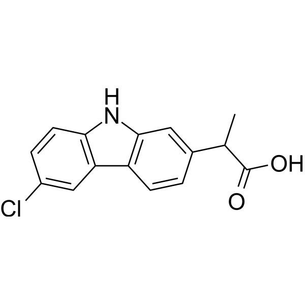
HY-B1227
CarprofenCOX Inhibitor -

HY-14648
DexamethasoneGR Agonist -
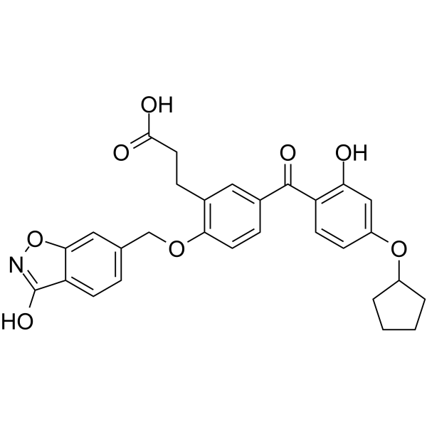
HY-12270
T-5224c-Fos/AP-1 Inhibitor -
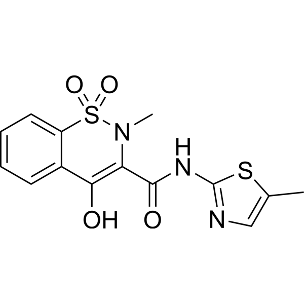
HY-B0261
MeloxicamCOX Inhibitor -

HY-14648C
Dexamethasone (Water Soluble)Muscular Atrophy/Hypertension/Depression Modeling Reagent -
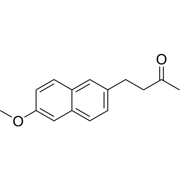
HY-B0559
NabumetoneCOX-2 Inhibitor -
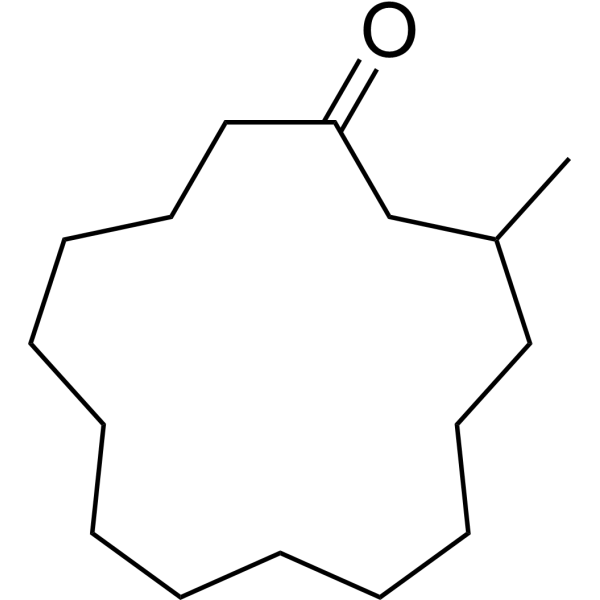
HY-N0633
MusconeTNF Receptor Inhibitor -
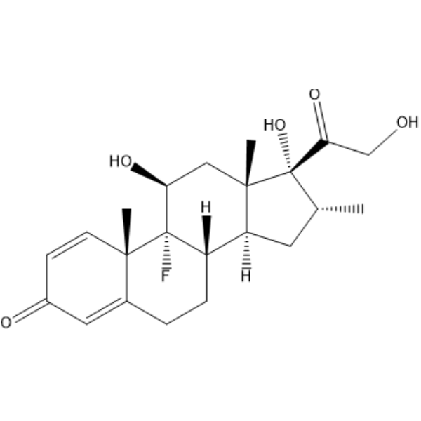
HY-14648R
Dexamethasone (Standard) -
HY-N6258
KahweolApoptosis Inducer -

HY-14648G
Dexamethasone (GMP)GR Agonist


Biological Activity
Description
IC50 & Target
COX, Reactive oxygen species, MMP[1]
In Vitro
Feprazone (2.5-10 μM; 48 h) rescues cell viability of FFAs-stimulated human aortic endothelial cells (HAECs)[1].
Feprazone (5, 10 μM; 24 h) reduces ROS production in HAECs to only 2.4- and 1.6-fold at 5 and 10 μM, respectively, while 300 μM FFA increases ROS production by 3.4-fold; also decreases the mRNA expression and secretion of cytokines CCL5, IL-6, and IL-8, as well as MMP-2 and MMP-9[1].
Feprazone (5, 10 μM; 6 h) decreases TLR4 and MyD88 activities, as well as reduces the phosphorylation of p65 and subsequent activation of NF-κB[1].
Feprazone (30 and 60 μM; 7 days) suppresses the adipogenesis in differentiating 3 T3-L1 cells; reduced the triglyceride content and increased lipolysis during 3 T3-L1 adipogenesis[3].
MedChemExpress (MCE) has not independently confirmed the accuracy of these methods. They are for reference only. Further protocols information, click here.
-
Cell Line:HAECs (stimulated with 300 μM FFAs)
-
Concentration:2.5, 5 and 10 μM
-
Incubation Time:48 h
-
Result:Rescued cell viability to 81 and 93% of baseline at 5 and 10 μM, while FFAs reduced the cell viability to 63% of baseline.
-
Cell Line:HAECs (stimulated with 300 μM FFAs)
-
Concentration:5 and 10 μM
-
Incubation Time:24 h
-
Result:Decreased the mRNA expression and secretion of cytokines CCL5, IL-6, and IL-8 in a dose-dependent manner.
Dose-dependently mitigated the VCAM-1 and ICAM-1 expression to only 1.7- and 1.8-fold, respectively, while FFA increased to 2.8- and 3.4-fold, respectively.
-
Cell Line:HAECs (stimulated with 300 μM FFAs)
-
Concentration:5 and 10 μM
-
Incubation Time:6 h
-
Result:Decreased TLR4 and MyD88 expression, as well as reduced the phosphorylation of p65 and subsequent activation of NF-κB.
In Vivo
MedChemExpress (MCE) has not independently confirmed the accuracy of these methods. They are for reference only.
-
Animal Model:Male C57BL/6 N mice [high-fat diet (HFD) induced obesity model][3]
-
Dosage:75 mg/kg
-
Administration:(no described in the research)
-
Result:The visceral adipocyte tissue weights of mice in the control, HFD, and HFD + Feprazone groups were 0.38, 3.51, and 2.37 g, respectively.
The average bodyweights of mice in the control, HFD, and HFD + Feprazone groups were 29.6, 41.3, and 34.1 g, respectively.


Chemical Information
-
CAS No. 30748-29-9
-
Appearance Solid
-
Molecular Weight 320.39
-
Formula C20H20N2O2
-
Color White to off-white
-
SMILES
O=C(N(N(C1=O)C2=CC=CC=C2)C3=CC=CC=C3)C1C/C=C(C)\C
-
Synonyms
DA2370; Prenazone; Zepelin
-
Shipping
Room temperature in continental US; may vary elsewhere.
-
Storage
4°C, protect from light
* In solvent : -80°C, 6 months; -20°C, 1 month (protect from light)


Solvent & Solubility
In Vitro:
DMSO : 12.5 mg/mL (39.01 mM; ultrasonic and warming and heat to 60°C; Hygroscopic DMSO has a significant impact on the solubility of product, please use newly opened DMSO)
Please refer to the solubility information to select the appropriate solvent. Once prepared, please aliquot and store the solution to prevent product inactivation from repeated freeze-thaw cycles.
Storage method and period of stock solution: -80°C, 6 months; -20°C, 1 month (protect from light). When stored at -80°C, please use it within 6 months. When stored at -20°C, please use it within 1 month.
Please refer to the solubility information to select the appropriate solvent. Once prepared, please aliquot and store the solution to prevent product inactivation from repeated freeze-thaw cycles.
Storage method and period of stock solution: -80°C, 6 months; -20°C, 1 month (protect from light). When stored at -80°C, please use it within 6 months. When stored at -20°C, please use it within 1 month.
Concentration (start) × Volume (start) = Concentration (final) × Volume (final)

Protocols
-
RNA extraction experimental
By lysing cells, releasing RNA, and removing impurities such as proteins and DNA, high-purity RNA products are finally obtained. The commonly used traditional method is the guanidine isothiocyanate/phenol/chloroform method (Trizol), which is suitable for a variety of animal materials including animal tissues, microorganisms, cultured cells, etc., and most plant materials.
-
3T3-L1 preadipocyte-to-adipocyte differentiation
3T3-L1 preadipocytes are induced to differentiate after growth arrest using adipogenic media containing insulin, dexamethasone, and IBMX; differentiation is assessed by lipid-droplet accumulation, triglyceride increase, Oil Red O staining, and adipocyte-marker induction such as PPARγ and C/EBPα.
-
Mitochondrial membrane-potential fluorescent assay
Mitochondrial membrane potential fluorescent assays estimate ΔΨm in living cells using lipophilic cationic dyes such as TMRM, TMRE, rhodamine 123, and JC-1, which accumulate in mitochondria according to membrane polarization; loss of signal after FCCP or CCCP treatment is interpreted as mitochondrial depolarization. TMRM/TMRE and rhodamine 123 are commonly used for semi-quantitative live-cell microscopy or flow cytometry, while JC-1 can report a shift from red aggregate fluorescence to green monomer fluorescence during depolarization; interpretation requires controls because dye concentration, quenching mode, cell type, dye efflux, and mitochondrial mass can affect fluorescence independently of ΔΨm.
-
ROS/oxidative-stress fluorescent staining
ROS/oxidative-stress fluorescent staining uses cell-permeant fluorogenic probes that become fluorescent after oxidation inside cells or tissues; commonly used examples include DCFH-DA/DCFDA for broad cellular oxidant detection, DHE for superoxide-related signal detection, MitoSOX for mitochondrial superoxide-related signal detection, and CellROX probes for oxidative-stress-associated fluorescence readouts. The assay detects probe oxidation rather than a single ROS species unless the probe and analysis method have been chemically validated for that species. DCFH-DA enters cells, is deacetylated by intracellular esterases to DCFH, and produces fluorescent DCF after oxidation, so the readout is used as an operational measure of total cellular oxidative stress rather than a species-specific ROS measurement. DHE and MitoSOX can report superoxide-related oxidation, but red fluorescence alone can include non-specific ethidium-like oxidation products; HPLC or optimized spectral approaches are
-
Mitochondrial membrane-potential and mitochondrial mass staining
Mitochondrial membrane potential staining measures the electrochemical polarization across the mitochondrial inner membrane in live cells using lipophilic cationic fluorescent probes; early rhodamine-based work showed that selective mitochondrial dye accumulation is lost when the mitochondrial transmembrane potential is dissipated. JC-1 reports mitochondrial polarization by shifting from green monomer fluorescence to red J-aggregate fluorescence as dye concentration increases within energized mitochondria; therefore, the red/green fluorescence ratio is used as a relative readout of mitochondrial membrane potential. TMRE or TMRM staining provides a single-channel relative readout because these cationic rhodamine esters accumulate in polarized mitochondria, and lower fluorescence indicates reduced mitochondrial polarization when acquisition and dye-loading conditions are controlled. Mitochondrial mass staining is commonly performed with MitoTracker Green FM or related MitoTracker dyes as
-
Research Protocol for Cardiovascular Diseases
Cardiovascular disease can be modeled as maladaptive cardiac remodeling, where ischemic injury or pressure overload activates inflammatory signaling, fibroblast activation, extracellular-matrix deposition, cardiomyocyte hypertrophy, vascular remodeling, and progressive ventricular dysfunction. The TGF-β/SMAD axis is a central profibrotic pathway after myocardial injury and pressure overload, while innate immune and cytokine pathways regulate leukocyte recruitment, scar formation, and adverse remodeling. Key unresolved questions include which inflammatory signals are reparative versus harmful, when fibrosis is protective versus maladaptive, and whether pathway inhibition improves function without weakening necessary infarct healing or compensatory remodeling.
-
Research Protocol for Inflammation-related Diseases
The NLRP3 inflammasome is a cytosolic innate immune signaling platform that integrates priming signals and danger-signal activation to promote caspase-1 activation, maturation of IL-1β and IL-18, and gasdermin D-mediated pyroptotic cell death. The core experimental logic is to determine whether inflammatory disease phenotypes are driven by increased NLRP3 expression, ASC-containing inflammasome assembly, caspase-1 cleavage, GSDMD cleavage, and extracellular release of IL-1β/IL-18 rather than by nonspecific cell injury alone. The pathway is strongly linked to inflammation-related disease phenotypes because monosodium urate crystals activate NALP3/NLRP3 inflammasome signaling in gout-like crystal inflammation, cholesterol crystals activate NLRP3 inflammasomes in atherogenesis models, and DSS-induced intestinal inflammation has been reported to involve NLRP3 inflammasome activity. However, experimental colitis studies also show context-dependent protective effects of NLRP3 inflammasome co
-
Research Protocol for Metabolic Diseases
AMP-activated protein kinase, AMPK, is a conserved cellular energy sensor that responds to reduced cellular energy status and coordinates metabolism by increasing ATP-generating catabolic pathways while suppressing ATP-consuming anabolic processes. In metabolic disease research, the AMPK pathway is experimentally relevant because it regulates hepatic lipid synthesis, fatty acid oxidation, glucose production, skeletal-muscle glucose disposal, mTORC1-linked biosynthesis, autophagy, mitochondrial homeostasis, and whole-body energy balance. The central pathway logic is that energy stress, metformin, exercise-like stimulation, or direct AMPK activators increase AMPKα Thr172 phosphorylation and downstream substrate phosphorylation, including ACC and RAPTOR. Phosphorylation of ACC suppresses lipogenesis and supports fatty acid oxidation, whereas phosphorylation of RAPTOR suppresses mTORC1 signaling and links cellular energy status to growth and protein synthesis control. The pathway is linked
-
Lipid Droplets: Oil Red O/Sudan Dye Lipid Staining
Lipid droplets are intracellular organelles with a neutral-lipid core that stores triacylglycerols and sterol esters, and Oil Red O or Sudan dyes detect these hydrophobic lipid deposits by partitioning into retained lipids in fresh or frozen specimens. Oil Red O stains neutral triglycerides and lipids in frozen tissue sections or air-dried cytologic preparations, while Sudan Black B has also been used as a histochemical fat stain for lipid-rich tissue structures.


Purity & Documentation
-
Data Sheet (282 KB)
-
SDS (254 KB)
- English - EN (254 KB)
- Français - FR (254 KB)
- Deutsch - DE (254 KB)
- Norwegian - NO (254 KB)
- Español - ES (254 KB)
- Swedish - SV (254 KB)
- Italian - IT (254 KB)
- Korean - KR (254 KB)
- Portuguese - PT (254 KB)
-
Handling Instructions (2659 KB)
References
[1]. Song M, et al. Feprazone Prevents Free Fatty Acid (FFA)-Induced Endothelial Inflammation by Mitigating the Activation of the TLR4/MyD88/NF-κB Pathway. ACS Omega. 2021 Feb 9;6(7):4850-4856. [Content Brief]
[2]. Fletcher MR, et al. Feprazone, a new anti-inflammatory agent. Studies of potency and gastrointestinal tolerance. Ann Rheum Dis. 1975 Apr;34(2):190-4. [Content Brief]
[3]. Che L, et al. Feprazone Displays Antiadipogenesis and Antiobesity Capacities in in Vitro 3 T3-L1 Cells and in Vivo Mice. ACS Omega. 2021 Mar 7;6(10):6674-6680. [Content Brief]


Complete Stock Solution Preparation Table
Please refer to the solubility information to select the appropriate solvent. Once prepared, please aliquot and store the solution to prevent product inactivation from repeated freeze-thaw cycles.
Storage method and period of stock solution: -80°C, 6 months; -20°C, 1 month (protect from light). When stored at -80°C, please use it within 6 months. When stored at -20°C, please use it within 1 month.
| Optional Solvent | Concentration Solvent Mass | 1 mg | 5 mg | 10 mg | 25 mg |
|---|---|---|---|---|---|
| DMSO | 1 mM | 3.1212 mL | 15.6060 mL | 31.2120 mL | 78.0299 mL |
| 5 mM | 0.6242 mL | 3.1212 mL | 6.2424 mL | 15.6060 mL | |
| 10 mM | 0.3121 mL | 1.5606 mL | 3.1212 mL | 7.8030 mL | |
| 15 mM | 0.2081 mL | 1.0404 mL | 2.0808 mL | 5.2020 mL | |
| 20 mM | 0.1561 mL | 0.7803 mL | 1.5606 mL | 3.9015 mL | |
| 25 mM | 0.1248 mL | 0.6242 mL | 1.2485 mL | 3.1212 mL | |
| 30 mM | 0.1040 mL | 0.5202 mL | 1.0404 mL | 2.6010 mL |


Feprazone Related Classifications
HY-114911 Related Classifications
 Powered by Bioz