



Pyrazofurin
Based on 5 publication(s) in Google Scholar
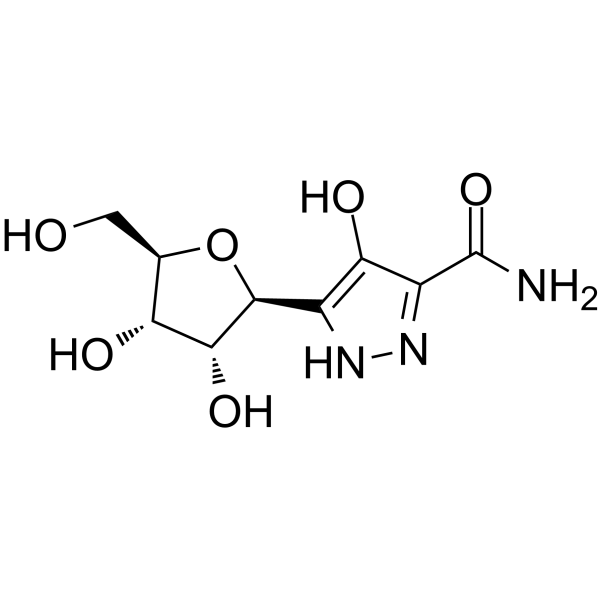
Pyrazofurin is an antitumor pyrimidine nucleoside analogue and a orotate-phosphoribosyltransferase inhibitor. Pyrazofurin inhibits cell proliferation and intracellular DNA synthesis by inhibiting uridine 5'-phosphate synthase. Pyrazofurin is also an antibiotic with a broad spectrum of antiviral activity.
Para uso exclusivo en investigación. No vendemos a pacientes.
- Pureza : 99.00%
- No. CAS: 30868-30-5
- Fòrmula: C9H13N3O6
- Peso molecular:259.22
-
Almacenamiento:
-20°C, stored under nitrogen
* In solvent : -80°C, 6 months; -20°C, 1 month (stored under nitrogen)

To place orders, for customer services and technical support, please contact: MedChemExpress USA
Tel: 609-228-6898 E-mail: [email protected] [email protected]


-
 Actividad biológica
-
Chemical Information
-
Solvente y solubilidad
- Protocolo
- Pureza y Documentación
- Referencias
-
 Help & FAQs


Publications Citing Use of MedChemExpress (MCE) Pyrazofurin
MoreVer todos los productos específicos de isoformas DNA/RNA Synthesis
MoreVer todos los productos específicos de isoformas Antibiotic
More

Actividad biológica
Descripciòn
In Vitro
Pyrazofurin (75 μM; 52 h) inhibits DNA synthesis in guinea pig lymphocytes treated with Canavagin A(HY-P2149), but is reversed by exogenous uridine[1].
Pyrazofurin (0.01-200 μM; 24-96 h) inhibits the growth of head and neck cancer cell lines HEP-2, UMSCC-14B and UMSCC-14C with the IC50 between 0.06 and 0.37 μM, and decreases the levels of intracellular UTP and CTP and increases the levels of ATP and GTP[2].
Pyrazofurin (0.1-100 μM; 72 h) inhibits parainfluenza type 3, measles, vaccinia, and herpes simplex type 2 viruses in Vero cells with the ED50 between 2.8 and 20 μM [3].
MedChemExpress (MCE) has not independently confirmed the accuracy of these methods. They are for reference only.
In Vivo
MedChemExpress (MCE) has not independently confirmed the accuracy of these methods. They are for reference only.
-
Animal Model:Rift Valley fever virus-infected female Swiss Webster mice aged 3-4 weeks old[4]
-
Dosage:0.25, 2.5 and 10 mg/kg
-
Administration:Subcutaneous injection (s.c.); 5 days
-
Result:Increased survival rate and generally prolonged life at 0.25 mg/kg dose.
Was uniformly ineffective at protecting mice from death but did increase the time to death at 2.5 mg/kg dose.
Had toxic to mice at high doses (10 mg/kg).
- Research Protocol for Infectious Diseases
- BrdU Incorporation Assay
- Somatic Cell Culture
- CFSE Dye Dilution Proliferation Assay
- Dye-dilution cell tracking and proliferation staining
- CCK-8/WST-8 Cell Proliferation Assay
- Protocol for Cell Cycle
- Cell Counting-Based Growth Curve Assay
- Colony Formation (Clonogenic) Assay
- Cell Viability Determination by MTT Colorimetric Assay
- EdU Incorporation Assay (Click Chemistry-Based DNA Synthesis Measurement)
- Ki-67 Immunostaining Proliferation Assay
- PCNA Immunodetection Proliferation Assay
- Protocol for Cell Counting and Cell Density Analysis
- MTT Cell Proliferation Assay


Chemical Information
-
No. CAS 30868-30-5
-
Appearance Solid
-
Peso molecular 259.22
-
Fòrmula C9H13N3O6
-
Color White to off-white
-
SMILES
O=C(N)C1=NNC([C@@H]2O[C@@H]([C@H]([C@H]2O)O)CO)=C1O
-
Synonyms
Pirazofurin
-
Envío
Room temperature in continental US; may vary elsewhere.
-
Almacenamiento
-20°C, stored under nitrogen
* In solvent : -80°C, 6 months; -20°C, 1 month (stored under nitrogen)


Publications (5)
-
Journal Impact Factor
-
Most Recent
-
Nat Metab
Uridine-sensitized screening identifies demethoxy-coenzyme Q and NUDT5 as regulators of nucleotide synthesis. [Abstract]2025 Nov 13. PMID: 41233602 -
Autophagy
2026 Jul;22(7):1657-1678. PMID: 41968644 -
-
Oncogene
LAMP5 modulates IRF4 stability and nuclear transport: a critical mechanism in myeloma progression and therapy. [Abstract]2025 Jul 28. PMID: 40721659 -


Solvente y solubilidad
In Vitro:
DMSO : 50 mg/mL (192.89 mM; Need ultrasonic; Hygroscopic DMSO has a significant impact on the solubility of product, please use newly opened DMSO)
H2O : 5 mg/mL (19.29 mM; ultrasonic and warming and heat to 60°C)
Please refer to the solubility information to select the appropriate solvent. Once prepared, please aliquot and store the solution to prevent product inactivation from repeated freeze-thaw cycles.
Storage method and period of stock solution: -80°C, 6 months; -20°C, 1 month (stored under nitrogen). When stored at -80°C, please use it within 6 months. When stored at -20°C, please use it within 1 month.
* Note: If you choose water as the stock solution, please dilute it to the working solution, then filter and sterilize it with a 0.22 μm filter before use.
Please refer to the solubility information to select the appropriate solvent. Once prepared, please aliquot and store the solution to prevent product inactivation from repeated freeze-thaw cycles.
Storage method and period of stock solution: -80°C, 6 months; -20°C, 1 month (stored under nitrogen). When stored at -80°C, please use it within 6 months. When stored at -20°C, please use it within 1 month.
* Note: If you choose water as the stock solution, please dilute it to the working solution, then filter and sterilize it with a 0.22 μm filter before use.
Concentration (start) × Volume (start) = Concentration (final) × Volume (final)
In Vivo:
Select the appropriate dissolution method based on your experimental animal and administration route.
- For the following dissolution methods, please ensure to first prepare a clear stock solution using an In Vitro approach and then sequentially add co-solvents:
- To ensure reliable experimental results, the clarified stock solution can be appropriately stored based on storage conditions. As for the working solution for In Vivo experiments, it is recommended to prepare freshly and use it on the same day.
- The percentages shown for the solvents indicate their volumetric ratio in the final prepared solution. If precipitation or phase separation occurs during preparation, heat and/or sonication can be used to aid dissolution.
Add each solvent one by one: 10% DMSO 40% PEG300 5% Tween-80 45% Saline
Solubility: ≥ 2.5 mg/mL (9.64 mM); Clear solution
This protocol yields a clear solution of ≥ 2.5 mg/mL (saturation unknown).
Taking 1 mL working solution as an example, add 100 μL DMSO stock solution (25.0 mg/mL) to 400 μL PEG300, and mix evenly; then add 50 μL Tween-80 and mix evenly; then add 450 μL Saline to adjust the volume to 1 mL.
Preparation of Saline: Dissolve 0.9 g sodium chloride in ddH₂O and dilute to 100 mL to obtain a clear Saline solution.
Add each solvent one by one: 10% DMSO 90% (20% SBE-β-CD in Saline)
Solubility: ≥ 2.5 mg/mL (9.64 mM); Clear solution
This protocol yields a clear solution of ≥ 2.5 mg/mL (saturation unknown).
Taking 1 mL working solution as an example, add 100 μL DMSO stock solution (25.0 mg/mL) to 900 μL 20% SBE-β-CD in Saline, and mix evenly.
Preparation of 20% SBE-β-CD in Saline (4°C, storage for one week): 2 g SBE-β-CD powder is dissolved in 10 mL Saline, completely dissolve until clear.
In Vivo Dissolution Calculator
Please enter the basic information of animal experiments:
-
-
-
-
Recommended: Prepare an additional quantity of animals to account for potential losses during experiments.
Please enter your animal formula composition:
-
%DMSO +
Recommended: Keep the proportion of DMSO in working solution below 2% if your animal is weak.
-
%+
-
+%Tween-80 + +
-
%Saline +
The co-solvents required include: DMSO, . All of co-solvents are available by MedChemExpress (MCE). , Tween 80. All of co-solvents are available by MedChemExpress (MCE).
Working solution concentration: 0.22 mg/mL
Method for preparing stock solution: mg drug dissolved in μL DMSO. Stock solution concentration: mg/mL. * In solvent : -80°C, 6 months; -20°C, 1 month (stored under nitrogen)
1. Take μL DMSO stock solution;
2. Add μL .
μL , mix evenly;
3. Then add μL Tween 80, mix evenly;
4. Then add μL
Please ensure that the stock solution in the first step is dissolved to a clear state, and add co-solvents in sequence. You can use ultrasonic heating (ultrasonic cleaner, recommended frequency 20-40 kHz), vortexing, etc. to assist dissolution.


Pureza y Documentación
-
Ficha de datos (277 KB)
-
SDS (252 KB)
- English - EN (252 KB)
- Français - FR (252 KB)
- Deutsch - DE (252 KB)
- Norwegian - NO (252 KB)
- Español - ES (252 KB)
- Swedish - SV (252 KB)
- Italian - IT (252 KB)
- Korean - KR (252 KB)
- Portuguese - PT (252 KB)
-
Instrucciones de manejo (2659 KB)
Referencias
[1]. Ringer DP, et al. Alteration in de novo pyrimidine biosynthesis during uridine reversal of pyrazofurin-inhibited DNA synthesis. Neuropsychopharmacology. J Biochem Toxicol. 1991 Spring;6(1):19-27. [Content Brief]
[2]. Peters GJ, et al. Antipyrimidine effects of five different pyrimidine de novo synthesis inhibitors in three head and neck cancer cell lines. Nucleosides Nucleotides Nucleic Acids. 2018;37(6):329-339. [Content Brief]
[3]. Petrie CR 3rd, et al. Synthesis and biological activity of certain nucleoside and nucleotide derivatives of pyrazofurin. J Med Chem. 1986 Feb;29(2):268-78. [Content Brief]
[4]. Canonico PG, et al. Antiviral efficacy of pyrazofurin against selected RNA viruses. Antiviral Res. 1982 Dec;2(6):331-7. [Content Brief]


Complete Stock Solution Preparation Table
Please refer to the solubility information to select the appropriate solvent. Once prepared, please aliquot and store the solution to prevent product inactivation from repeated freeze-thaw cycles.
Storage method and period of stock solution: -80°C, 6 months; -20°C, 1 month (stored under nitrogen). When stored at -80°C, please use it within 6 months. When stored at -20°C, please use it within 1 month.
| Optional Solvent | Concentration Solvent Mass | 1 mg | 5 mg | 10 mg | 25 mg |
|---|---|---|---|---|---|
| H2O / DMSO | 1 mM | 3.8577 mL | 19.2886 mL | 38.5773 mL | 96.4432 mL |
| 5 mM | 0.7715 mL | 3.8577 mL | 7.7155 mL | 19.2886 mL | |
| 10 mM | 0.3858 mL | 1.9289 mL | 3.8577 mL | 9.6443 mL | |
| 15 mM | 0.2572 mL | 1.2859 mL | 2.5718 mL | 6.4295 mL | |
| DMSO | 20 mM | 0.1929 mL | 0.9644 mL | 1.9289 mL | 4.8222 mL |
| 25 mM | 0.1543 mL | 0.7715 mL | 1.5431 mL | 3.8577 mL | |
| 30 mM | 0.1286 mL | 0.6430 mL | 1.2859 mL | 3.2148 mL | |
| 40 mM | 0.0964 mL | 0.4822 mL | 0.9644 mL | 2.4111 mL | |
| 50 mM | 0.0772 mL | 0.3858 mL | 0.7715 mL | 1.9289 mL | |
| 60 mM | 0.0643 mL | 0.3215 mL | 0.6430 mL | 1.6074 mL | |
| 80 mM | 0.0482 mL | 0.2411 mL | 0.4822 mL | 1.2055 mL | |
| 100 mM | 0.0386 mL | 0.1929 mL | 0.3858 mL | 0.9644 mL |
* Note: If you choose water as the stock solution, please dilute it to the working solution, then filter and sterilize it with a 0.22 μm filter before use.


Pyrazofurin Related Classifications
HY-122502 Related Classifications
 Powered by Bioz