



SKF-96365
Based on 29 publication(s) in Google Scholar
SKF-96365 is a TRPC channel antagonist and store-operated calcium entry (SOCE) inhibitor. SKF-96365 reduces calcium ion influx by inhibiting the activity and expression of TRPC6, STIM1 and Orai1. SKF-96365 inhibits voltage-gated sodium current (cardiac INa/NaV1.5) and slows myocardial conduction. SKF-96365 inhibits phosphorylation/activation of CaMKIIγ and suppresses the downstream AKT signaling pathway. SKF-96365 induces G2/M phase cell cycle arrest, apoptosis and cytoprotective autophagy in colorectal cancer cells. SKF-96365 alleviates allergic rhinitis symptoms by reducing inflammatory cytokine levels. SKF-96365 reduces intracellular calcium overload, inhibits Homer1 expression, prevents nuclear damage and suppresses apoptosis. SKF-96365 inhibits the growth of colorectal cancer xenografts in nude mice. SKF-96365 is applicable to research related to allergic rhinitis, colorectal cancer, Parkinson's disease, persistent spontaneous nociception and hyperalgesia.
商品は「研究用試薬」です。人や動物の医療用・臨床診断用・食品用の製品ではありません。
研究用途以外に使用した場合、当社は一切の責任を負いかねます。
- 純度 : 99.97%
- CAS 番号: 162849-90-3
- 分子式: C22H26N2O3
- 分子量:366.45
-
保管条件:Pure form -20°C, 3 years , 4°C, 2 years ; In solvent -80°C, 6 months , -20°C, 1 month


To place orders, for customer services and technical support, please contact: MedChemExpress USA
Tel: 609-228-6898 E-mail: [email protected] [email protected]


-
 生物活性
生物活性
-
化学情報
-
溶剤 & 溶解度
- プロトコル
- 純度とドキュメンテーション
- 参考文献
-
 Help & FAQs


MedChemExpress(MCE)の使用を引用している文献 SKF-96365
More- Nat Immunol. 2026 May;27(5):949-960. [Abstract]
- Bioact Mater. 2021 Apr 21;6(11):4073-4082. [Abstract]
- Autophagy. 2021 Nov;17(11):3592-3606. [Abstract]
- ACS Nano. 2026 Jul 7.
- Theranostics. 2021 May 25;11(15):7379-7390. [Abstract]
- Theranostics. 2021 Mar 5;11(10):5045-5060. [Abstract]
- Theranostics. 2020 May 16;10(14):6483-6499. [Abstract]
- Adv Sci (Weinh). 2025 Dec 22:e09056. [Abstract]
- Adv Sci (Weinh). 2022 Oct 19;e2202857. [Abstract]
- Cell Death Differ. 2025 Nov 10. [Abstract]
- Stem Cell Res Ther. 2025 Apr 24;16(1):206. [Abstract]
- Cell Rep. 2024 Apr 23;43(4):114095. [Abstract]
- Br J Pharmacol. 2021 Jan;178(2):346-362. [Abstract]
- J Agric Food Chem. 2024 Jan 10;72(1):140-152. [Abstract]
- Cells. 2023 May 24;12(11):1461. [Abstract]
- Eur J Pharmacol. 2025 Sep 15:1003:177900. [Abstract]
- Acta Physiol. 2023 Apr;237(4):e13926. [Abstract]
- Front Pharmacol. 2021 Jul 14:12:684538. [Abstract]
- Chem Biol Interact. 2025 Sep 5:418:111577. [Abstract]
- J Neuroendocrinol. 2020 Jun;32(6):e12876. [Abstract]
- Mol Cell Biochem. 2024 Sep;479(9):2365-2379. [Abstract]
- Acta Biochim Biophys Sin (Shanghai). 2019 Aug 5;51(8):767-777. [Abstract]
- Front Physiol. 2021 Mar 9:12:639857. [Abstract]
- FASEB J. 2019 Sep;33(9):9775-9784. [Abstract]
- J Pharm Pharmacol. 2023 Dec 8;75(12):1530-1543. [Abstract]
- Biochem Biophys Res Commun. 2025 Sep 19:785:152676. [Abstract]
- Physiol Rep. 2026 Jun;14(11):e70975. [Abstract]
- Res Sq. 2024 Jun 11.
- Heliyon. 2024 Jul 6;10(14):e33994. [Abstract]
 Customer Validation & Images

-
WB
-
IF
-
RT-PCR
-
WB
-
Cell Migration/Invasion Assay
Calcium Channel アイソフォーム固有の製品をすべて表示
More

生物活性
製品説明
体外実験
SKF-96365 (0-10 μM; applied until steady-state inhibition is reached) inhibits hNaV1.5 currents stably expressed in HEK 293 cells, with an IC50 of 0.94 μM[2].
SKF-96365 (0-20 μM) dose-dependently inhibits store-operated calcium entry in human colorectal cancer cell lines HCT116 and HT29[3].
SKF-96365 (0-40 μM, 48-72 h) inhibits the growth of human colorectal cancer cells HCT116 (IC50 = 10.88 μM) and HT29 (IC50 = 14.56 μM), and exhibits significantly lower toxicity against normal colonic epithelial cells NCM460[3].
SKF-96365 (0-20 μM, 48 h) induces G2/M cell cycle arrest in human colorectal cancer cells HCT116 and HT29 by regulating key G2/M transition proteins, including upregulating p21waf/Cip1 and downregulating p-Cdc25c, Cdc25c, and Cyclin B[3].
SKF-96365 (10 μM, 0-24 h) induces apoptosis in human colorectal cancer cell lines HCT116 and HT29 via the endogenous mitochondrial pathway, a process characterized by early (12 h) mitochondrial membrane depolarization, Bax translocation and cytochrome c release, followed by caspase activation and marked apoptosis (24 h)[3].
SKF-96365 (0-20 μM, 12 h) induces cytoprotective autophagy in human colorectal cancer cells HCT116 and HT29. This effect appears as early as 12 h (prior to apoptosis) and antagonizes apoptosis by sequestering mitochondria into autophagosomes to reduce cytosolic cytochrome c levels[3].
SKF-96365 (0-20 μM, 24 h) inhibits the AKT/mTOR signaling pathway and the calcium/Ca2+/CaMKIIγ/AKT signaling pathway in human colorectal cancer cell lines HCT116 and HT29[3].
SKF-96365 (1-50 μM, 24 h) significantly increases the survival rate of PC12 cells treated with MPP+, reduces LDH release, alleviates nuclear damage, membrane damage, and late apoptotic cell death, mitigates intracellular calcium overload, and decreases the mRNA and protein expression levels of Homer1[4].
SKF-96365 (1-10 µM) inhibits melittin-induced inward currents in acutely isolated small and medium-sized dorsal root ganglion cells of rats in a dose-dependent manner[5].
SKF-96365 (10 µM) inhibits melittin-induced intracellular Ca2+ elevation, and the proportion of cells exhibiting this inhibitory effect reaches 46.5% in acutely isolated rat dorsal root ganglion cells sensitive to melittin[5].
MedChemExpress (MCE) has not independently confirmed the accuracy of these methods. They are for reference only. Further protocols information, click here.
-
Cell Line:PC12 (adrenal gland pheochromocytoma) cells
-
Concentration:1 μM, 10 μM, 50 μM
-
Incubation Time:30 min pretreatment before MPP+ insult; 24 h post-MPP+ measurement
-
Result:Significantly inhibited the MPP+-induced decrease in cell viability at 10 μM and 50 μM, with viability levels significantly higher than the MPP+-treated vehicle group.
Did not show a significant effect on cell viability compared to the vehicle group at 1 μM.
Did not affect cell viability in untreated normal PC12 cells at all tested concentrations.
-
Cell Line:PC12 cells
-
Concentration:50 μM
-
Incubation Time:30 min pretreatment before MPP+ insult; 24 h post-MPP+ flow cytometry
-
Result:Increased the percentage of normal (AV-/PI-) cells compared to the MPP+-treated vehicle group.
Decreased the percentage of late apoptotic (AV+/PI+) cells compared to the MPP+-treated vehicle group.
Showed no significant effect on necrotic (AV-/PI+) cells.
-
Cell Line:PC12 cells
-
Concentration:50 μM
-
Incubation Time:30 min pretreatment before MPP+ insult; measured at 3 h, 6 h, 12 h post-MPP+
-
Result:Significantly decreased Homer1 mRNA expression in a time-dependent manner, with significant reductions observed at 3 h, 6 h, and 12 h post-MPP+ insult compared to the MPP+-treated vehicle group.
MPP+ alone did not alter Homer1 mRNA expression compared to untreated control cells.
-
Cell Line:PC12 cells
-
Concentration:50 μM
-
Incubation Time:30 min pretreatment before MPP+ insult; measured at 3 h, 6 h, 12 h post-MPP+
-
Result:Significantly decreased Homer1 protein expression in a time-dependent manner, with significant reductions observed at 3 h, 6 h, and 12 h post-MPP+ insult compared to the MPP+-treated vehicle group.
MPP+ alone did not alter Homer1 protein expression compared to untreated control cells.
体内実験
SKF-96365 (20 mg/kg; i.p.; once daily for 14 consecutive days) inhibits the growth of colorectal cancer xenografts in athymic BALB/c nude mice by inducing cell cycle arrest, apoptosis and protective autophagy via suppressing the CaMKIIγ/AKT-mediated pathway[3].
MedChemExpress (MCE) has not independently confirmed the accuracy of these methods. They are for reference only.
-
Animal Model:athymic BALB/c nude (5-6-week-old female; subcutaneous xenograft model via HCT116 cell inoculation)[3]
-
Dosage:20 mg/kg
-
Administration:i.p.; daily; 14 days
-
Result:Reduced tumor growth compared to vehicle control.
Decreased tumor tissue expression of phosphorylated AKT (p-AKT) and proliferating cell nuclear antigen (PCNA).
Increased expression of cleaved caspase-3 and LC3-II.
Decreased levels of phosphorylated CaMKII (p-CaMKII) and p-AKT in tumor lysates via western blot analysis.
Increased levels of LC3-II, cleaved PARP, cleaved caspase-3, and cleaved caspase-9 in tumor lysates via western blot analysis.
Caused no significant weight loss or liver/kidney injury.
-
Animal Model:BALB/c (female; allergic rhinitis model via intraperitoneal sensitization and intranasal challenge)[1]
-
Dosage:200 μg; 400 μg
-
Administration:i.n.; once daily; 8 days (1 hour before ovalbumin challenge)
-
Result:Significantly reduced frequencies of sneezing and nasal rubbing compared to untreated allergic rhinitis mice, with the 400 μg dose being more effective than the 200 μg dose.
Significantly decreased serum levels of OVA-specific IgE, histamine, and LTC4 compared to untreated allergic rhinitis mice, with greater reductions observed in the 400 μg group.
Significantly decreased nasal lavage fluid levels of OVA-specific IgE, IL-4, IL-5, IL-6, IL-13, and IL-33 compared to untreated allergic rhinitis mice, with greater reductions observed in the 400 μg group.
Significantly decreased counts of total inflammatory cells, eosinophils, macrophages, neutrophils, and lymphocytes in nasal lavage fluid compared to untreated allergic rhinitis mice, with greater reductions observed in the 400 μg group.
Reduced eosinophil counts in nasal mucosa compared to untreated allergic rhinitis mice, with the 400 μg dose producing more pronounced effects than the 200 μg dose.
Downregulated TRPC6 immunolabeling in nasal mucosa compared to untreated allergic rhinitis mice, with the 400 μg dose producing more pronounced effects than the 200 μg dose.
Decreased nasal mucosa mRNA levels of TRPC6, STIM1, and Orai1 compared to untreated allergic rhinitis mice, with the 400 μg dose producing more pronounced effects than the 200 μg dose.


化学情報
-
CAS 番号 162849-90-3
-
性状 Oil
-
分子量 366.45
-
分子式 C22H26N2O3
-
Color Colorless to light yellow
-
SMILES
COC1=CC=C(CCCOC(C2=CC=C(OC)C=C2)CN3C=CN=C3)C=C1
-
輸送条件
Room temperature in continental US; may vary elsewhere.
-
保管条件
Pure form -20°C 3 years 4°C 2 years In solvent -80°C 6 months -20°C 1 month


Publications (29)
-
Journal Impact Factor
-
Most Recent
-
Nat Immunol
Lipid asymmetry disruption by XKR8 orchestrates neutrophil extracellular trap formation and inhibits fungal infection. [Abstract]2026 May;27(5):949-960. PMID: 41781710 -
Bioact Mater
Improved activity of MC3T3-E1 cells by the exciting piezoelectric BaTiO3/TC4 using low-intensity pulsed ultrasound. [Abstract]2021 Apr 21;6(11):4073-4082. PMID: 33997494
SKF-96365 purchased from MedChemExpress. Usage Cited in: Bioact Mater. 2021 Apr 21;6(11):4073-4082. [Abstract]
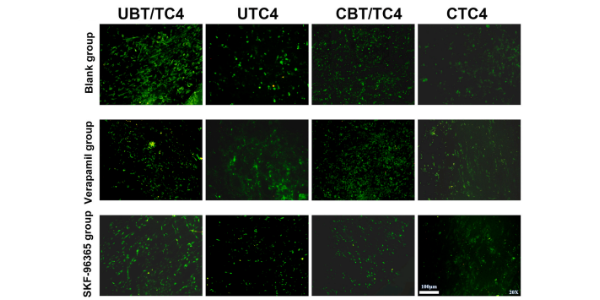
Intracellular calcium fluorescence staining of MC3T3-E1 cells on day 1 in a blank and samples inhibited by verapamil (10 μmol/L, 24 h) or SKF-96365 (10 μmol/L, 24 h).
SKF-96365 purchased from MedChemExpress. Usage Cited in: Bioact Mater. 2021 Apr 21;6(11):4073-4082. [Abstract]
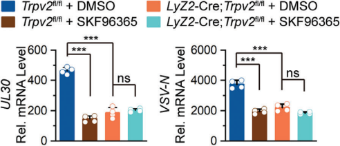
SKF-96365 (100 μM; 2 h) significantly inhibits the mRNA levels of the HSV-1 UL30 gene in Trpv2fl/fl BMDCs but not in Lyz2-Cre;Trpv2fl/fl BMDCs.
-
Autophagy
Cannabidiol inhibits human glioma by induction of lethal mitophagy through activating TRPV4. [Abstract]2021 Nov;17(11):3592-3606. PMID: 33629929 -
-
Theranostics
TRPV2-spike protein interaction mediates the entry of SARS-CoV-2 into macrophages in febrile conditions. [Abstract]2021 May 25;11(15):7379-7390. PMID: 34158856 -
Theranostics
FGF19/SOCE/NFATc2 signaling circuit facilitates the self-renewal of liver cancer stem cells. [Abstract]2021 Mar 5;11(10):5045-5060. PMID: 33754043
SKF-96365 purchased from MedChemExpress. Usage Cited in: Theranostics. 2021 Mar 5;11(10):5045-5060. [Abstract]
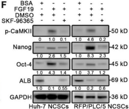
WB were applied to measure levels of Nanog, Oct-4, Sox2 and ALB in FGF19 (100 ng/ml) treated NCSCs, in the presence of SKF-96365 (5 µM) or not.
-
Theranostics
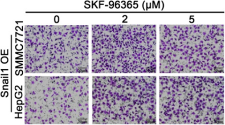
STIM1 is a metabolic checkpoint regulating the invasion and metastasis of hepatocellular carcinoma. [Abstract]2020 May 16;10(14):6483-6499. PMID: 32483465
SKF-96365 purchased from MedChemExpress. Usage Cited in: Theranostics. 2020 May 16;10(14):6483-6499. [Abstract]
Transwell assays were performed to detect the effects of different concentrations SKF-96365 on the invasion ability of Snail1 OE-SMMC7721 and HepG2 cells. The results show that SKF-96365 (2-5 μM) enhances the invasive ability of hepatocellular carcinoma cells with Snail1 overexpression.
-
Adv Sci (Weinh)
Hyperviscous Diabetic Bone Marrow Niche Impairs BMSCs Osteogenesis via TRPV2-Mediated Cytoskeletal-Nuclear Mechanotransduction. [Abstract]2025 Dec 22:e09056. PMID: 41431162 -
Adv Sci (Weinh)
The Transient Receptor Potential Vanilloid 2 (TRPV2) Channel Facilitates Virus Infection Through the Ca2+ -LRMDA Axis in Myeloid Cells. [Abstract]2022 Oct 19;e2202857. PMID: 36261399 -
Cell Death Differ
OR2T6 modulates autophagy through the PPP3CA-mediated pathways to suppress gastric cancer. [Abstract]2025 Nov 10. PMID: 41214150 -
Stem Cell Res Ther
2025 Apr 24;16(1):206. PMID: 40275329
SKF-96365 purchased from MedChemExpress. Usage Cited in: Stem Cell Res Ther. 2025 Apr 24;16(1):206. [Abstract]
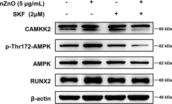
Western blot analysis of CaMKK2, AMPK activation and RUNX2 in MSCs after osteogenic differentiation for 72 h treated with TRP channel blocker SKF-96,365 hydrochloride (SKF, 2 µM) under control and nZnO (5 µg/mL) conditions.
-
Cell Rep
Viral infection and spread are inhibited by the polyubiquitination and downregulation of TRPV2 channel by the interferon-stimulated gene TRIM21. [Abstract]2024 Apr 23;43(4):114095. PMID: 38613787 -
Br J Pharmacol
Small intestinal glucose and sodium absorption through calcium-induced calcium release and store-operated Ca2+ entry mechanisms. [Abstract]2021 Jan;178(2):346-362. PMID: 33080043 -
J Agric Food Chem
miR-2765 Modulates the Seasonal Polyphenism in Cacopsylla chinensis by Targeting a Novel Cold Rreceptor CcTRPC3. [Abstract]2024 Jan 10;72(1):140-152. PMID: 38118125 -
Cells
Impact of Short-Term (+)-JQ1 Exposure on Mouse Aorta: Unanticipated Inhibition of Smooth Muscle Contractility. [Abstract]2023 May 24;12(11):1461. PMID: 37296583 -
Eur J Pharmacol
Novel mechanisms of metformin-induced vasorelaxation of mesenteric arterioles via endothelium-dependent hyperpolarization to treat murine colitis. [Abstract]2025 Sep 15:1003:177900. PMID: 40617384 -
Acta Physiol
2023 Apr;237(4):e13926. PMID: 36606511 -
Front Pharmacol
Role of Serosal TRPV4-Constituted SOCE Mechanism in Secretagogues-Stimulated Intestinal Epithelial Anion Secretion. [Abstract]2021 Jul 14:12:684538. PMID: 34335254 -
Chem Biol Interact
Platycodin D reverses tumor necrosis factor-α-induced endothelial dysfunction by increasing nitric oxide through G protein-coupled estrogen receptor-mediated eNOS activity. [Abstract]2025 Sep 5:418:111577. PMID: 40447174 -
J Neuroendocrinol
The caudal neurosecretory system: A novel thermosensitive tissue and its signal pathway in olive flounder (Paralichthys olivaceus). [Abstract]2020 Jun;32(6):e12876. PMID: 32542811 -
Mol Cell Biochem
Inhibition of STIM1 alleviates high glucose-induced proliferation and fibrosis by inducing autophagy in mesangial cells. [Abstract]2024 Sep;479(9):2365-2379. PMID: 37736800 -
Acta Biochim Biophys Sin (Shanghai)
Combined bone marrow stromal cells and oxiracetam treatments ameliorates acute cerebral ischemia/reperfusion injury through TRPC6. [Abstract]2019 Aug 5;51(8):767-777. PMID: 31236585 -
Front Physiol
Cyclopiazonic Acid-Induced Ca2+ Store Depletion Initiates Endothelium-Dependent Hyperpolarization-Mediated Vasorelaxation of Mesenteric Arteries in Healthy and Colitis Mice. [Abstract]2021 Mar 9:12:639857. PMID: 33767636 -
FASEB J
2019 Sep;33(9):9775-9784. PMID: 31166814 -
J Pharm Pharmacol
Homoplantaginin attenuates high glucose-induced vascular endothelial dysfunction via inhibiting store-operated calcium entry channel and endoplasmic reticulum stress. [Abstract]2023 Dec 8;75(12):1530-1543. PMID: 37774413 -
Biochem Biophys Res Commun
Glucose transports in the ileum: mechanism, regulation and physiological role of ileal glucose absorption. [Abstract]2025 Sep 19:785:152676. PMID: 41005286 -
Physiol Rep
Monosodium glutamate-mediated Ca2+-dependent intestinal epithelial ion transports in health and IBS-D in male mice. [Abstract]2026 Jun;14(11):e70975. PMID: 42281391 -
-
Heliyon
N,N,N',N'-Tetrakis(2-pyridylmethyl)ethylenediamine induces endothelium-dependent hyperpolarization-mediated vasorelaxation via store-operated calcium entry mechanism in healthy and intestinal inflammatory mice. [Abstract]2024 Jul 6;10(14):e33994. PMID: 39108891


溶剤 & 溶解度
体外:
DMSO : 100 mg/mL (272.89 mM; Need ultrasonic; Hygroscopic DMSO has a significant impact on the solubility of product, please use newly opened DMSO)
Please refer to the solubility information to select the appropriate solvent. Once prepared, please aliquot and store the solution to prevent product inactivation from repeated freeze-thaw cycles.
Storage method and period of stock solution: -80°C, 6 months; -20°C, 1 month. When stored at -80°C, please use it within 6 months. When stored at -20°C, please use it within 1 month.
Please refer to the solubility information to select the appropriate solvent. Once prepared, please aliquot and store the solution to prevent product inactivation from repeated freeze-thaw cycles.
Storage method and period of stock solution: -80°C, 6 months; -20°C, 1 month. When stored at -80°C, please use it within 6 months. When stored at -20°C, please use it within 1 month.
濃度 (開始) × 体積 (開始) = 濃度 (終了) × 体積 (終了)
体内:
Select the appropriate dissolution method based on your experimental animal and administration route.
- For the following dissolution methods, please ensure to first prepare a clear stock solution using an In Vitro approach and then sequentially add co-solvents:
- To ensure reliable experimental results, the clarified stock solution can be appropriately stored based on storage conditions. As for the working solution for In Vivo experiments, it is recommended to prepare freshly and use it on the same day.
- The percentages shown for the solvents indicate their volumetric ratio in the final prepared solution. If precipitation or phase separation occurs during preparation, heat and/or sonication can be used to aid dissolution.
Add each solvent one by one: 10% DMSO 40% PEG300 5% Tween-80 45% Saline
Solubility: ≥ 5 mg/mL (13.64 mM); Clear solution
This protocol yields a clear solution of ≥ 5 mg/mL (saturation unknown).
Taking 1 mL working solution as an example, add 100 μL DMSO stock solution (50.0 mg/mL) to 400 μL PEG300, and mix evenly; then add 50 μL Tween-80 and mix evenly; then add 450 μL Saline to adjust the volume to 1 mL.
Preparation of Saline: Dissolve 0.9 g sodium chloride in ddH₂O and dilute to 100 mL to obtain a clear Saline solution.
In Vivo Dissolution Calculator
Please enter the basic information of animal experiments:
-
-
-
-
Recommended: Prepare an additional quantity of animals to account for potential losses during experiments.
Please enter your animal formula composition:
-
%DMSO +
Recommended: Keep the proportion of DMSO in working solution below 2% if your animal is weak.
-
%+
-
+%Tween-80 + +
-
%Saline +
The co-solvents required include: DMSO, . All of co-solvents are available by MedChemExpress (MCE). , Tween 80. All of co-solvents are available by MedChemExpress (MCE).
Working solution concentration: 0.22 mg/mL
Method for preparing stock solution: mg drug dissolved in μL DMSO. Stock solution concentration: mg/mL.
1. Take μL DMSO stock solution;
2. Add μL .
μL , mix evenly;
3. Then add μL Tween 80, mix evenly;
4. Then add μL
Please ensure that the stock solution in the first step is dissolved to a clear state, and add co-solvents in sequence. You can use ultrasonic heating (ultrasonic cleaner, recommended frequency 20-40 kHz), vortexing, etc. to assist dissolution.

プロトコル
-
RNA extraction experimental
By lysing cells, releasing RNA, and removing impurities such as proteins and DNA, high-purity RNA products are finally obtained. The commonly used traditional method is the guanidine isothiocyanate/phenol/chloroform method (Trizol), which is suitable for a variety of animal materials including animal tissues, microorganisms, cultured cells, etc., and most plant materials.
-
Western Blot
Western blotting (WB) is a commonly used experimental method in molecular biology, biochemistry, and immunogenetics for identifying and quantifying target proteins. It combines gel electrophoresis with immunoassay, enabling researchers to analyze protein expression, post-translational modifications, and molecular weight.
-
Annexin V plus membrane-impermeant dye apoptosis staining
Annexin V-based apoptosis assays rely on the detection of phosphatidylserine (PS) externalization from the inner leaflet of the plasma membrane to the outer leaflet, an early biochemical hallmark of apoptosis. Fluorescently labeled Annexin V binds PS in a calcium-dependent manner, enabling identification of early apoptotic cells by flow cytometry or fluorescence microscopy. When combined with a membrane-impermeant DNA-binding dye (e. g. , propidium iodide), this approach allows discrimination between viable (Annexin V−/dye−), early apoptotic (Annexin V+/dye−), and late apoptotic or necrotic (Annexin V+/dye+) cell populations by assessing membrane integrity and PS exposure.
-
BrdU Incorporation Assay
Bromodeoxyuridine (BrdU) incorporation assay is based on the principle that BrdU, a thymidine analog, is incorporated into newly synthesized DNA during the S phase of the cell cycle, thereby serving as a marker of DNA replication and cellular proliferation. Incorporated BrdU can be detected using anti-BrdU antibodies following DNA denaturation, enabling visualization or quantification of proliferating cells through immunochemical detection methods such as immunofluorescence or immunohistochemistry.
-
Protocol for Cell Cycle
Cell-cycle analysis by flow cytometry measures DNA content in single cells to estimate the fraction of cells in G0/G1, S, and G2/M phases. Propidium iodide intercalates into DNA, and after RNA removal with RNase, fluorescence intensity reflects cellular DNA content: 2N cells are assigned to G0/G1, cells between 2N and 4N to S phase, and 4N cells to G2/M. DNA-content analysis alone cannot reliably separate G0 from G1 or G2 from M. Ki-67 can distinguish quiescent G0 cells from cycling cells, EdU or BrdU incorporation marks active DNA synthesis in S phase, and phospho-histone H3 staining identifies mitotic cells within the 4N population.
-
Research Protocol for Inflammation-related Diseases
The NLRP3 inflammasome is a cytosolic innate immune signaling platform that integrates priming signals and danger-signal activation to promote caspase-1 activation, maturation of IL-1β and IL-18, and gasdermin D-mediated pyroptotic cell death. The core experimental logic is to determine whether inflammatory disease phenotypes are driven by increased NLRP3 expression, ASC-containing inflammasome assembly, caspase-1 cleavage, GSDMD cleavage, and extracellular release of IL-1β/IL-18 rather than by nonspecific cell injury alone. The pathway is strongly linked to inflammation-related disease phenotypes because monosodium urate crystals activate NALP3/NLRP3 inflammasome signaling in gout-like crystal inflammation, cholesterol crystals activate NLRP3 inflammasomes in atherogenesis models, and DSS-induced intestinal inflammation has been reported to involve NLRP3 inflammasome activity. However, experimental colitis studies also show context-dependent protective effects of NLRP3 inflammasome co
-
Ca2+ Staining Technique
Ca2+ staining is an experimental technique that utilizes specific fluorescent probes (such as Fluo-4 AM, Fura-2, etc.) to qualitatively or quantitatively detect dynamic changes in intracellular Ca2+ concentrations; this is achieved by monitoring the changes in fluorescent signals generated when these probes bind to free intracellular calcium ions. The underlying principle relies primarily on the presence of chelating groups within the probe's molecular structure that possess high affinity for calcium ions.
-
Kinase activity and phosphorylation assays
Kinase activity assays measure the ability of kinases to transfer phosphate groups from ATP to specific substrates, while phosphorylation assays detect the presence and levels of phosphorylated proteins. Common methods include radiolabeled ATP incorporation (e. g. ,), ADP release detection via bioluminescence (e. g. ,[3]), enzyme-linked immunosorbent assays (ELISA) for phospho-specific epitopes (e. g. ,[6]), and microtiter-based formats for high-throughput screening (e. g. ,[8]). The ADP-Glo assay quantifies kinase activity by measuring ADP produced during phosphorylation using a luciferase-based system. Radiometric assays involve autoradiography or scintillation counting after incorporation of 32P-labeled ATP into substrate proteins. ELISA-based approaches rely on phospho-specific antibodies to detect activated kinases in cell lysates or purified samples.
-
Apoptosis
Apoptosis, also called programmed cell death, is generally characterized by distinct morphological characteristics.
-
TUNEL staining for apoptotic DNA fragmentation
TUNEL staining detects DNA strand breaks by using terminal deoxynucleotidyl transferase to add labeled nucleotides to exposed 3′-OH DNA termini, generating either microscopic staining in fixed cells or tissue sections, or fluorescence/cytometric signal in cell suspensions. TUNEL positivity reflects DNA fragmentation but should not be interpreted alone as definitive apoptosis, because TUNEL can also label necrotic, autolytic, mechanically damaged, or DNA-repair-associated DNA breaks.
-
Flow cytometric DNA-content cell-cycle staining
Flow cytometric DNA-content cell-cycle staining measures the fluorescence intensity of DNA-bound fluorochromes in single cells or nuclei to estimate DNA content distributions, allowing assignment of populations to G0/G1, S, and G2/M phases by DNA histogram deconvolution. Propidium iodide (PI) intercalates into DNA, and PI fluorescence is proportional to cellular DNA content when staining is performed under conditions that make DNA accessible and minimize non-DNA signal. Cells with G2/M DNA content are expected to show approximately twice the fluorescence intensity of G0/G1 cells, while S-phase cells occupy intermediate fluorescence values. PI-based DNA-content analysis can also detect cells with fractional DNA content, often reported as sub-G1, when DNA fragmentation and extraction during staining reduce retained DNA signal in apoptotic cells. DAPI is an alternative DNA fluorochrome for univariate DNA-content analysis, while bivariate approaches combining DNA content with proliferation
-
Human pluripotent stem cell midbrain dopaminergic neuron differentiation
Human pluripotent stem cells are directed toward midbrain dopaminergic neurons by first inducing a neural floor-plate-like progenitor state, then patterning cells with ventralizing SHH signaling and midbrain/WNT-FGF cues, and finally maturing progenitors into neurons expressing dopaminergic markers such as TH, NURR1/NR4A2, PITX3, DAT/SLC6A3, VMAT2/SLC18A2, GIRK2/KCNJ6, FOXA2, LMX1A, and EN1. The main readouts are loss of pluripotency, acquisition of FOXA2+/LMX1A+ midbrain floor-plate progenitors, emergence of βIII-tubulin+/MAP2+ neurons, and production of TH+ dopaminergic neurons with molecular, dopamine-release, and electrophysiological features of midbrain dopaminergic identity.
-
Autophagy
Autophagy is a process in which eukaryotic cells use lysosomes to degrade their own cytoplasmic proteins and damaged organelles under the regulation of autophagy related gene (Atg). Microtubule-associated proteins light chain 3 (LC3) is recognized as autophagy marker, which transfers from cytoplasmic LC3 (LC3-I) to membrane type (LC3-II). LC3-II/I ratio could be detected by Western Blot and fluorescence microscopy.
-
Apoptosis Solutions
Apoptosis is a regulated, generally non-lytic cell-death pathway that removes unwanted, damaged, infected, or abnormal cells through coordinated morphological changes, caspase activation, DNA fragmentation, and membrane remodeling. The intrinsic apoptosis pathway is controlled mainly by mitochondrial outer membrane permeabilization, BCL-2 family proteins, cytochrome c release, apoptosome formation, caspase-9 activation, and downstream executioner caspase-3/7 activation. The extrinsic apoptosis pathway is initiated by death receptors such as Fas, TNFR, and TRAIL receptors, which recruit adaptor proteins and activate caspase-8 before engaging executioner caspases or mitochondrial amplification through BID cleavage. Apoptosis is linked to many phenotypes, including cancer cell killing, tissue homeostasis, immune regulation, neurodegeneration, infection response, and treatment-induced cytotoxicity; unresolved questions include how apoptosis interacts with necroptosis, pyroptosis, ferroptos
-
Calcium Spark Assay
Calcium sparks are localized, transient increases in intracellular calcium concentration ([Ca2+]i) that occur in cardiac myocytes and represent elementary events underlying excitation-contraction coupling. These events are generated by the coordinated opening of clusters of ryanodine receptors (RyRs) on the sarcoplasmic reticulum membrane, leading to a brief release of Ca2+ into the cytosol. The detection and analysis of calcium sparks provide insights into the mechanisms of calcium handling and signaling in cardiac cells. Imaging techniques using fluorescent calcium indicators such as Fluo-3 are employed to visualize these subcellular calcium transients with high spatial and temporal resolution. The protocol is based on established methodologies described in primary literature for both experimental measurement and automated analysis of calcium sparks.
-
Protocol for Kinase activity and phosphorylation assays
Kinase activity assays measure transfer of phosphate from ATP to a protein or peptide substrate, generating phosphorylated substrate, ADP, or incorporated radiolabeled phosphate as the readout; phosphorylation assays measure site-specific phosphorylation in cells or tissues as a proxy for kinase-pathway activation, inhibition, or substrate regulation. Phosphorylation can be detected by phospho-specific Western blot, immunoprecipitation kinase assay, phospho-immunofluorescence, phospho-flow cytometry, luminescent ADP detection, radiolabeled ATP incorporation, or reporter-based pathway assays, and these readouts can be applied to cancer cells, primary neurons, mouse tumors, organoids, inflammatory macrophages, ferroptosis studies, and mitophagy studies when the kinase target is biologically relevant.
-
Lysosome and acidic-vesicle live-cell staining
Lysosome and acidic-vesicle live-cell staining detects acidic intracellular compartments by using membrane-permeant acidotropic probes that accumulate in low-pH vesicles, including lysosomes, late endosomes, autolysosomes, and acidic phagosomes. LysoTracker staining is commonly used as an intensity-based readout of acidic lysosomal compartment abundance or enlargement, while acridine orange produces green fluorescence in less concentrated compartments and red fluorescence after concentration-dependent accumulation in acidic vesicular organelles. Loss or reduction of acridine-orange red signal can be used as a readout of lysosomal membrane permeabilization or reduced acidic-vesicle integrity. This protocol is designed for live cultured cells and can be adapted for fluorescence microscopy, high-content imaging, plate-reader readout, or flow cytometry when the selected literature supports the readout. Because these dyes report acidotropic accumulation rather than lysosome identity alone,
-
Macroautophagy Solutions
Macroautophagy is a conserved lysosome-dependent degradation pathway in which cytoplasmic material is sequestered into double-membrane autophagosomes and delivered to lysosomes for degradation and recycling. The pathway supports cellular homeostasis during nutrient limitation, organelle stress, protein-aggregate accumulation, infection, differentiation, and tissue remodeling by coupling cargo sequestration, autophagosome maturation, lysosomal fusion, and degradation of cargo-derived macromolecules. The core molecular sequence includes initiation by nutrient- and stress-regulated autophagy machinery, autophagosome nucleation, LC3/ATG8-family conjugation to autophagosomal membranes, cargo selection through receptors such as SQSTM1/p62, autophagosome-lysosome fusion, and lysosomal degradation. LC3 was identified as a mammalian homolog of yeast Atg8 that localizes to autophagosomal membranes after processing, and p62/SQSTM1 was shown to connect ubiquitinated cargo with autophagic degradati


純度とドキュメンテーション
-
データシート (286 KB)
-
SDS (251 KB)
- English - EN (251 KB)
- Français - FR (251 KB)
- Deutsch - DE (251 KB)
- Norwegian - NO (251 KB)
- Español - ES (251 KB)
- Swedish - SV (251 KB)
- Italian - IT (251 KB)
- Korean - KR (251 KB)
- Portuguese - PT (251 KB)
-
取扱説明書 (2659 KB)
参考文献
[1]. Ba G, et al. Therapeutic effects of SKF-96365 on murine allergic rhinitis induced by OVA. Int J Immunopathol Pharmacol. 2021;35:20587384211015054. [Content Brief]
[2]. Chen KH, et al. SKF-96365 strongly inhibits voltage-gated sodium current in rat ventricular myocytes. Pflugers Arch. 2015;467(6):1227-1236. [Content Brief]
[3]. Jing Z, et al. SKF-96365 activates cytoprotective autophagy to delay apoptosis in colorectal cancer cells through inhibition of the calcium/CaMKIIγ/AKT-mediated pathway. Cancer Lett. 2016;372(2):226-238. [Content Brief]
[4]. Chen T, et al. Protective effects of SKF-96365, a non-specific inhibitor of SOCE, against MPP+-induced cytotoxicity in PC12 cells: potential role of Homer1. PLoS One. 2013;8(1):e55601. [Content Brief]
[5]. Ding J, et al. Effects of SKF-96365, a TRPC inhibitor, on melittin-induced inward current and intracellular Ca2+ rise in primary sensory cells. Neurosci Bull. 2011;27(3):135-142. [Content Brief]


Complete Stock Solution Preparation Table
Please refer to the solubility information to select the appropriate solvent. Once prepared, please aliquot and store the solution to prevent product inactivation from repeated freeze-thaw cycles.
Storage method and period of stock solution: -80°C, 6 months; -20°C, 1 month. When stored at -80°C, please use it within 6 months. When stored at -20°C, please use it within 1 month.
| Optional Solvent | Concentration Solvent Mass | 1 mg | 5 mg | 10 mg | 25 mg |
|---|---|---|---|---|---|
| DMSO | 1 mM | 2.7289 mL | 13.6444 mL | 27.2889 mL | 68.2221 mL |
| 5 mM | 0.5458 mL | 2.7289 mL | 5.4578 mL | 13.6444 mL | |
| 10 mM | 0.2729 mL | 1.3644 mL | 2.7289 mL | 6.8222 mL | |
| 15 mM | 0.1819 mL | 0.9096 mL | 1.8193 mL | 4.5481 mL | |
| 20 mM | 0.1364 mL | 0.6822 mL | 1.3644 mL | 3.4111 mL | |
| 25 mM | 0.1092 mL | 0.5458 mL | 1.0916 mL | 2.7289 mL | |
| 30 mM | 0.0910 mL | 0.4548 mL | 0.9096 mL | 2.2741 mL | |
| 40 mM | 0.0682 mL | 0.3411 mL | 0.6822 mL | 1.7056 mL | |
| 50 mM | 0.0546 mL | 0.2729 mL | 0.5458 mL | 1.3644 mL | |
| 60 mM | 0.0455 mL | 0.2274 mL | 0.4548 mL | 1.1370 mL | |
| 80 mM | 0.0341 mL | 0.1706 mL | 0.3411 mL | 0.8528 mL | |
| 100 mM | 0.0273 mL | 0.1364 mL | 0.2729 mL | 0.6822 mL |


SKF-96365 関連分類
HY-125942 関連分類
 Powered by Bioz