



Oleuropein Aglycone
Based on 1 publication(s) in Google Scholar
Oleuropein Aglycone (3,4-DHPEA-EA) is a secoiridoid that exerts antioxidant, anti-inflammatory, and anti-apoptotic effects by promoting ERK/AMPK/eNOS phosphorylation and inhibiting JNK/p38 MAPK and inflammation-related signaling pathways. Oleuropein Aglycone is used in research on acute kidney injury induced by ischemia-reperfusion injury, pleurisy, osteoporosis, Alzheimer's disease, neurodegenerative diseases, and triple-negative breast cancer.
For research use only. We do not sell to patients.
- Purity : 98.46%
- CAS No.: 31773-95-2
- Formula: C19H22O8
- Molecular Weight:378.37
-
Storage:
-20°C, sealed storage, away from moisture
* In solvent : -80°C, 6 months; -20°C, 1 month (sealed storage, away from moisture)


To place orders, for customer services and technical support, please contact: MedChemExpress USA
Tel: 609-228-6898 E-mail: [email protected] [email protected]


-
 Biological Activity
-
Chemical Information
-
Solvent & Solubility
- Protocols
- Purity & Documentation
- References
-
 Help & FAQs


-
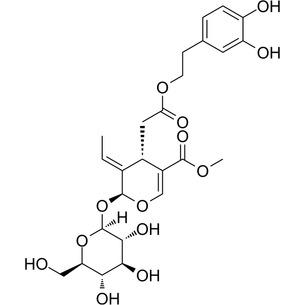
HY-N0292
OleuropeinApoptosis Inducer -

HY-P4146
SurvodutideGCGR/GLP-1R Agonist -
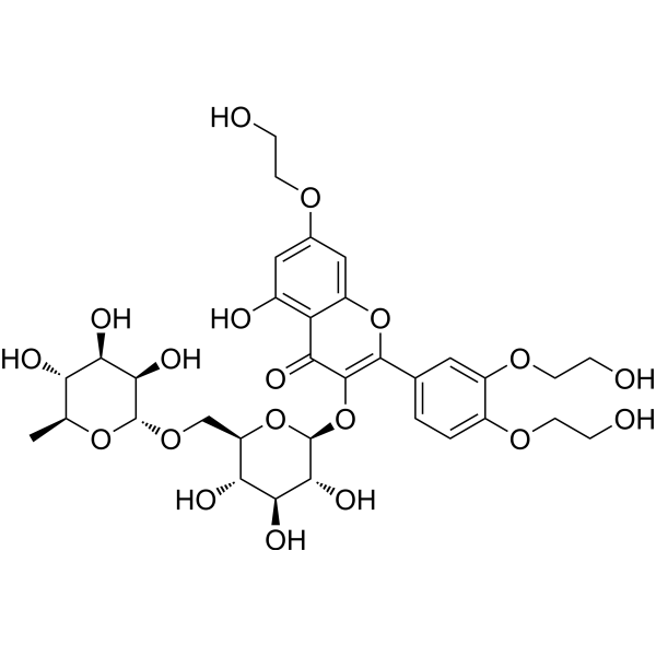
HY-N0139
TroxerutinNOD-like Receptor (NLR) Inhibitor -
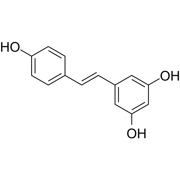
HY-16561
ResveratrolPolyphenolic Phytoalexin -
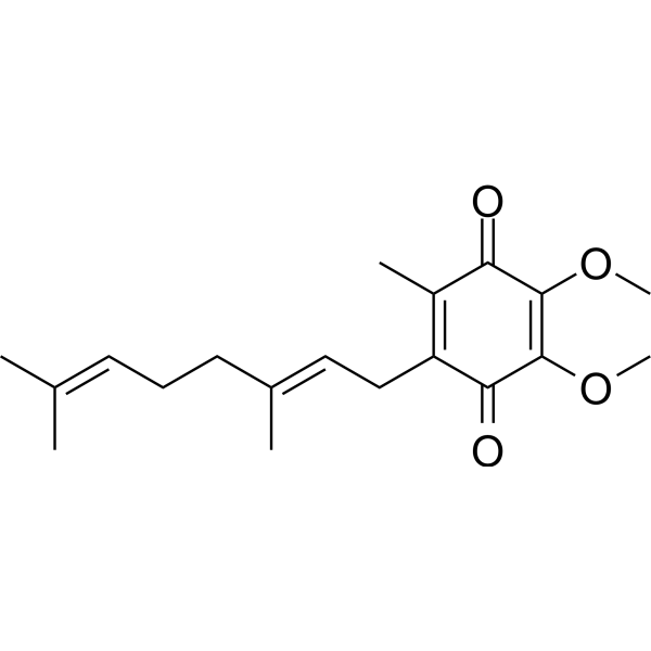
HY-118075
Coenzyme Q2Apoptosis Inducer -
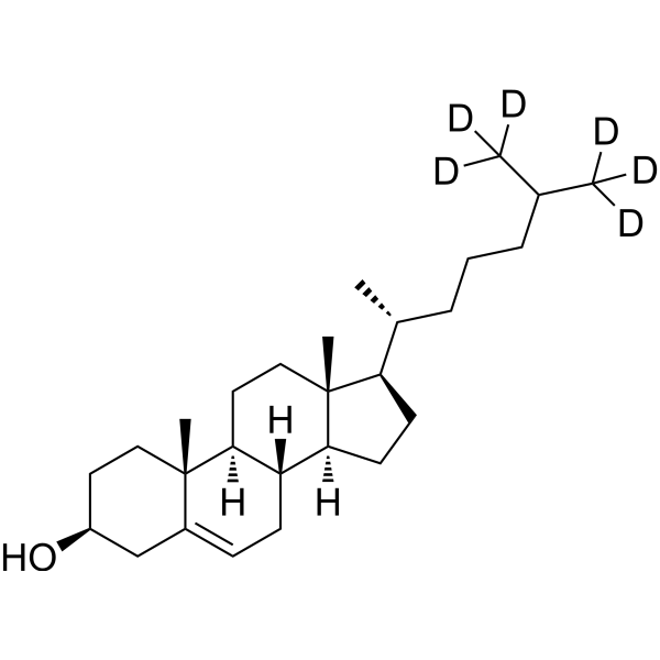
HY-N0322S1
Cholesterol-d6Stable Isotope -
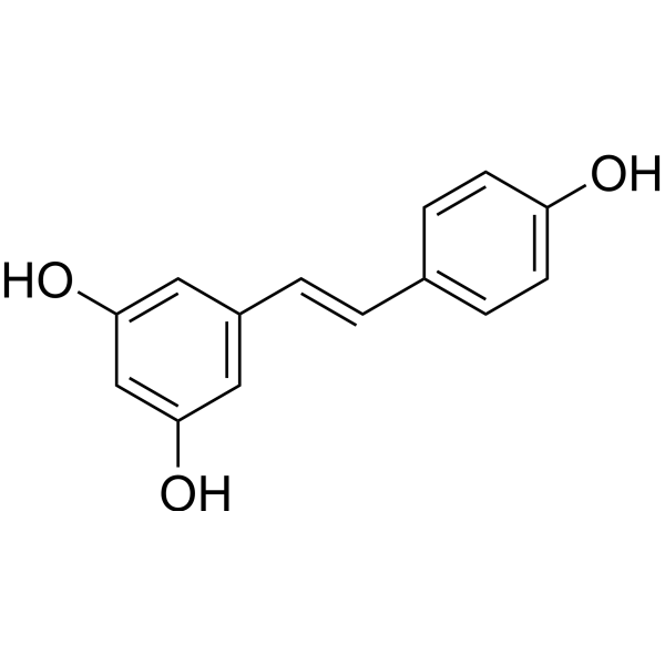
HY-16561R
Resveratrol (Standard) -
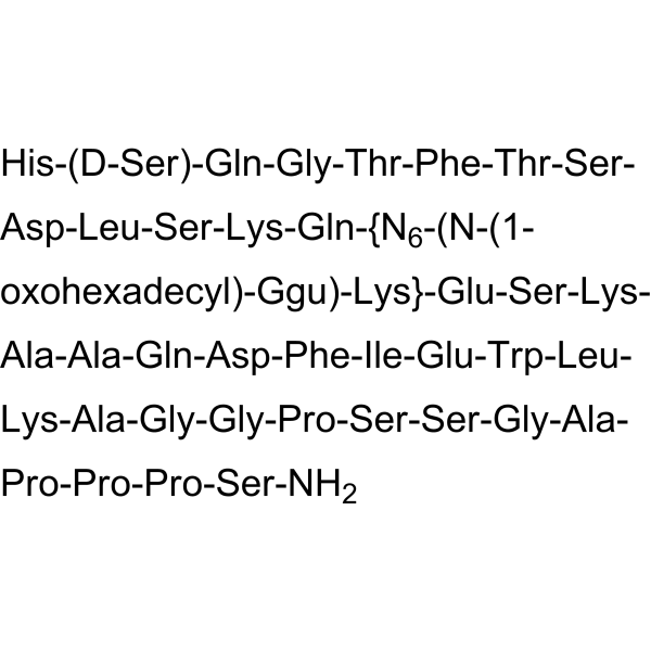
HY-P10018
BamadutideGLP-1R/GCGR Agonist -

HY-12294B
(Rac)-PEAQXNMDA Receptor Antagonist -
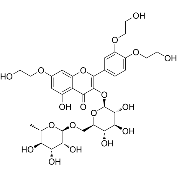
HY-N0139R
Troxerutin (Standard) -

HY-16561G
Resveratrol (GMP)Polyphenolic Phytoalexin -

HY-124920
MethiocarbInsecticide
Publications Citing Use of MedChemExpress (MCE) Oleuropein Aglycone
MoreAll AMPK Isoforms
More

Biological Activity
Description
In Vitro
Oleuropein Aglycone (3,4-DHPEA-EA) (1:0-1:10; 50 h) significantly inhibits S100A9 amyloid formation by reducing the effective rate of amyloid growth and the overall amyloid burden[4].
Oleuropein aglycone (1:0-1:10; 6-48 h) reduces the length and number of S100A9 amyloid fibrils and protein oligomerization in a concentration-dependent manner[4].
Oleuropein aglycone (137 μM) depolymerizes preformed S100A9 fibrils and converts them into non-fibrillar aggregates[4].
Oleuropein aglycone inhibits the development of S100A9 amyloid and reduces the β-sheet content in preformed fibrils[4].
Oleuropein aglycone interacts with both native and amyloid S100A9, with apparent dissociation constants of 0.5 and 200 μM[4].
Oleuropein aglycone (1:2; 1-48 h) depolymerizes preformed S100A9 fibrils into non-fibrillar aggregates that are no longer toxic[4].
Oleuropein aglycone (1:1-1:10; 10-60 min) attenuates the S100A9 amyloid-induced increase in intracellular free Ca2+ levels in SH-SY5Y cells, returning to baseline levels at S100A9 to OleA molar ratios of 1:5 and 1:10[4].
Oleuropein aglycone (1:1-1:10; 24 h) targets S100A9 amyloid on the cell membrane and reduces its accumulation in GM1-containing membrane rafts[4].
Oleuropein Aglycone (5-100 μM; 24 h) is well tolerated by human BMSCs, and a concentration of 5 μM maintains at least 85% cell viability[3].
Oleuropein Aglycone (180 °C; 3 h) is mostly degraded in heated extra virgin olive oil, with only 6% remaining[6].
Oleuropein aglycone (1:1-1:10; 24 h) attenuates S100A9-induced cytotoxicity in SH-SY5Y cells, achieving complete recovery at S100A9 to Oleuropein aglycone molar ratios of 1:5 and 1:10[4].
Oleuropein aglycone (1:1-1:10; 24 h) reduces ROS production in S100A9-induced SH-SY5Y cells, returning to baseline levels at S100A9 to OleA molar ratios of 1:5 and 1:10[4].
Oleuropein aglycone (0.78-100 mM; 40 min) exhibits antioxidant activity in the DPPH assay with an IC50 of 25.23 μM[5].
Oleuropein Aglycone (compound 4) (10 μM) exhibits peroxyl radical scavenging capacity, with an ORACFL value of 60.1 μM[10].
Oleuropein Aglycone (100 μM; 24 h) significantly reduces H2O2-induced intracellular ROS generation in HeLa cells[10].
Oleuropein Aglycone (1-100 µM; 24 h) exhibits low cytotoxicity in young HUVECs, RS-HUVECs, THP-1, and hSAECs, with cell viability remaining at least 85% at concentrations up to 5 µM[7].
Oleuropein Aglycone (IC50; 24 h) significantly increases γH2A.X levels in MDA-MB-231 cells to approximately 165% of the control, exhibiting the greatest effect among the tested olive oil phenolics[9].
Oleuropein Aglycone (5 µM; 2 h) reduces IL-1β and IL-8 mRNA expression and IL-6 secretion in LPS-stimulated young HUVECs[7].
Oleuropein Aglycone (5 µM; 2 h) downregulates TNF-α and IL-8 mRNA expression and reduces IL-6 production in LPS-stimulated THP-1 cells[7].
Oleuropein Aglycone (5 µM; 24 h) exerts anti-SASP activity by reducing the expression of IL-1β, TNF-α, ICAM-1, and VCAM, and decreasing IL-6 release in RS-HUVECs[7].
Oleuropein Aglycone (5 µM; 2 h) decreases IL-1β and SOD2 mRNA expression, reduces IL-6 release, lowers ACE2 mRNA, and downregulates P-NF-kB and ACE2 protein levels in Polyinosinic-polycytidylic acid (Poly (I:C)) (HY-107202)-stimulated hSAECs[7].
MedChemExpress (MCE) has not independently confirmed the accuracy of these methods. They are for reference only. Further protocols information, click here.
-
Cell Line:Human bone marrow mesenchymal stromal cells (BMSCs)
-
Concentration:5, 10, 25, 50, 100 μM; 5 μM (selected for further experiments)
-
Incubation Time:24 h
-
Result:Determined the concentration of oleuropein aglycone that would ensure at least 85% cell viability to be 5 μM.
-
Cell Line:Human bone marrow mesenchymal stromal cells (BMSCs)
-
Concentration:5 μM
-
Incubation Time:14 days
-
Result:Induced a more modest reduction in intracellular lipid droplet accumulation.
Showed a reduction in the percentage of stained area in OA-treated ADMSCs compared to untreated ADMSCs.
-
Cell Line:SH-SY5Y cells
-
Concentration:S100A9 to OleA molar ratio of 1:2
-
Incubation Time:1 to 48 h
-
Result:A decrease of the cytotoxic effect of the preformed S100A9 amyloids was observed when they were treated with OleA.
-
Cell Line:SH-SY5Y
-
Concentration:0.5, 1, 10, 100 μM
-
Incubation Time:24 h
-
Result:At a concentration of 0.1 μM, the percentage of control cell growth was almost 100%.
-
Cell Line:Young HUVECs, Replicative Senescent HUVECs (RS-HUVECs), THP-1 monocytic cells, and primary human small airway epithelial cells (hSAECs)
-
Concentration:5, 10, 25, 50, 100 μM (0.36-36 µg/mL)
-
Incubation Time:24 h
-
Result:Maintained at least 85% cell viability at concentrations up to 5 µM.
-
Cell Line:Young HUVECs
-
Concentration:5 µM
-
Incubation Time:2 h (pre-treatment); 3 h (LPS stimulation)
-
Result:Inhibited IL-1β and IL-8 mRNA expression.
Exerted a significant reduction in IL-6 secretion.
-
Cell Line:THP-1 monocytic cells
-
Concentration:5 µM
-
Incubation Time:2 h (pre-treatment); 3 h (LPS stimulation)
-
Result:Significantly reduced TNF-α and IL-8 mRNA expression.
Significantly reduced the production of IL-6 evaluated in cell lysates.
-
Cell Line:Replicative senescent HUVECs (RS-HUVECs)
-
Concentration:5 µM
-
Incubation Time:24 h
-
Result:Significantly reduced the mRNA expression of IL-1β, TNF-α, ICAM-1, and VCAM.
Efficiently reduced IL-6 release in the conditioned medium.
Observed a slight, but not significant, increase in SOD2 expression.
-
Cell Line:Human small airway epithelial cells (hSAECs)
-
Concentration:5 µM
-
Incubation Time:2 h (pre-treatment); 24 h (Poly(I:C) treatment)
-
Result:Significantly lowered IL-1β and SOD2 mRNA expression.
Significantly reduced IL-6 release.
Significantly downregulated P-NF-kB protein expression.
Significantly reduced ACE2 mRNA and downregulated ACE2 protein level.
Did not affect TMPRSS2 protein expression.
In Vivo
Oleuropein aglycone induces tumor regression in mice within 9-12 days[6].
Oleuropein aglycone (12.5 mg/kg; i.p.; three times per week; until day 35) reduces endpoint tumor burden by 22.0% in a TNBC xenograft mouse model [9].
Oleuropein aglycone (6.25 mg/kg; i.p.; three times per week; until day 35) and 2.5 mg/kg OLC retain antitumor activity, reducing endpoint tumor burden by 23.7% in a TNBC xenograft mouse model[9].
MedChemExpress (MCE) has not independently confirmed the accuracy of these methods. They are for reference only.
-
Animal Model:CD mice (male, 20-25 g)[2]
-
Dosage:100 μM/kg; 40 μM/kg
-
Administration:i.p.; 30 min after carrageenan challenge
-
Result:Reduced the degree of lung injury.
Attenuated neutrophil infiltration into the lung tissue.
No positive staining for ICAM-1 or P-selectin was observed in the lungs.
Attenuated the release of TNF-α and IL-1β in pleural exudates.
Significantly reduced NO exudate levels.
No positive staining for nitrotyrosine was found in the lungs.
Significantly attenuated lipid peroxidation in the lungs.
Reduced the degree of PARP activation.
-
Animal Model:CB17-SCID (cr-Prkdcscid/scid/Rj) mice[9]
-
Dosage:12.5 mg/kg
-
Administration:i.p.; three times per week; until Day 35
-
Result:Reduced endpoint tumor weight to 0.23 g, corresponding to a 22.0% decrease compared with the vehicle control group.
-
Animal Model:CB17-SCID (cr-Prkdcscid/scid/Rj) mice[9]
-
Dosage:6.25 mg/kg
-
Administration:i.p.; three times per week; until Day 35
-
Result:Decreased endpoint tumor weight to 0.225 g, corresponding to a 23.7% reduction compared with the vehicle control group.


Chemical Information
-
CAS No. 31773-95-2
-
Appearance Oil
-
Molecular Weight 378.37
-
Formula C19H22O8
-
Color Off-white to light yellow
-
SMILES
O=C(OCCC1=CC=C(O)C(O)=C1)C[C@H]2/C([C@H](O)OC=C2C(OC)=O)=C\C
-
Synonyms
3,4-DHPEA-EA
-
Shipping
Room temperature in continental US; may vary elsewhere.
-
Storage
-20°C, sealed storage, away from moisture
* In solvent : -80°C, 6 months; -20°C, 1 month (sealed storage, away from moisture)


Publications (1)
-
Journal Impact Factor
-
Most Recent


Solvent & Solubility
In Vitro:
DMSO : 50 mg/mL (132.15 mM; Need ultrasonic; Hygroscopic DMSO has a significant impact on the solubility of product, please use newly opened DMSO)
Please refer to the solubility information to select the appropriate solvent. Once prepared, please aliquot and store the solution to prevent product inactivation from repeated freeze-thaw cycles.
Storage method and period of stock solution: -80°C, 6 months; -20°C, 1 month (sealed storage, away from moisture). When stored at -80°C, please use it within 6 months. When stored at -20°C, please use it within 1 month.
Please refer to the solubility information to select the appropriate solvent. Once prepared, please aliquot and store the solution to prevent product inactivation from repeated freeze-thaw cycles.
Storage method and period of stock solution: -80°C, 6 months; -20°C, 1 month (sealed storage, away from moisture). When stored at -80°C, please use it within 6 months. When stored at -20°C, please use it within 1 month.
Concentration (start) × Volume (start) = Concentration (final) × Volume (final)

Protocols
-
Kinase activity and phosphorylation assays
Kinase activity assays measure the ability of kinases to transfer phosphate groups from ATP to specific substrates, while phosphorylation assays detect the presence and levels of phosphorylated proteins. Common methods include radiolabeled ATP incorporation (e. g. ,), ADP release detection via bioluminescence (e. g. ,[3]), enzyme-linked immunosorbent assays (ELISA) for phospho-specific epitopes (e. g. ,[6]), and microtiter-based formats for high-throughput screening (e. g. ,[8]). The ADP-Glo assay quantifies kinase activity by measuring ADP produced during phosphorylation using a luciferase-based system. Radiometric assays involve autoradiography or scintillation counting after incorporation of 32P-labeled ATP into substrate proteins. ELISA-based approaches rely on phospho-specific antibodies to detect activated kinases in cell lysates or purified samples.
-
Western Blot
Western blotting (WB) is a commonly used experimental method in molecular biology, biochemistry, and immunogenetics for identifying and quantifying target proteins. It combines gel electrophoresis with immunoassay, enabling researchers to analyze protein expression, post-translational modifications, and molecular weight.
-
Apoptosis
Apoptosis, also called programmed cell death, is generally characterized by distinct morphological characteristics.
-
Nephrotoxicity Study
This protocol assesses nephrotoxicity by combining functional kidney injury readouts, urinary/tissue injury biomarkers, and renal histopathology. Serum creatinine and BUN reflect impaired kidney function, while KIM-1, NGAL, clusterin, osteopontin, IL-18, cystatin C, nephrin, Oat5, urinary protein, glucose, and alkaline phosphatase have been used to detect tubular injury in cisplatin-, gentamicin-, and acetaminophen-induced nephrotoxicity models.
-
Apoptosis Solutions
Apoptosis is a regulated, generally non-lytic cell-death pathway that removes unwanted, damaged, infected, or abnormal cells through coordinated morphological changes, caspase activation, DNA fragmentation, and membrane remodeling. The intrinsic apoptosis pathway is controlled mainly by mitochondrial outer membrane permeabilization, BCL-2 family proteins, cytochrome c release, apoptosome formation, caspase-9 activation, and downstream executioner caspase-3/7 activation. The extrinsic apoptosis pathway is initiated by death receptors such as Fas, TNFR, and TRAIL receptors, which recruit adaptor proteins and activate caspase-8 before engaging executioner caspases or mitochondrial amplification through BID cleavage. Apoptosis is linked to many phenotypes, including cancer cell killing, tissue homeostasis, immune regulation, neurodegeneration, infection response, and treatment-induced cytotoxicity; unresolved questions include how apoptosis interacts with necroptosis, pyroptosis, ferroptos
-
Research Protocol for Inflammation-related Diseases
The NLRP3 inflammasome is a cytosolic innate immune signaling platform that integrates priming signals and danger-signal activation to promote caspase-1 activation, maturation of IL-1β and IL-18, and gasdermin D-mediated pyroptotic cell death. The core experimental logic is to determine whether inflammatory disease phenotypes are driven by increased NLRP3 expression, ASC-containing inflammasome assembly, caspase-1 cleavage, GSDMD cleavage, and extracellular release of IL-1β/IL-18 rather than by nonspecific cell injury alone. The pathway is strongly linked to inflammation-related disease phenotypes because monosodium urate crystals activate NALP3/NLRP3 inflammasome signaling in gout-like crystal inflammation, cholesterol crystals activate NLRP3 inflammasomes in atherogenesis models, and DSS-induced intestinal inflammation has been reported to involve NLRP3 inflammasome activity. However, experimental colitis studies also show context-dependent protective effects of NLRP3 inflammasome co
-
Protocol for Kinase activity and phosphorylation assays
Kinase activity assays measure transfer of phosphate from ATP to a protein or peptide substrate, generating phosphorylated substrate, ADP, or incorporated radiolabeled phosphate as the readout; phosphorylation assays measure site-specific phosphorylation in cells or tissues as a proxy for kinase-pathway activation, inhibition, or substrate regulation. Phosphorylation can be detected by phospho-specific Western blot, immunoprecipitation kinase assay, phospho-immunofluorescence, phospho-flow cytometry, luminescent ADP detection, radiolabeled ATP incorporation, or reporter-based pathway assays, and these readouts can be applied to cancer cells, primary neurons, mouse tumors, organoids, inflammatory macrophages, ferroptosis studies, and mitophagy studies when the kinase target is biologically relevant.
-
Breast Cancer Modeling
Breast cancer is a heterogeneous cancer, and it has been distinguished into four subtypes: luminal A, luminal B, HER2-positive and basal-like. Molecular mutations, epigenetic alterations, hormone exposure and immune microenvironment are related to the progression of breast cancer.
-
Alzheimer’s Disease Modeling
Alzheimer’s Disease (AD) is a neurodegenerative disorder characterized by a progressive decline in cognitive functions and loss of specific types of neurons and synapses. Alzheimer's symptoms can be simulated in mice by injecting drugs (such as Aβ) or genetically modified.


Purity & Documentation
-
Data Sheet (297 KB)
-
SDS (394 KB)
- English - EN (394 KB)
- Français - FR (394 KB)
- Deutsch - DE (394 KB)
- Norwegian - NO (394 KB)
- Español - ES (394 KB)
- Swedish - SV (394 KB)
- Italian - IT (394 KB)
- Korean - KR (394 KB)
- Portuguese - PT (394 KB)
-
Handling Instructions (2659 KB)
References


Complete Stock Solution Preparation Table
Please refer to the solubility information to select the appropriate solvent. Once prepared, please aliquot and store the solution to prevent product inactivation from repeated freeze-thaw cycles.
Storage method and period of stock solution: -80°C, 6 months; -20°C, 1 month (sealed storage, away from moisture). When stored at -80°C, please use it within 6 months. When stored at -20°C, please use it within 1 month.
| Optional Solvent | Concentration Solvent Mass | 1 mg | 5 mg | 10 mg | 25 mg |
|---|---|---|---|---|---|
| DMSO | 1 mM | 2.6429 mL | 13.2146 mL | 26.4292 mL | 66.0729 mL |
| 5 mM | 0.5286 mL | 2.6429 mL | 5.2858 mL | 13.2146 mL | |
| 10 mM | 0.2643 mL | 1.3215 mL | 2.6429 mL | 6.6073 mL | |
| 15 mM | 0.1762 mL | 0.8810 mL | 1.7619 mL | 4.4049 mL | |
| 20 mM | 0.1321 mL | 0.6607 mL | 1.3215 mL | 3.3036 mL | |
| 25 mM | 0.1057 mL | 0.5286 mL | 1.0572 mL | 2.6429 mL | |
| 30 mM | 0.0881 mL | 0.4405 mL | 0.8810 mL | 2.2024 mL | |
| 40 mM | 0.0661 mL | 0.3304 mL | 0.6607 mL | 1.6518 mL | |
| 50 mM | 0.0529 mL | 0.2643 mL | 0.5286 mL | 1.3215 mL | |
| 60 mM | 0.0440 mL | 0.2202 mL | 0.4405 mL | 1.1012 mL | |
| 80 mM | 0.0330 mL | 0.1652 mL | 0.3304 mL | 0.8259 mL | |
| 100 mM | 0.0264 mL | 0.1321 mL | 0.2643 mL | 0.6607 mL |


Oleuropein Aglycone Related Classifications
HY-135115 Related Classifications
 Powered by Bioz