
1935 Results for "

red LED light

" in MedChemExpress (MCE) Product Catalog:
Products (1935)

1935 Results for "red LED light" in MCE Product Catalog:

375
375 Publications Verification
Cat. No.: HY-D1055
CAS No.: 1003197-00-9
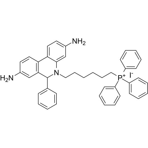
MitoSOX Red is a live cell fluorescent probe that specifically targets mitochondria and is cell membrane permeable. MitoSOX Red enters mitochondria and is oxidized by superoxide but not by other ROS or RNS generating systems. The oxidized MitoSOX Red then binds to nucleic acids in mitochondria/nucleus, producing strong red fluorescence. MitoSOX Red can be used as a fluorescent indicator to specifically detect superoxide. In addition, superoxide dismutase (SOD) can prevent the oxidation of MitoSOX Red.
Excitation/emission wavelength: 510/580 nm.
loading...
    loading...
272
272 Cited Publications
Cat. No.: HY-15534
CAS No.: 3520-43-2
Pureté:  98.71%
Synonyms: CBIC2
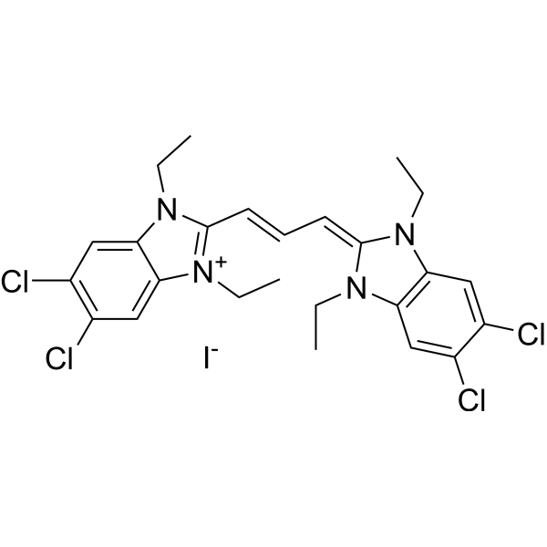
JC-1 (CBIC2) is an ideal fluorescent probe widely used to detect mitochondrial membrane potential. JC-1 accumulates in mitochondria in a potential dependent manner and can be used to detect the membrane potential of cells or purified mitochondria. In normal mitochondria, JC-1 aggregates in the mitochondrial matrix to form a polymer, which emits strong red fluorescence (Ex=585 nm, Em=590 nm); When the mitochondrial membrane potential is low, JC-1 cannot aggregate in the matrix of mitochondria and produce green fluorescence (ex=510 nm, em= 527 nm) .
loading...
    loading...
209
209 Cited Publications
Cat. No.: HY-D0079
CAS No.: 104821-25-2
Synonyms: Hydroethidine; PD-MY 003
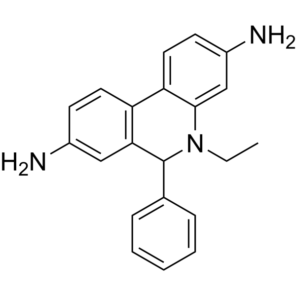
Dihydroethidium, also known as DHE, is a peroxide indicator. Dihydroethidium penetrates cell membranes to form a fluorescent protein complex with blue fluoresces. After entering the cells, Dihydroethidium is mainly localized in the cell membrane, cytoplasm and nucleus, and the staining effect is the strongest in the nucleus. Dihydroethidium produces inherent blue fluorescence with a maximum excitation wavelength of 370 nm and a maximum emission wavelength of 420 nm; after dehydrogenation, Dihydroethidium combines with RNA or DNA to produce red fluorescence with a maximum excitation wavelength of 300 nm and a maximum emission wavelength of 610 nm. 535 nm can also be used as the excitation wavelength for actual observation .
loading...
    loading...
179
179 Cited Publications
Cat. No.: HY-D0815
CAS No.: 25535-16-4
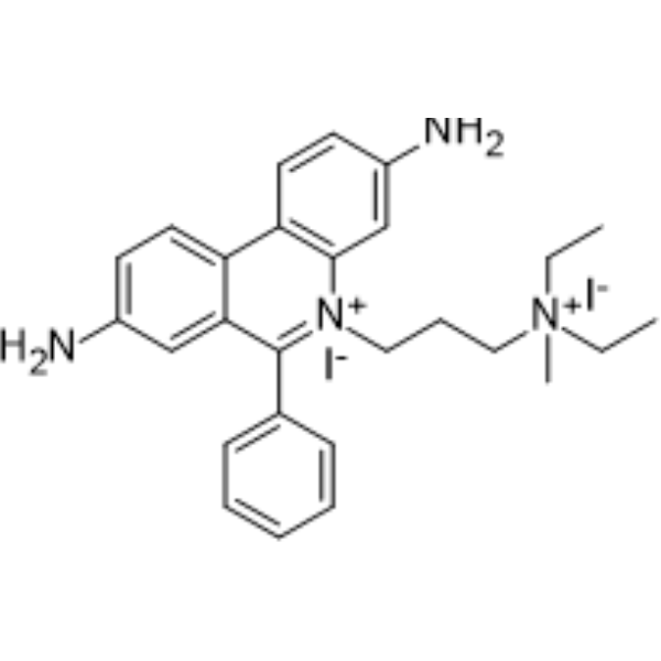
Propidium Iodide (PI) is a nuclear staining agent that stains DNA. Propidium Iodide is an analogue of ethidine bromide that emits red fluorescence upon embedding in double-stranded DNA. Propidium Iodide cannot pass through living cell membranes, but it can pass through damaged cell membranes to stain the nucleus. Propidium Iodide has a fluorescence wavelength of 493/617 nm and a wavelength of 536/635 nm after Mosaic with DNA. Propidium Iodide is commonly used in the detection of apoptosis (apoptosis) or necrosis (necrosis), and is often used in flow cytometry analysis.
loading...
    loading...
178
178 Cited Publications
Cat. No.: HY-D1301
CAS No.: 217075-36-0
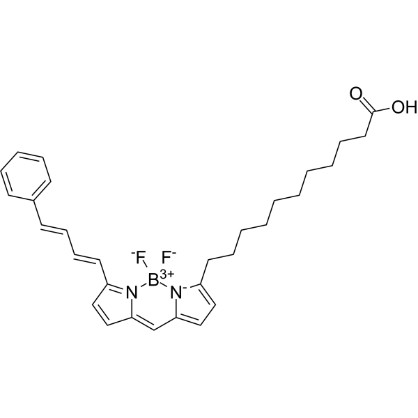
BODIPY 581/591 C11 is a BODIPY borofluoroprene derivative with good light stability and low fluorescence artifacts. BODIPY 581/591 C11 can be used for study lipid peroxidation and antioxidant properties in living cells, or detect ferroptosis by reaction with hydroxyl radicals. BODIPY 581/591 C11 is emitted at 591 nm (reduced prototype), or redshifted to 510 nm (oxidized type). The excitation wavelengths were 581 nm (reduced prototype) and 500 nm (oxidized type) .
loading...
    loading...
154
154 Cited Publications
Cat. No.: HY-12591
CAS No.: 103404-75-7
Pureté:  99.92%
Synonyms: D-(-)-Luciferin sodium; Firefly luciferin sodium; Beetle Luciferin sodium
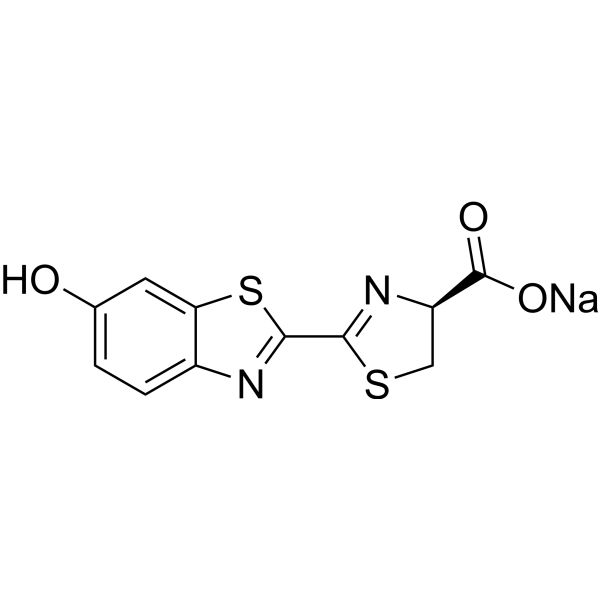
D-luciferin sodium is the natural substrate of the enzyme luciferase (Luc) that catalyzes the production of the typical yellowgreen light of fireflies. The 560 nm chemiluminescence from this reaction peaks within seconds, with light output that is proportional to luciferase concentration when the substrate luciferin is present in excess. The luciferase (luc) gene is a popular reporter gene for research and agent screening. Chemiluminescent techniques are virtually background-free, making the luc reporter gene ideal for detecting low-level gene expression. As little as 0.02 pg of luciferase can be reliably measured in a standard scintillation counter. In addition to its role as a reporter of gene expression, luciferase is commonly used in an extremely sensitive assay for ATP . We of er the firefly luciferase (HY-P1004A), luciferin free acid (HY-12591A), as well as its water-soluble sodium salts (HY-12591) and potassium salts (HY-12591B) .
loading...
    loading...
154
154 Cited Publications
Cat. No.: HY-12591A
CAS No.: 2591-17-5
Pureté:  99.54%
Synonyms: D-(-)-Luciferin; Firefly luciferin; Beetle Luciferin
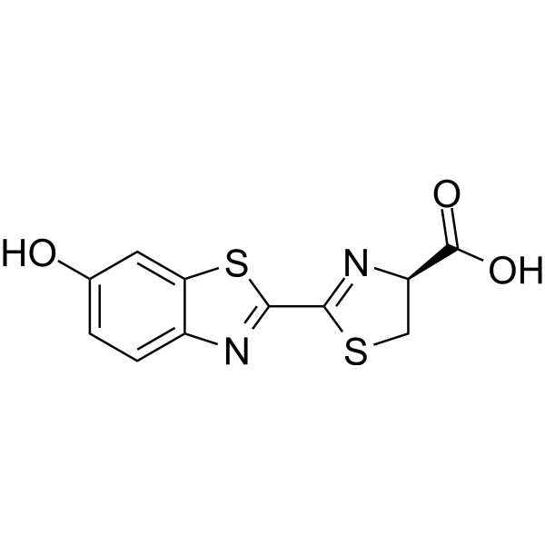
D-luciferin is the natural substrate of the enzyme luciferase (Luc) that catalyzes the production of the typical yellowgreen light of fireflies. The 560 nm chemiluminescence from this reaction peaks within seconds, with light output that is proportional to luciferase concentration when the substrate luciferin is present in excess. The luciferase (luc) gene is a popular reporter gene for research and agent screening. Chemiluminescent techniques are virtually background-free, making the luc reporter gene ideal for detecting low-level gene expression. As little as 0.02 pg of luciferase can be reliably measured in a standard scintillation counter. In addition to its role as a reporter of gene expression, luciferase is commonly used in an extremely sensitive assay for ATP . We of er the firefly luciferase (HY-P1004A), luciferin free acid (HY-12591A), as well as its water-soluble sodium salts (HY-12591) and potassium salts (HY-12591B) .
loading...
    loading...
154
154 Cited Publications
Cat. No.: HY-12591B
CAS No.: 115144-35-9
Pureté:  99.97%
Synonyms: D-(-)-Luciferin potassium; Firefly luciferin potassium; Beetle Luciferin potassium
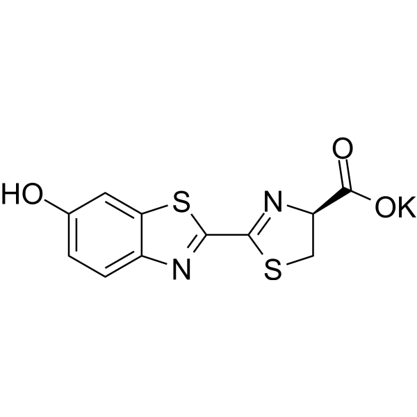
D-luciferin potassium is the natural substrate of the enzyme luciferase (Luc) that catalyzes the production of the typical yellowgreen light of fireflies. The 560 nm chemiluminescence from this reaction peaks within seconds, with light output that is proportional to luciferase concentration when the substrate luciferin is present in excess. The luciferase (luc) gene is a popular reporter gene for research and agent screening. Chemiluminescent techniques are virtually background-free, making the luc reporter gene ideal for detecting low-level gene expression. As little as 0.02 pg of luciferase can be reliably measured in a standard scintillation counter. In addition to its role as a reporter of gene expression, luciferase is commonly used in an extremely sensitive assay for ATP . We offer the firefly luciferase (HY-P1004A), luciferin free acid (HY-12591A), as well as its water-soluble sodium salts (HY-12591) and potassium salts (HY-12591B) .
loading...
    loading...
146
146 Cited Publications
Cat. No.: HY-D0718
CAS No.: 7385-67-3
Synonyms: Nile Blue A oxazone; Phenoxazone 9
Target:  

Fluorescent Dye

Domaines de recherche:  

Others

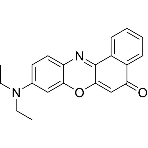
Nile red (Nile blue oxazone) is a lipophilic stain. Nile red has environment-sensitive fluorescence. Nile red is intensely fluorescent in a lipid-rich environment while it has minimal fluorescence in aqueous media. Nile red is an excellent vital stain for the detection of intracellular lipid droplets by fluorescence microscopy and flow cytof uorometry. Nile red stains intracellular lipid droplets red. The fluorescence wavelength is 543/598 nm .
loading...
    loading...
134
134 Cited Publications
Cat. No.: HY-D0938
CAS No.: 150347-59-4
Synonyms: CFSE; 5(6)-Carboxyfluorescein diacetate succinimidyl ester; 5(6)-CFDA N-succinmidyl ester
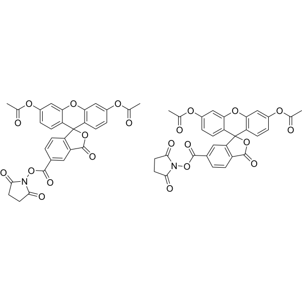
CFDA-SE is a fluorescent dye that can penetrate the cell membrane. It can react with the free amine group in the cytoskeleton protein inside the cell, and finally form a protein complex with fluorescence. After entering the cell, CFDA-SE locates in the cell membrane, cytoplasm and nucleus, and the fluorescence staining is strongest in the nucleus . CFDA-SE dye can be uniformly inherited by the cells with cell division and proliferation, and its attenuation is proportional to the number of cell divisions. This phenomenon can be detected and analyzed by flow cytometry under the excitation light of 488 nm, and can be used to detect the proliferation of cells .
loading...
    loading...
100
100 Cited Publications
Cat. No.: HY-K1073

MCE Annexin V-FITC/PI Apoptosis Detection Kit provides a rapid and convenient method to detect cell apoptosis and necrosis. After staining, live cells show little or no fluorescence (Annexin V-/PI-), early apoptosis cells show green fluorescence(Annexin V+/PI-), late apoptosis cells and necrosis cells show red and green fluorescence (Annexin V+/PI+).


loading...
    loading...
56
56 Cited Publications
Cat. No.: HY-D1451
CAS No.: 154214-55-8
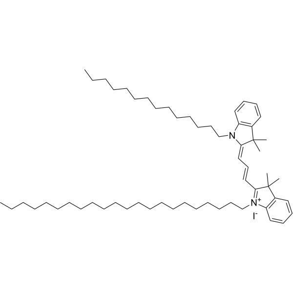
PKH 26 is a red fluorescent dye, PKH 26 can stably bind to the lipid region of cell membrane and emit red fluorescence (Ex/Em=551/567 nm), which is mainly used for in vitro cell labeling, in vitro cell proliferation studies and in vivo and in vitro cell tracing studies .
loading...
    loading...
52
52 Cited Publications
Cat. No.: HY-D1374
CAS No.: 1144107-82-3
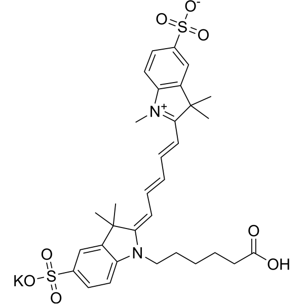
Sulfo-Cy5 carboxylic acid potassium is a Cy5 derivative that can be used for chemical synthesis. Cy5 is a red fluorescent label dye for imaging (Ex/Em=642/670 nm) .
loading...
    loading...
52
52 Cited Publications
Cat. No.: HY-D1533
CAS No.: 1447815-38-4
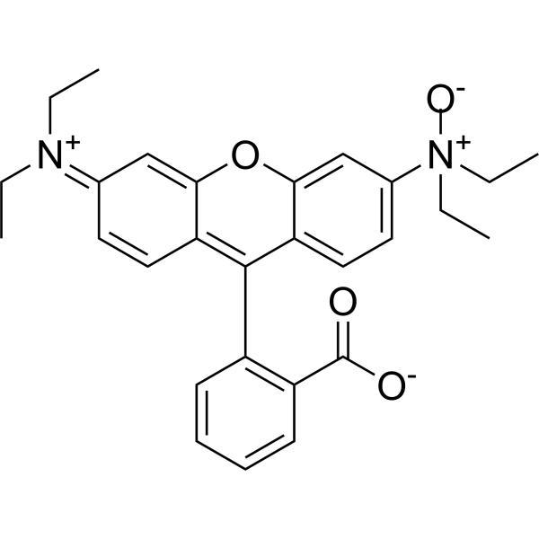
RhoNox-1 is a fluorescent probe for the specific detection of divalent iron ions, and when RhoNox-1 reacts with Fe 2+. RhoNox-1 can generate an irreversible orange (red) fluorescent product (Ex/Em:540/575 nm) . FeRhoNox-1 can enter the cell well, suitable for the detection of Fe 2+ in living cells, and tends to be localized in the Golgi apparatus .
loading...
    loading...
48
48 Cited Publications
Cat. No.: HY-D0989
CAS No.: 145037-81-6
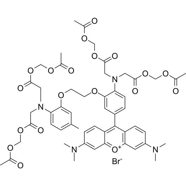
Rhod-2 is a high-affinity visible light excitation wavelength Ca 2+ fluorescent probe, Rhod-2, AM is an acetyl methyl ester derivative of Rhod-2, which has cell membrane permeability and can easily enter cells with simple culture. Once it enters the cell, it is sheared by its lactesterase to produce Rhod-2 without membrane permeability, which remains in the cell to perform the corresponding physiological functions. Maximum excitation/emission wavelength: 549/578 nm .
loading...
    loading...
45
45 Cited Publications
Cat. No.: HY-101879
CAS No.: 65-61-2
Pureté:  99.65%
Target:  

Fluorescent Dye

Domaines de recherche:  

Others

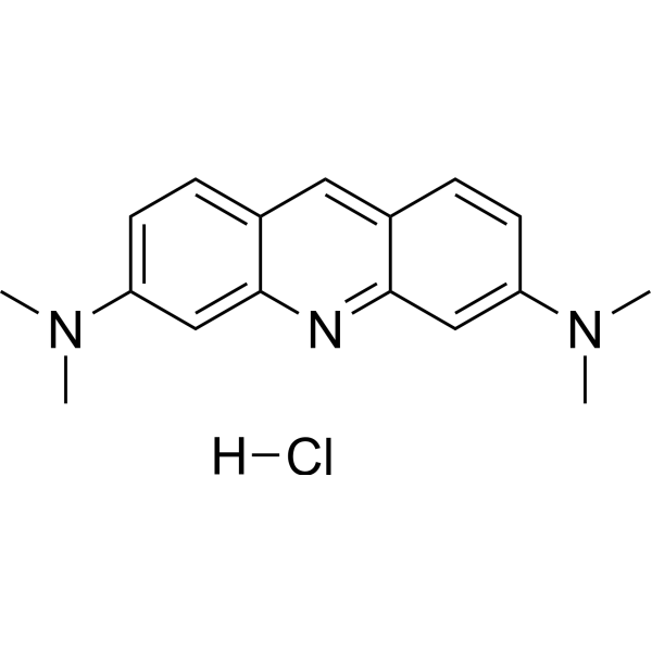
Acridine Orange hydrochloride is a cell-permeable fluorescent nucleic acid dye and lysosomal probe with a pKa of 9.65. Acridine Orange hydrochloride enters cells via passive diffusion, is protonated and accumulates in acidic lysosomes, and forms aggregates at high concentrations, producing orange-red fluorescence; when the lysosomal proton gradient or membrane integrity is impaired, Acridine Orange hydrochloride is released into the cytoplasm, green fluorescence increases and red fluorescence decreases, so it can be used to monitor lysosomal acidification status and changes in membrane permeability. Acridine Orange hydrochloride binds to nucleic acids through intercalation or electrostatic interactions, exhibiting mainly green fluorescence when bound to double-stranded DNA and mainly red fluorescence when bound to RNA or single-stranded DNA, and can be used for nucleic acid staining and differential DNA/RNA detection. The Ex/Em of Acridine Orange hydrochloride monomer is approximately 492/525 nm; when bound to double-stranded DNA it is approximately 488/520-524 nm, and when bound to RNA or single-stranded DNA it is approximately 457/630-650 nm .
loading...
    loading...
45
45 Cited Publications
Cat. No.: HY-D0942
CAS No.: 10127-02-3
Synonyms: Euchrysine 3RX
Target:  

Fluorescent Dye

Domaines de recherche:  

Others

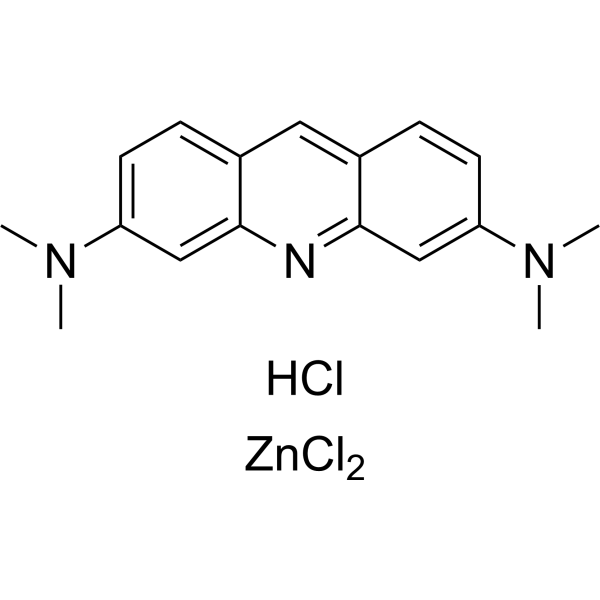
Acridine Orange zinc chloride salt is a cell-permeable fluorescent nucleic acid dye and lysosomal probe with a pKa of 9.65. Acridine Orange zinc chloride salt enters cells via passive diffusion, is protonated and accumulates in acidic lysosomes, and forms aggregates at high concentrations, producing orange-red fluorescence; when the lysosomal proton gradient or membrane integrity is impaired, Acridine Orange zinc chloride salt is released into the cytoplasm, green fluorescence increases and red fluorescence decreases, so it can be used to monitor lysosomal acidification status and changes in membrane permeability. Acridine Orange zinc chloride salt binds to nucleic acids through intercalation or electrostatic interactions, exhibiting mainly green fluorescence when bound to double-stranded DNA and mainly red fluorescence when bound to RNA or single-stranded DNA, and can be used for nucleic acid staining and differential DNA/RNA detection. The Ex/Em of Acridine Orange zinc chloride salt monomer is approximately 492/525 nm; when bound to double-stranded DNA it is approximately 488/520-524 nm, and when bound to RNA or single-stranded DNA it is approximately 457/630-650 nm .
loading...
    loading...
45
45 Cited Publications
Cat. No.: HY-D0952
CAS No.: 494-38-2
Target:  

Fluorescent Dye

Domaines de recherche:  

Others

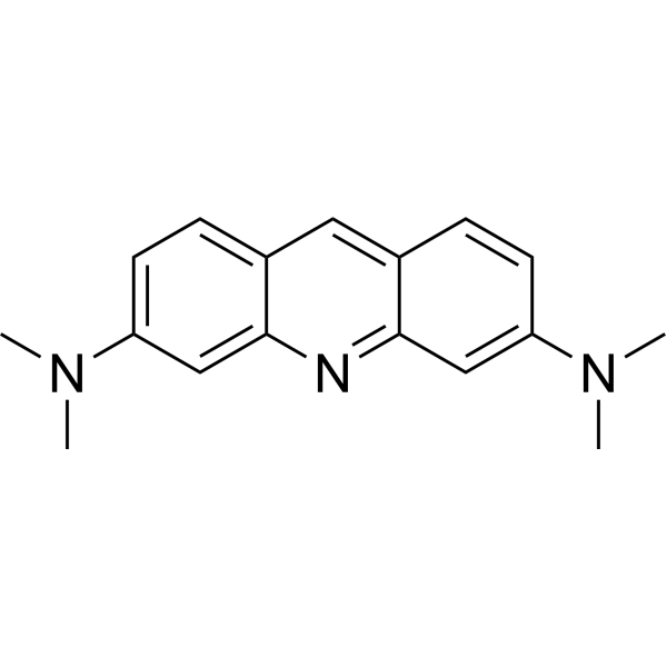
Acridine Orange base is a cell-permeable fluorescent nucleic acid dye and lysosomal probe with a pKa of 9.65. Acridine Orange base enters cells via passive diffusion, is protonated and accumulates in acidic lysosomes, and forms aggregates at high concentrations, producing orange-red fluorescence; when the lysosomal proton gradient or membrane integrity is impaired, Acridine Orange base is released into the cytoplasm, green fluorescence increases and red fluorescence decreases, so it can be used to monitor lysosomal acidification status and changes in membrane permeability. Acridine Orange base binds to nucleic acids through intercalation or electrostatic interactions, exhibiting mainly green fluorescence when bound to double-stranded DNA and mainly red fluorescence when bound to RNA or single-stranded DNA, and can be used for nucleic acid staining and differential DNA/RNA detection. The Ex/Em of Acridine Orange base monomer is approximately 492/525 nm; when bound to double-stranded DNA it is approximately 488/520-524 nm, and when bound to RNA or single-stranded DNA it is approximately 457/630-650 nm .
loading...
    loading...
30
30 Cited Publications
Cat. No.: HY-N0729
CAS No.: 60-33-3
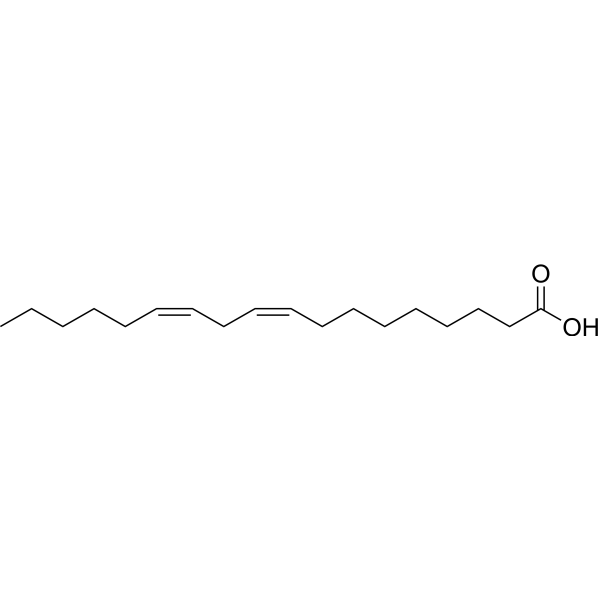
Linoleic acid is a common polyunsaturated (PUFA) found in plant-based oils, nuts and seeds. Linoleic acid is a part of membrane phospholipids, and functions as a structural component to maintain a certain level of membrane fluidity of the transdermal water barrier of the epidermis. Linoleic acid induces red blood cells and hemoglobin damage via oxidative mechanism .
loading...
    loading...
25
25 Cited Publications
Cat. No.: HY-B1247
CAS No.: 553-12-8
Synonyms: PPIX
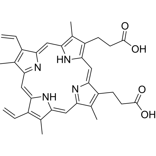
Protoporphyrin IX is a final intermediate in the heme biosynthetic pathway, which acts as a radiation sensitizer enhancing ROS generation even in a hypoxic state and inducing DNA damage. Protoporphyrin IX also acts as a photo sensitizer undergoing photobleaching that occurs through direct degradation by light irradiation. Protoporphyrin IX is formed and accumulated following 5-aminolevulinic acid (5-ALA) (HY-W000450) administration in the tumor cells of rats. Protoporphyrin IX causes selective improvement of basal cell carcinoma when activated red fluorescence of a peak wavelength at 405 nm. Protoporphyrin IX is promising for research of sonodynamic and photodynamic agents for a wide range of cancers, such as bladder cancer and nodular basal cell carcinoma .
loading...
    loading...