



Taurohyodeoxycholic acid
Based on 2 publication(s) in Google Scholar
Taurohyodeoxycholic acid is an orally active 6 alpha-hydroxylated bile acid. Taurohyodeoxycholic acid decreases colonic MPO activity, TNF-α, lL-6 serum levels and the expression of COX-2. Taurohyodeoxycholic acid alleviates trinitrobenzene sulfonic acid induced ulcerative colitis via regulating Th1/Th2 and Th17/Treg cells balance. Taurohyodeoxycholic acid ameliorates high-fat diet-induced nonalcoholic fatty liver disease in mice. Taurohyodeoxycholic acid prevents Taurochenodeoxycholic acid (HY-N2027)-induced hepatotoxicity in bile fistula rats. Taurohyodeoxycholic acid can be used for the study of nonalcoholic fatty liver disease (NAFLD), colitis and biliary fistula.
For research use only. We do not sell to patients.
- Purity : 99.73%
- CAS No.: 2958-04-5
- Formula: C26H45NO6S
- Molecular Weight:499.70
-
Storage:Powder -20°C, 3 years , 4°C, 2 years ; In solvent -80°C, 6 months , -20°C, 1 month

To place orders, for customer services and technical support, please contact: MedChemExpress USA
Tel: 609-228-6898 E-mail: [email protected] [email protected]


-
 Biological Activity
-
Chemical Information
-
Solvent & Solubility
- Protocols
- Purity & Documentation
- References
-
 Help & FAQs


-
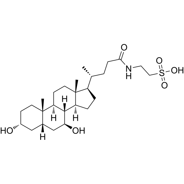
HY-19696
TauroursodeoxycholateEndoplasmic Reticulum Stress Inhibitor -
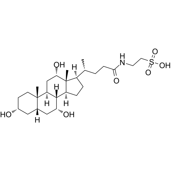
HY-B1788
Taurocholic acidImmunoregulation Agent -
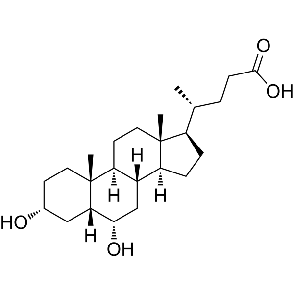
HY-N0169
Hyodeoxycholic acidGPBAR1 Agonist -
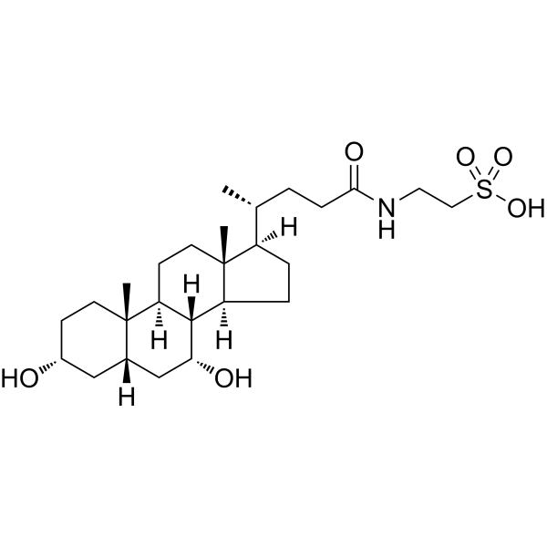
HY-N2027
Taurochenodeoxycholic acidEndogenous Metabolite -
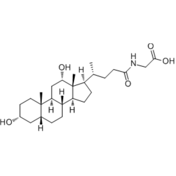
HY-125731
Glycodeoxycholic acidEndogenous Metabolite -
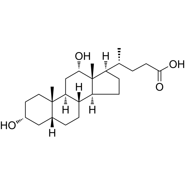
HY-N0593
Deoxycholic acidBile Acid Receptor Activator -
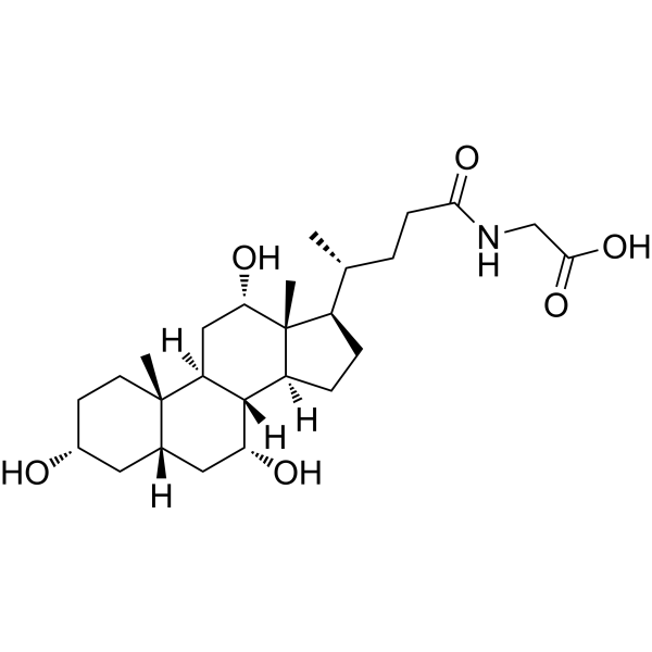
HY-N1423
Glycocholic acidBile Acid Derivative -
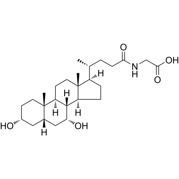
HY-N2334
Glycochenodeoxycholic acidEndogenous Metabolite -
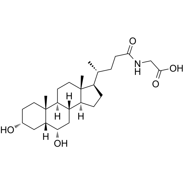
HY-126995
Glycohyodeoxycholic acidHyodeoxycholic acid Metabolite -
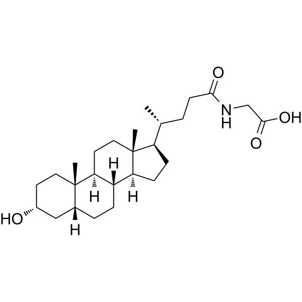
HY-116374
Glycolithocholic acidSecondary Bile Acid -
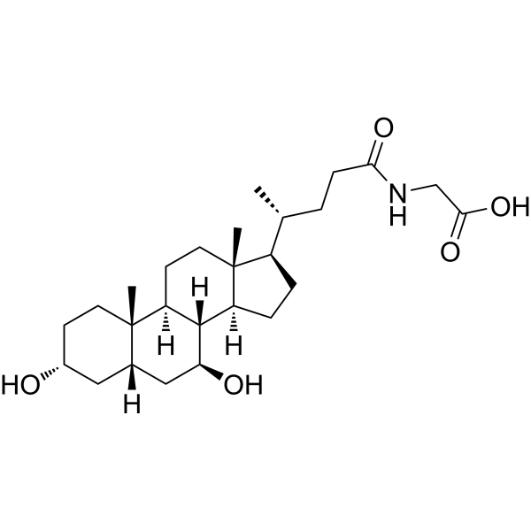
HY-N1424
Glycoursodeoxycholic acidEndogenous Metabolite -
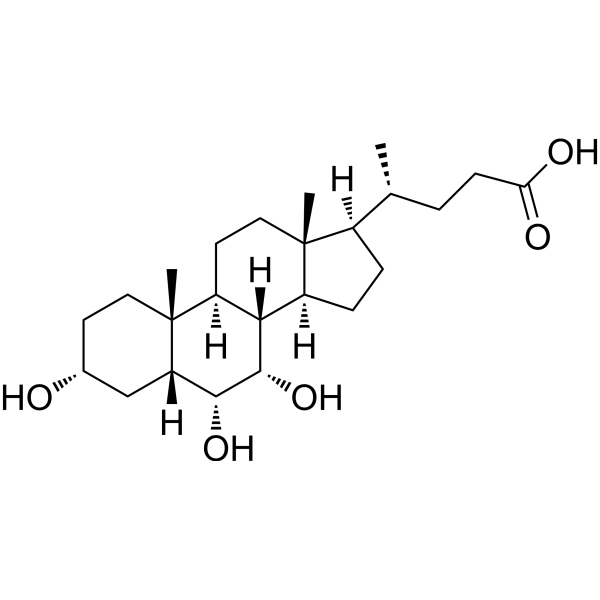
HY-121238
Hyocholic acidAnti-diabete Agent
Publications Citing Use of MedChemExpress (MCE) Taurohyodeoxycholic acid
MoreAll Endogenous Metabolite Isoforms
More

Biological Activity
Description
Cellular Effect
|
Cell Line

|
Type | Value | Description | References |
|---|---|---|---|---|
| CHO | EC50 |
24.2 μM
Compound: 10a, tauro
|
Agonist activity at human TGR5 expressed in CHO cells by luciferase assay
Agonist activity at human TGR5 expressed in CHO cells by luciferase assay
|
[PMID: 18307294] |
In Vitro
Taurohyodeoxycholic acid (25-100 μM, 12 h) dose-dependently decreases triglyceride levels in Palmitic acid (HY-N08307)/Oleic acid (HY-N1446)-treated AML-12 cells[1].
MedChemExpress (MCE) has not independently confirmed the accuracy of these methods. They are for reference only. Further protocols information, click here.
In Vivo
Taurohyodeoxycholic acid (20-100 mg/kg, p.o. or i.g., daily, 7 days) alleviates trinitrobenzene sulfonic acid (TNBS) -induced colitis in mice[2].
Taurohyodeoxycholic acid (4 mg/kg/min (8 μmol/min/kg), i.v., 1 h) prevents Taurochenodeoxycholic acid (HY-N2027)-induced hepatotoxicity in bile fistula rats[3].
MedChemExpress (MCE) has not independently confirmed the accuracy of these methods. They are for reference only.
-
Animal Model:High-fat diet was fed to 4-week-old male C57BL/6 mice to induce nonalcoholic fatty liver disease[1].
-
Dosage:200 mg/kg
-
Administration:p.o. daily for 17 days
-
Result:Reduced body weight, liver index, and epididymal white adipose tissue index in high-fat diet-fed mice.
Decreased serum triglyceride, total cholesterol, and hepatic triglyceride levels.
Improved hepatic steatosis and glucose homeostasis (enhanced glucose tolerance and insulin sensitivity).
Upregulated hepatic CYP7B1 protein expression and downregulated hepatic CYP7A1 and CD36 protein expressions.
Dose-dependently decreased triglyceride levels in palmitic acid/oleic acid-treated AML-12 cells.
-
Animal Model:Trinitrobenzene sulfonic acid (100 μL of 2.0 mg TNBS in 50% EtOH) was intrarectally administered to 7-8-week-old male SPF Kunming mice to induce ulcerative colitis[2].
-
Dosage:20, 40, 80 mg/kg
-
Administration:p.o. daily for 7 days
-
Result:Alleviated TNBS-induced colitis by improving body weight, colon length, spleen weight, histological characteristics, and reducing MPO activity in mice.
Reduced Th1-/Th17-related cytokines (IFN-γ, IL-12, p70, IL-6, IL-17A, IL-21, IL-22, TNF-α) and transcription factors (T-bet, STAT4, RORγt, STAT3) in the colon.
Increased Th2-/Treg-related cytokines (IL-4, IL-10, TGF-β1) and transcription factors (GATA3, STAT6, Foxp3, Smad3) in the colon.
Inhibited IFN-γ, IL-17A, T-bet, RORγt expressions and improved IL-4, IL-10, GATA3, Foxp3 expressions in the spleen.
Restored the proportion of Th1, Th2, Th17, Treg cells and balanced Th1/Th2 and Th17/Treg immune response in colitis mice.
-
Animal Model:Taurochenodeoxycholic acid was intravenously infused into bile fistula Sprague-Dawley rats (200-250 g)[3].
-
Dosage:4 mg/kg/min (8 μmol/min/kg)
-
Administration:i.v. for 1 h
-
Result:Increased bile flow, with maximum flow rate twice that of the control group after 60 minutes.
Enhanced biliary secretion of total bile acids, reaching a maximum rate of 17.5 μmol/min/kg at 60 minutes; Increased phospholipid secretion, with a peak of 0.65 μmol/min/kg.
Restored biliary calcium secretion; reduced biliary leakage of alkaline phosphatase to levels similar to the control group and decreased lactate dehydrogenase leakage.
Increased the maximum biliary secretion rate of taurochenodeoxycholic acid to 7.6 μmol/min/kg, with its own maximum secretion rate reaching 7.9 μmol/min/kg.
-
Animal Model:Trinitrobenzene sulfonic acid (TNBS, 0.5 mg in 0.1 ml of 50% ethanol) was intrarectally administered into the colon of 22-25 g male Balb/c mice via a 3.5F catheter inserted 4 cm proximal to the anus to induce ulcerative colitis model[4].
-
Dosage:25, 50, 100 mg/kg
-
Administration:i.g., daily for 7 days
-
Result:Alleviated TNBS-induced colitis by improving body weight, reducing macroscopic colonic damage and histopathological changes, and decreasing colonic MPO activity in mice.
Reduced serum levels of pro-inflammatory cytokines (TNF-α, IL-6) and decreased the expression of COX-2 in the colon.


Chemical Information
-
CAS No. 2958-04-5
-
Appearance Solid
-
Molecular Weight 499.70
-
Formula C26H45NO6S
-
Color White to off-white
-
SMILES
C[C@@]12[C@]3([H])[C@](C[C@@H]([C@]1([H])C[C@@H](CC2)O)O)([H])[C@@]4([H])[C@](CC3)([C@@](CC4)([H])[C@H](C)CCC(NCCS(=O)(O)=O)=O)C
-
Structure Classification
-
Initial Source
-
Shipping
Room temperature in continental US; may vary elsewhere.
-
Storage
Powder -20°C 3 years 4°C 2 years In solvent -80°C 6 months -20°C 1 month


Publications (2)
-
Journal Impact Factor
-
Most Recent
-
Cell Host Microbe
2025 Aug 19:S1931-3128(25)00291-4. PMID: 40848719 -
Phytomedicine
Geniposide alleviated bile acid-associated NLRP3 inflammasome activation by regulating SIRT1/FXR signaling in bile duct ligation-induced liver fibrosis. [Abstract]2023 Sep:118:154971. PMID: 37494875


Solvent & Solubility
In Vitro:
DMSO : 100 mg/mL (200.12 mM; Need ultrasonic; Hygroscopic DMSO has a significant impact on the solubility of product, please use newly opened DMSO)
Please refer to the solubility information to select the appropriate solvent. Once prepared, please aliquot and store the solution to prevent product inactivation from repeated freeze-thaw cycles.
Storage method and period of stock solution: -80°C, 6 months; -20°C, 1 month. When stored at -80°C, please use it within 6 months. When stored at -20°C, please use it within 1 month.
Please refer to the solubility information to select the appropriate solvent. Once prepared, please aliquot and store the solution to prevent product inactivation from repeated freeze-thaw cycles.
Storage method and period of stock solution: -80°C, 6 months; -20°C, 1 month. When stored at -80°C, please use it within 6 months. When stored at -20°C, please use it within 1 month.
Concentration (start) × Volume (start) = Concentration (final) × Volume (final)
In Vivo:
Select the appropriate dissolution method based on your experimental animal and administration route.
- For the following dissolution methods, please ensure to first prepare a clear stock solution using an In Vitro approach and then sequentially add co-solvents:
- To ensure reliable experimental results, the clarified stock solution can be appropriately stored based on storage conditions. As for the working solution for In Vivo experiments, it is recommended to prepare freshly and use it on the same day.
- The percentages shown for the solvents indicate their volumetric ratio in the final prepared solution. If precipitation or phase separation occurs during preparation, heat and/or sonication can be used to aid dissolution.
Add each solvent one by one: 10% DMSO 40% PEG300 5% Tween-80 45% Saline
Solubility: ≥ 2.5 mg/mL (5.00 mM); Clear solution
This protocol yields a clear solution of ≥ 2.5 mg/mL (saturation unknown).
Taking 1 mL working solution as an example, add 100 μL DMSO stock solution (25.0 mg/mL) to 400 μL PEG300, and mix evenly; then add 50 μL Tween-80 and mix evenly; then add 450 μL Saline to adjust the volume to 1 mL.
Preparation of Saline: Dissolve 0.9 g sodium chloride in ddH₂O and dilute to 100 mL to obtain a clear Saline solution.
Add each solvent one by one: 10% DMSO 90% (20% SBE-β-CD in Saline)
Solubility: ≥ 2.5 mg/mL (5.00 mM); Clear solution
This protocol yields a clear solution of ≥ 2.5 mg/mL (saturation unknown).
Taking 1 mL working solution as an example, add 100 μL DMSO stock solution (25.0 mg/mL) to 900 μL 20% SBE-β-CD in Saline, and mix evenly.
Preparation of 20% SBE-β-CD in Saline (4°C, storage for one week): 2 g SBE-β-CD powder is dissolved in 10 mL Saline, completely dissolve until clear.
In Vivo Dissolution Calculator
Please enter the basic information of animal experiments:
-
-
-
-
Recommended: Prepare an additional quantity of animals to account for potential losses during experiments.
Please enter your animal formula composition:
-
%DMSO +
Recommended: Keep the proportion of DMSO in working solution below 2% if your animal is weak.
-
%+
-
+%Tween-80 + +
-
%Saline +
The co-solvents required include: DMSO, . All of co-solvents are available by MedChemExpress (MCE). , Tween 80. All of co-solvents are available by MedChemExpress (MCE).
Working solution concentration: 0.22 mg/mL
Method for preparing stock solution: mg drug dissolved in μL DMSO. Stock solution concentration: mg/mL.
1. Take μL DMSO stock solution;
2. Add μL .
μL , mix evenly;
3. Then add μL Tween 80, mix evenly;
4. Then add μL
Please ensure that the stock solution in the first step is dissolved to a clear state, and add co-solvents in sequence. You can use ultrasonic heating (ultrasonic cleaner, recommended frequency 20-40 kHz), vortexing, etc. to assist dissolution.

Protocols
-
RNA extraction experimental
By lysing cells, releasing RNA, and removing impurities such as proteins and DNA, high-purity RNA products are finally obtained. The commonly used traditional method is the guanidine isothiocyanate/phenol/chloroform method (Trizol), which is suitable for a variety of animal materials including animal tissues, microorganisms, cultured cells, etc., and most plant materials.
-
Naïve CD4+ T-cell subset differentiation/polarization
Naïve CD4+ T-cell subset differentiation/polarization is an in vitro assay in which purified naïve CD4+ T cells are activated through TCR and CD28 costimulation and cultured with defined cytokines and neutralizing antibodies to generate Th0, Th1, Th2, Th17, or induced Treg-like populations. Differentiation is detected by subset-associated cytokines and transcription factors: IFN-γ/T-bet for Th1, IL-4/GATA3 for Th2, IL-17A/RORγt for Th17, and Foxp3 for induced Treg cells. The assay readout is usually generated by intracellular cytokine staining after restimulation, transcription-factor staining by flow cytometry, ELISA of secreted cytokines, or gene-expression analysis. The result reflects cytokine-directed lineage commitment or polarization rather than antigen-specific immune protection by itself.
-
DSS-Induced Colitis
Dextran sulfate sodium (DSS)-induced colitis is generated by administering DSS in mouse drinking water, producing epithelial injury, barrier disruption, weight loss, diarrhea, fecal blood, colon shortening, histologic mucosal damage, and inflammatory mediator changes; the model is mainly used to study acute or chronic intestinal inflammation resembling selected features of ulcerative colitis. DSS injury is interpreted through clinical and tissue readouts rather than a single molecular endpoint: daily body weight, stool consistency, and bleeding are combined into a disease activity index, while colon length, histology, cytokines, myeloperoxidase activity, intestinal permeability, and tight-junction markers provide complementary measures of inflammation and barrier damage.
-
TNBS-Induced Colitis
TNBS-induced colitis is produced by intrarectal delivery of 2,4,6-trinitrobenzene sulfonic acid in ethanol, where ethanol disrupts the mucosal barrier and TNBS haptenates colonic proteins, generating immune-mediated colonic inflammation with weight loss, diarrhea, ulceration, transmural injury, inflammatory-cell infiltration, and cytokine responses. The model is used as an experimental intestinal inflammation model with Crohn’s disease–like features, especially when Th1-type responses, IL-12–dependent inflammation, chronic relapsing inflammation, or fibrosis-related endpoints are studied.
-
Hepatotoxicity Study
This protocol evaluates hepatotoxicity using complementary in vivo mouse APAP acute liver injury and in vitro hepatocyte-based cytotoxicity readouts. In vivo APAP injury is assessed by serum ALT/AST, liver histology, hepatic glutathione, protein adducts, necrosis, inflammation, and regeneration-related endpoints. In vitro hepatotoxicity is assessed by loss of viability, leakage of ALT/AST/LDH, oxidative-stress markers, mitochondrial function, nuclear morphology, intracellular calcium, and high-content imaging endpoints.
-
Research Protocol for Metabolic Diseases
AMP-activated protein kinase, AMPK, is a conserved cellular energy sensor that responds to reduced cellular energy status and coordinates metabolism by increasing ATP-generating catabolic pathways while suppressing ATP-consuming anabolic processes. In metabolic disease research, the AMPK pathway is experimentally relevant because it regulates hepatic lipid synthesis, fatty acid oxidation, glucose production, skeletal-muscle glucose disposal, mTORC1-linked biosynthesis, autophagy, mitochondrial homeostasis, and whole-body energy balance. The central pathway logic is that energy stress, metformin, exercise-like stimulation, or direct AMPK activators increase AMPKα Thr172 phosphorylation and downstream substrate phosphorylation, including ACC and RAPTOR. Phosphorylation of ACC suppresses lipogenesis and supports fatty acid oxidation, whereas phosphorylation of RAPTOR suppresses mTORC1 signaling and links cellular energy status to growth and protein synthesis control. The pathway is linked


Purity & Documentation
-
Data Sheet (282 KB)
-
SDS (419 KB)
- English - EN (419 KB)
- Français - FR (419 KB)
- Deutsch - DE (419 KB)
- Norwegian - NO (419 KB)
- Español - ES (419 KB)
- Swedish - SV (419 KB)
- Italian - IT (419 KB)
- Korean - KR (419 KB)
- Portuguese - PT (419 KB)
-
Handling Instructions (2659 KB)
References
[1]. Roda A, et, al. Taurohyodeoxycholic acid protects against taurochenodeoxycholic acid-induced cholestasis in the rat. Hepatology. 1998 Feb;27(2):520-5. [Content Brief]
[2]. Carubbi F, et, al. Comparative cytotoxic and cytoprotective effects of taurohyodeoxycholic acid (THDCA) and tauroursodeoxycholic acid (TUDCA) in HepG2 cell line. Biochim Biophys Acta. 2002 Jan 30;1580(1):31-9. [Content Brief]
[3]. Zheng N, et al. Astragalus polysaccharide attenuates nonalcoholic fatty liver disease through THDCA in high-fat diet-fed mice. J Ethnopharmacol. 2024 Feb 10;320:117401. [Content Brief]
[4]. Lv L, et al. Taurohyodeoxycholic acid alleviates trinitrobenzene sulfonic acid induced ulcerative colitis via regulating Th1/Th2 and Th17/Treg cells balance. Life Sci. 2023 Apr 1;318:121501. [Content Brief]
[5]. Roda A, et al. Taurohyodeoxycholic acid protects against taurochenodeoxycholic acid-induced cholestasis in the rat. Hepatology. 1998 Feb;27(2):520-5. [Content Brief]
[6]. He J, et al. Protective effect of taurohyodeoxycholic acid from Pulvis Fellis Suis on trinitrobenzene sulfonic acid induced ulcerative colitis in mice. Eur J Pharmacol. 2011 Nov 16;670(1):229-35. [Content Brief]


Complete Stock Solution Preparation Table
Please refer to the solubility information to select the appropriate solvent. Once prepared, please aliquot and store the solution to prevent product inactivation from repeated freeze-thaw cycles.
Storage method and period of stock solution: -80°C, 6 months; -20°C, 1 month. When stored at -80°C, please use it within 6 months. When stored at -20°C, please use it within 1 month.
| Optional Solvent | Concentration Solvent Mass | 1 mg | 5 mg | 10 mg | 25 mg |
|---|---|---|---|---|---|
| DMSO | 1 mM | 2.0012 mL | 10.0060 mL | 20.0120 mL | 50.0300 mL |
| 5 mM | 0.4002 mL | 2.0012 mL | 4.0024 mL | 10.0060 mL | |
| 10 mM | 0.2001 mL | 1.0006 mL | 2.0012 mL | 5.0030 mL | |
| 15 mM | 0.1334 mL | 0.6671 mL | 1.3341 mL | 3.3353 mL | |
| 20 mM | 0.1001 mL | 0.5003 mL | 1.0006 mL | 2.5015 mL | |
| 25 mM | 0.0800 mL | 0.4002 mL | 0.8005 mL | 2.0012 mL | |
| 30 mM | 0.0667 mL | 0.3335 mL | 0.6671 mL | 1.6677 mL | |
| 40 mM | 0.0500 mL | 0.2502 mL | 0.5003 mL | 1.2508 mL | |
| 50 mM | 0.0400 mL | 0.2001 mL | 0.4002 mL | 1.0006 mL | |
| 60 mM | 0.0334 mL | 0.1668 mL | 0.3335 mL | 0.8338 mL | |
| 80 mM | 0.0250 mL | 0.1251 mL | 0.2502 mL | 0.6254 mL | |
| 100 mM | 0.0200 mL | 0.1001 mL | 0.2001 mL | 0.5003 mL |


Taurohyodeoxycholic acid Related Classifications
HY-114360 Related Classifications
 Powered by Bioz