



Kenpaullone
Based on 18 publication(s) in Google Scholar
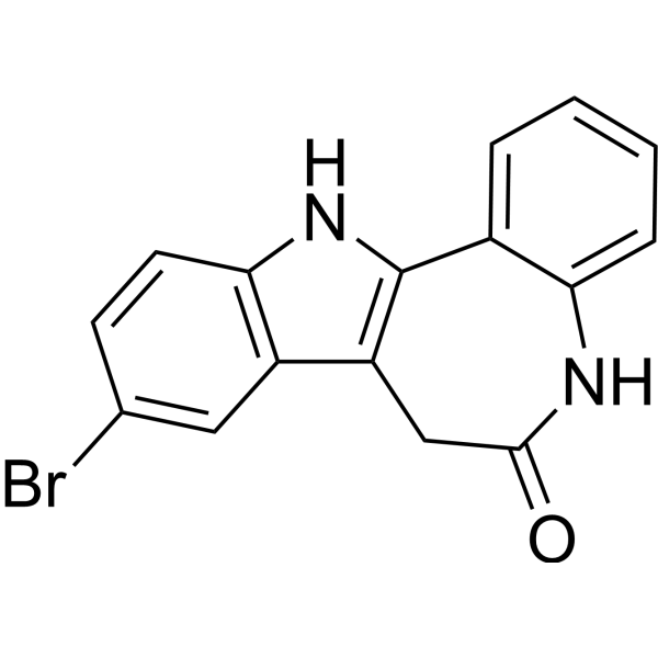
Kenpaullone is a potent inhibitor of CDK1/cyclin B and GSK-3β, with IC50s of 0.4 μM and 23 nM, and also inhibits CDK2/cyclin A, CDK2/cyclin E, and CDK5/p25 with IC50s of 0.68 μM, 7.5 μM, 0.85 μM, respectively. Kenpaullone, a small molecule inhibitor of KLF4, reduces self-renewal of breast cancer stem cells and cell motility in vitro.
For research use only. We do not sell to patients.
- Purity : 98.20%
- CAS No.: 142273-20-9
- Formula: C16H11BrN2O
- Molecular Weight:327.18
-
Storage:Powder -20°C, 3 years , 4°C, 2 years ; In solvent -80°C, 2 years , -20°C, 1 year

To place orders, for customer services and technical support, please contact: MedChemExpress USA
Tel: 609-228-6898 E-mail: [email protected] [email protected]


-
 Biological Activity
-
Chemical Information
-
Solvent & Solubility
- Protocols
- Purity & Documentation
- References
-
 Help & FAQs


-

HY-10182
LaduviglusibGSK-3 Inhibitor -
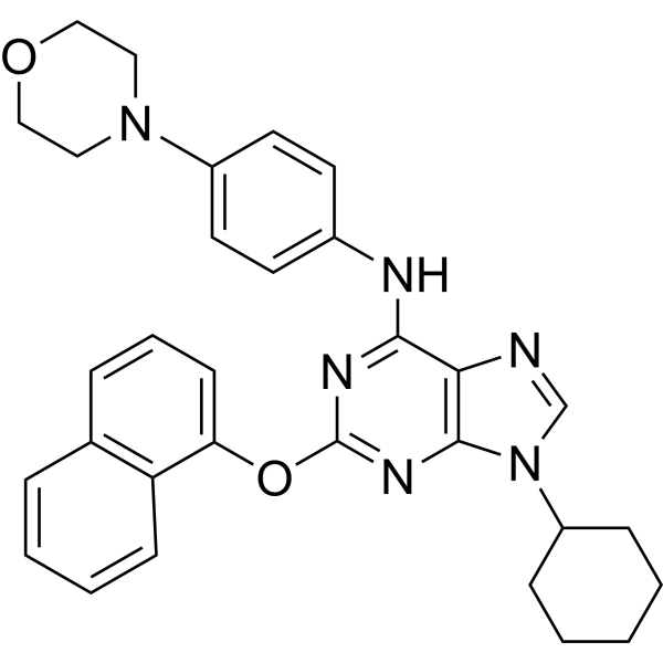
HY-15108
PurmorphamineSmoothened Receptor Agonist -

HY-15371
ForskolinAdenylate Cyclase Activator -
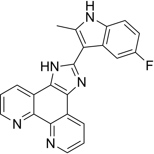
HY-16291
APTO-253 -
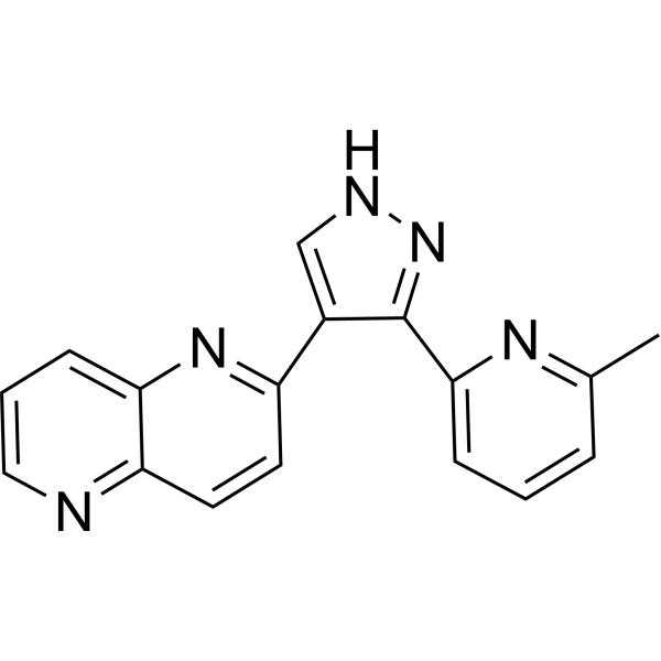
HY-13012
RepSoxTGFβR-1/ALK5 Inhibitor -
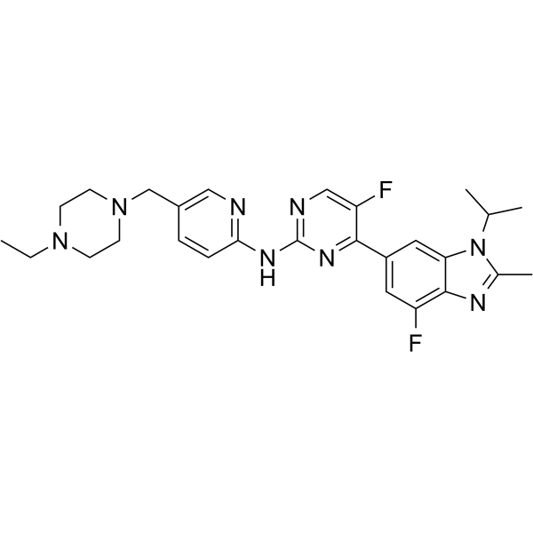
HY-16297A
AbemaciclibCDK4/6 Inhibitor -
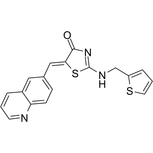
HY-12529
Ro-3306CDK1 Inhibitor -
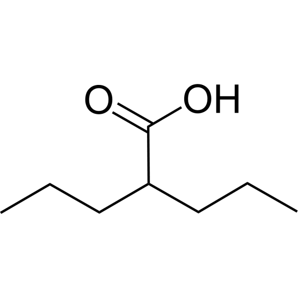
HY-10585
Valproic acidHDAC Inhibitor -
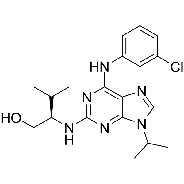
HY-18299A
Purvalanol ACDK Inhibitor -
HY-10580
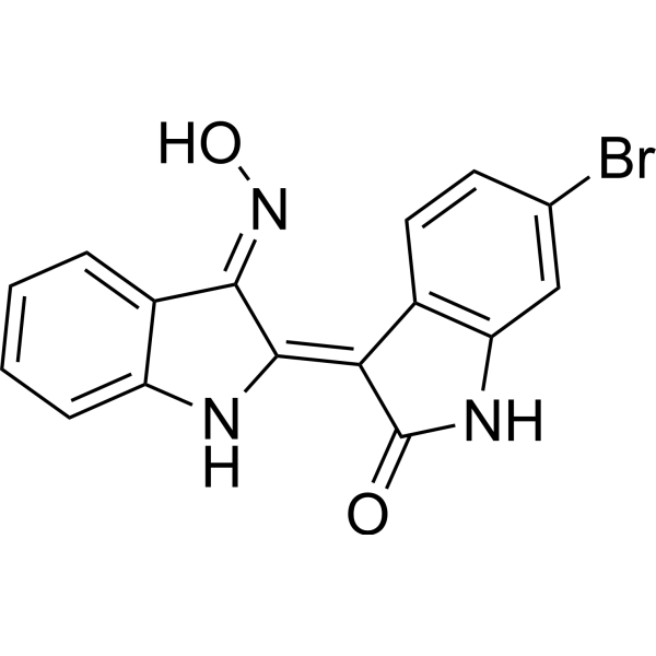
GSK 3 Inhibitor IXATP-Competitive Inhibitor -
HY-10475
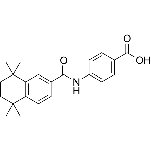
AM580RARα Agonist -
HY-12041
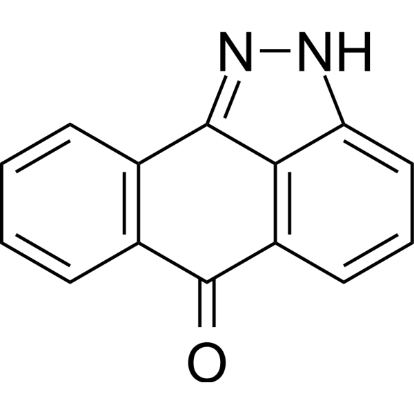
SP600125JNK Inhibitor
Publications Citing Use of MedChemExpress (MCE) Kenpaullone
More- Mil Med Res. 2020 Nov 1;7(1):52. [Abstract]
- Mil Med Res. 2020 Sep 6;7(1):42. [Abstract]
- J Biomed Sci. 2026 May 15;33(1):51. [Abstract]
- Adv Sci (Weinh). 2024 Mar;11(11):e2305260. [Abstract]
- Sci Adv. 2021 Apr 14;7(16):eabb2213. [Abstract]
- Int J Biol Sci. 2025 Sep 20;21(13):5802-5820. [Abstract]
- J Transl Med. 2024 Oct 10;22(1):922. [Abstract]
- Arch Pharm Res. 2026 Apr;49(4):554-574. [Abstract]
- Cell Rep. 2026 Mar 24;45(4):117201. [Abstract]
- Cell Rep. 2023 Nov 28;42(11):113442. [Abstract]
- Anal Chem. 2025 Jun 3;97(21):11099-11109. [Abstract]
- Life Sci. 2026 Oct 1:402:124596.
- Sci Rep. 2026 Jun 29.
- Cell Signal. 2026 Jul:143:112459. [Abstract]
- J Neurotrauma. 2026 Jan 29:8977151261415615. [Abstract]
- Eur J Integr Med. 2025 Aug 28;78:102545.
- bioRxiv. 2026 May 7.
- Res Sq. 2025 Apr 24.
 Customer Validation & Images

-
WB
-
WB
-
IF
-
Cell Proliferation/Viability Assay
-
RT-PCR


Biological Activity
Description
|
Cdk1/cyclin B 0.4 μM (IC50) |
cdk2/cyclin A 0.68 μM (IC50) |
Cdk5/p25 0.85 μM (IC50) |
CDK2/cyclinE 7.5 μM (IC50) |
GSK-3β 0.023 μM (IC50) |
erk1 20 μM (IC50) |
erk2 9 μM (IC50) |
c-raf 38 μM (IC50) |
Cellular Effect
|
Cell Line

|
Type | Value | Description | References |
|---|---|---|---|---|
| Fibroblast | IC50 |
10 μM
Compound: 3
|
Inhibition of human SOD1 expressed in mouse fibroblasts after 48 hrs by ELISA relative to control
Inhibition of human SOD1 expressed in mouse fibroblasts after 48 hrs by ELISA relative to control
|
[PMID: 24560539] |
| HCT-15 | IC50 |
23.86 μM
Compound: 4a
|
Cytotoxicity against human HCT15 cells after 48 hrs by SRB assay
Cytotoxicity against human HCT15 cells after 48 hrs by SRB assay
|
[PMID: 19217301] |
| K562 | IC50 |
>100 μM
Compound: 4a
|
Cytotoxicity against human K562 cells after 48 hrs by SRB assay
Cytotoxicity against human K562 cells after 48 hrs by SRB assay
|
[PMID: 19217301] |
| MCF7 | IC50 |
>100 μM
Compound: 4a
|
Cytotoxicity against human MCF7 cells after 48 hrs by SRB assay
Cytotoxicity against human MCF7 cells after 48 hrs by SRB assay
|
[PMID: 19217301] |
| Oocyte | IC50 |
0.4 μM
Compound: 4a
|
In vitro inhibitory activity against cyclin-dependent kinase 1-cyclin B (Cyclin-Dependent Kinase) harvested from starfish oocytes.
In vitro inhibitory activity against cyclin-dependent kinase 1-cyclin B (Cyclin-Dependent Kinase) harvested from starfish oocytes.
|
[PMID: 10425100] |
| Oocyte | IC50 |
0.4 μM
Compound: 1
|
Inhibition of Cyclin-dependent kinase 1-cyclin B from Starfish oocytes
Inhibition of Cyclin-dependent kinase 1-cyclin B from Starfish oocytes
|
[PMID: 10741555] |
| Oocyte | IC50 |
0.4 μM
Compound: Kenapaullone
|
Inhibition of Cyclin-dependent kinase 1-cyclin B from M phase starfish (Marthasterias glacialis) oocytes
Inhibition of Cyclin-dependent kinase 1-cyclin B from M phase starfish (Marthasterias glacialis) oocytes
|
[PMID: 12593668] |
| Oocyte | IC50 |
0.5 μM
Compound: 2
|
Inhibition of M-phase starfish oocytes CDK1/cyclin B assessed as incorporation of [gamma-33P]-ATP into histone H1 substrate after 30 mins by scintillation counting
Inhibition of M-phase starfish oocytes CDK1/cyclin B assessed as incorporation of [gamma-33P]-ATP into histone H1 substrate after 30 mins by scintillation counting
|
[PMID: 20621478] |
| PC-3 | IC50 |
>100 μM
Compound: 4a
|
Cytotoxicity against human PC3 cells after 48 hrs by SRB assay
Cytotoxicity against human PC3 cells after 48 hrs by SRB assay
|
[PMID: 19217301] |
| Sf9 | IC50 |
0.023 μM
Compound: 1a
|
Inhibition of GSK3-beta expressed in insect Sf9 cells
Inhibition of GSK3-beta expressed in insect Sf9 cells
|
[PMID: 19906467] |
| Sf9 | IC50 |
23 nM
Compound: 3
|
Inhibition of GSK3beta (unknown origin) expressed in sf9 cells after 30 mins by scintillation counting analysis in presence of [gamma-32P]ATP
Inhibition of GSK3beta (unknown origin) expressed in sf9 cells after 30 mins by scintillation counting analysis in presence of [gamma-32P]ATP
|
[PMID: 24560539] |
| U-251 | IC50 |
>100 μM
Compound: 4a
|
Cytotoxicity against human U251 cells after 48 hrs by SRB assay
Cytotoxicity against human U251 cells after 48 hrs by SRB assay
|
[PMID: 19217301] |
In Vitro
Kenpaullone shows much less effect on c-src (IC50, 15 μM), casein kinase 2 (IC50, 20 μM), erk 1 (IC50, 20 μM), and erk 2 (IC50, 9 μM). Kenpaullone acts by competitive inhibition of ATP binding, and the apparent Ki is 2.5 μM. Kenpaullone can inhibit the growth of tumor cells in culture (mean GI50, 43 μM) and causes altered cell cycle progression most clearly revealed under conditions of recovery from serum starvation[1]. Kenpaullone demonstrates a wide range of biological utility, extending from maintenance of pancreatic β cell survival and proliferation to the induction of apoptosis in cancer cells[2].
MedChemExpress (MCE) has not independently confirmed the accuracy of these methods. They are for reference only. Further protocols information, click here.


Chemical Information
-
CAS No. 142273-20-9
-
Appearance Solid
-
Molecular Weight 327.18
-
Formula C16H11BrN2O
-
Color Off-white to yellow
-
SMILES
O=C1NC2=CC=CC=C2C(NC3=C4C=C(Br)C=C3)=C4C1
-
Synonyms
9-Bromopaullone; NSC-664704
-
Shipping
Room temperature in continental US; may vary elsewhere.
-
Storage
Powder -20°C 3 years 4°C 2 years In solvent -80°C 2 years -20°C 1 year


Publications (18)
-
Journal Impact Factor
-
Most Recent
-
Mil Med Res
Direct conversion of human fibroblasts into dopaminergic neuron-like cells using small molecules and protein factors. [Abstract]2020 Nov 1;7(1):52. PMID: 33129359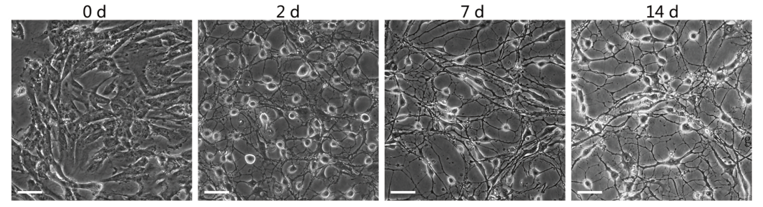
Kenpaullone purchased from MedChemExpress. Usage Cited in: Mil Med Res. 2020 Nov 1;7(1):52. [Abstract]
Kenpaullone (3 μmol/L,After 6–8 days replaced 1 μmol/L). Representative microscopy images depicting the morphological changes after chemical induction. Scale bars=100 μm.
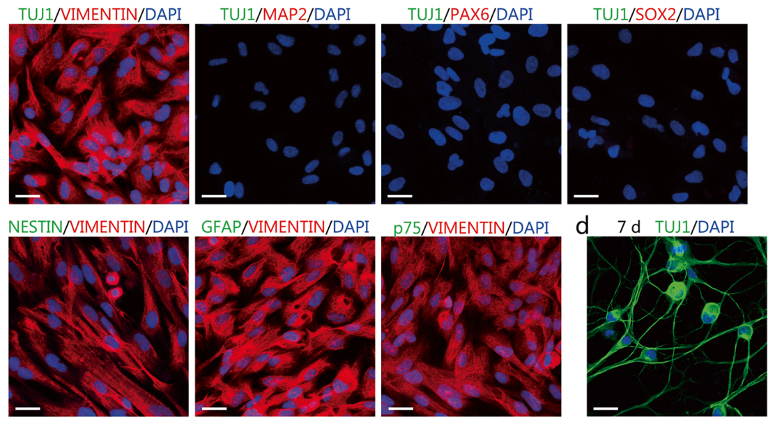
Kenpaullone purchased from MedChemExpress. Usage Cited in: Mil Med Res. 2020 Nov 1;7(1):52. [Abstract]
Kenpaullone (3 μmol/L,After 6–8 days replaced 1 μmol/L). Characterization of the cultured IMR-90 fibroblasts by immunostaining assay for the fibroblast marker VIMENTIN, neural markers Class III β-tubulin 1 (TUJ1) and microtubule-associated protein 2 (MAP2), neural progenitor/stem cell markers PAX6, SOX2, and NESTIN, glial cell marker GFAP, and neural crest cell marker p75. Scale bars=25 μm.
-
Mil Med Res
Efficient and rapid conversion of human astrocytes and ALS mouse model spinal cord astrocytes into motor neuron-like cells by defined small molecules. [Abstract]2020 Sep 6;7(1):42. PMID: 32892745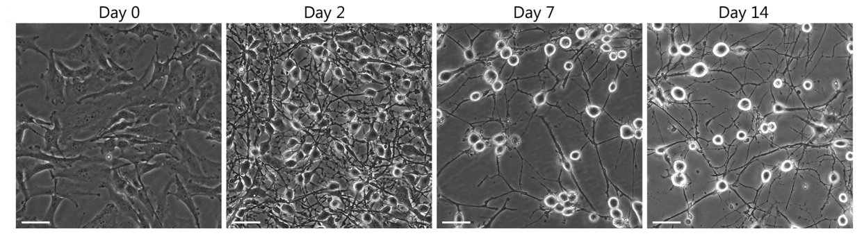
Kenpaullone purchased from MedChemExpress. Usage Cited in: Mil Med Res. 2020 Sep 6;7(1):42. [Abstract]
Kenpaullone (5 μM, refreshed every 2 days for 7 days). Microscopic images of the Human astrocytes (HA1800) cells morphological changes after chemical induction.
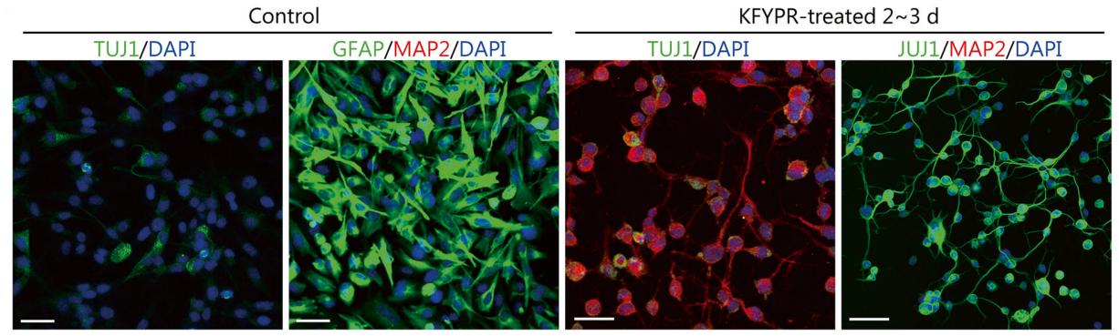
Kenpaullone purchased from MedChemExpress. Usage Cited in: Mil Med Res. 2020 Sep 6;7(1):42. [Abstract]
Kenpaullone (5 μM), Human astrocytes (HA1800) cells. Expression of class III β-tubulin 1 (TUJ1), microtubule-associated protein 2 (MAP2), and GFAP in the control group and the KFYPR-induced group.
-
J Biomed Sci
SARS-CoV-2 membrane protein recruits PP2A to dephosphorylate the nucleocapsid and promote virion production. [Abstract]2026 May 15;33(1):51. PMID: 42141450 -
Adv Sci (Weinh)
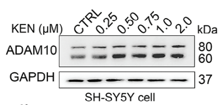
SHMT2 Mediates Small-Molecule-Induced Alleviation of Alzheimer Pathology Via the 5'UTR-dependent ADAM10 Translation Initiation. [Abstract]2024 Mar;11(11):e2305260. PMID: 38183387
Kenpaullone purchased from MedChemExpress. Usage Cited in: Adv Sci (Weinh). 2024 Mar;11(11):e2305260. [Abstract]
Chemical structure of KEN (top), immunoblots (middle), and quantification (bottom) of ADAM10 in SH‐SY5Y cells treated with KEN (0.25–2 µM for 36 h, n = 5).
Kenpaullone purchased from MedChemExpress. Usage Cited in: Adv Sci (Weinh). 2024 Mar;11(11):e2305260. [Abstract]
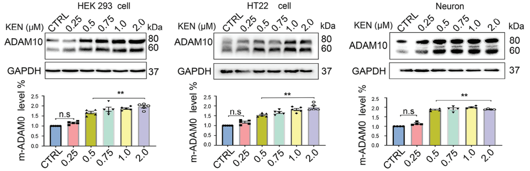
Immunoblots (top) and quantification (bottom) of ADAM10 treated with KEN (Kenpaullone, 0.25–2 µM for 36 h) in human HEK293 cell line, murine hippocampal cell line and primary cortical neurons, respectively (n = 5).
Kenpaullone purchased from MedChemExpress. Usage Cited in: Adv Sci (Weinh). 2024 Mar;11(11):e2305260. [Abstract]
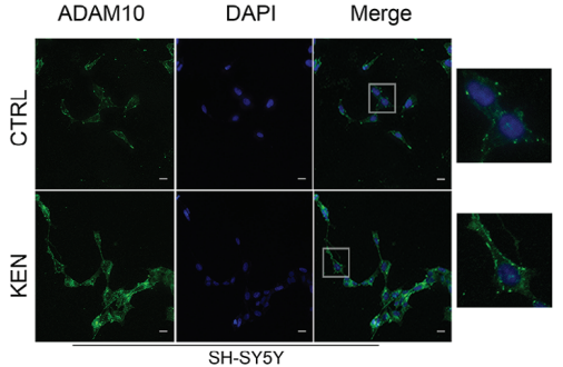
Immunofluorescent images (top) and quantification (bottom) of ADAM10 signals (green) in SH‐SY5Y cells treated with KEN (Kenpaullone, 0.75 µM for 36 h). DAPI is a nuclear marker (blue), scale bar = 25 µm (n > 10).
Kenpaullone purchased from MedChemExpress. Usage Cited in: Adv Sci (Weinh). 2024 Mar;11(11):e2305260. [Abstract]
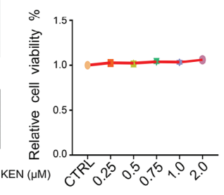
Cell viability of SH‐SY5Y cells treated with KEN (Kenpaullone, 0.25 to 2 µM for 36 h) measured by CCK‐8 kit (n = 5).
Kenpaullone purchased from MedChemExpress. Usage Cited in: Adv Sci (Weinh). 2024 Mar;11(11):e2305260. [Abstract]
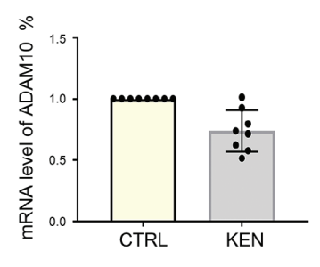
Relative mRNA levels of ADAM10 in SH‐SY5Y cells treated with KEN (Kenpaullone, 0.75 µM for 36 h, n = 8).
Kenpaullone purchased from MedChemExpress. Usage Cited in: Adv Sci (Weinh). 2024 Mar;11(11):e2305260. [Abstract]
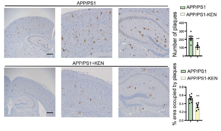
Kenpaullone (KEN, 7 mg/kg, every other day for 2 months, administered intraperitoneally). Immunohistochemical images (left) and quantification (right) of Aβ deposition in the brain of APP/PS1 and APP/PS1‐KEN, scale bar = 500 µm (n = 6).
-
Sci Adv
Chemical conversion of human epidermal stem cells into intestinal goblet cells for modeling mucus-microbe interaction and therapy. [Abstract]2021 Apr 14;7(16):eabb2213. PMID: 33853767 -
Int J Biol Sci
The KLF4/Galectin-3 cascade is a key determinant of tubular cell death and acute kidney injury. [Abstract]2025 Sep 20;21(13):5802-5820. PMID: 41079936 -
J Transl Med
KLF4 regulates trophoblast function and associates with unexplained recurrent spontaneous abortion. [Abstract]2024 Oct 10;22(1):922. PMID: 39390495 -
Arch Pharm Res
Tirzepatide mitigates atherosclerosis progression and modulates oxLDL-mediated proatherogenic effects in macrophages: evidence for M1/M2 homeostasis restoration. [Abstract]2026 Apr;49(4):554-574. PMID: 41904760 -
Cell Rep
SETDB2-mediated transcriptional repression of IDE in sensory neurons promotes migraine-like pain behaviors in mice. [Abstract]2026 Mar 24;45(4):117201. PMID: 41880325 -
Cell Rep
Hypoxia-sensitive cells trigger NK cell activation via the KLF4-ASH1L-ICAM-1 axis, contributing to impairment in the rat epididymis. [Abstract]2023 Nov 28;42(11):113442. PMID: 37952156 -
Anal Chem
Exposome-Scale Investigation of Cl-/Br-Containing Chemicals Using High-Resolution Mass Spectrometry, Multistage Machine Learning, and Cloud Computing. [Abstract]2025 Jun 3;97(21):11099-11109. PMID: 40401576 -
-
-
Cell Signal
Cell-type specific contribution of KLF4 to fibrotic nephropathy in tubular epithelium and fibroblast. [Abstract]2026 Jul:143:112459. PMID: 41785980 -
J Neurotrauma
Atorvastatin Regulates the Efferocytosis of Macrophages to Promote Hematoma Absorption and Inflammation Resolution in Experimental Subdural Hematoma. [Abstract]2026 Jan 29:8977151261415615. PMID: 41609140 -
-
-


Solvent & Solubility
In Vitro:
DMSO : 100 mg/mL (305.64 mM; Need ultrasonic; Hygroscopic DMSO has a significant impact on the solubility of product, please use newly opened DMSO)
Please refer to the solubility information to select the appropriate solvent. Once prepared, please aliquot and store the solution to prevent product inactivation from repeated freeze-thaw cycles.
Storage method and period of stock solution: -80°C, 2 years; -20°C, 1 year. When stored at -80°C, please use it within 2 years. When stored at -20°C, please use it within 1 year.
Please refer to the solubility information to select the appropriate solvent. Once prepared, please aliquot and store the solution to prevent product inactivation from repeated freeze-thaw cycles.
Storage method and period of stock solution: -80°C, 2 years; -20°C, 1 year. When stored at -80°C, please use it within 2 years. When stored at -20°C, please use it within 1 year.
Concentration (start) × Volume (start) = Concentration (final) × Volume (final)
In Vivo:
Select the appropriate dissolution method based on your experimental animal and administration route.
- For the following dissolution methods, please ensure to first prepare a clear stock solution using an In Vitro approach and then sequentially add co-solvents:
- To ensure reliable experimental results, the clarified stock solution can be appropriately stored based on storage conditions. As for the working solution for In Vivo experiments, it is recommended to prepare freshly and use it on the same day.
- The percentages shown for the solvents indicate their volumetric ratio in the final prepared solution. If precipitation or phase separation occurs during preparation, heat and/or sonication can be used to aid dissolution.
Add each solvent one by one: 10% DMSO 40% PEG300 5% Tween-80 45% Saline
Solubility: ≥ 2.5 mg/mL (7.64 mM); Suspended solution
This protocol yields a suspended solution of ≥ 2.5 mg/mL (saturation unknown). Suspended solution can be used for oral and intraperitoneal injection.
Taking 1 mL working solution as an example, add 100 μL DMSO stock solution (25.0 mg/mL) to 400 μL PEG300, and mix evenly; then add 50 μL Tween-80 and mix evenly; then add 450 μL Saline to adjust the volume to 1 mL.
Preparation of Saline: Dissolve 0.9 g sodium chloride in ddH₂O and dilute to 100 mL to obtain a clear Saline solution.
In Vivo Dissolution Calculator
Please enter the basic information of animal experiments:
-
-
-
-
Recommended: Prepare an additional quantity of animals to account for potential losses during experiments.
Please enter your animal formula composition:
-
%DMSO +
Recommended: Keep the proportion of DMSO in working solution below 2% if your animal is weak.
-
%+
-
+%Tween-80 + +
-
%Saline +
The co-solvents required include: DMSO, . All of co-solvents are available by MedChemExpress (MCE). , Tween 80. All of co-solvents are available by MedChemExpress (MCE).
Working solution concentration: 0.22 mg/mL
Method for preparing stock solution: mg drug dissolved in μL DMSO. Stock solution concentration: mg/mL.
1. Take μL DMSO stock solution;
2. Add μL .
μL , mix evenly;
3. Then add μL Tween 80, mix evenly;
4. Then add μL
Please ensure that the stock solution in the first step is dissolved to a clear state, and add co-solvents in sequence. You can use ultrasonic heating (ultrasonic cleaner, recommended frequency 20-40 kHz), vortexing, etc. to assist dissolution.

Protocols
-
Scratch/Wound-Healing Migration Assay
The scratch/wound-healing migration assay measures collective migration of adherent cells into an experimentally created cell-free gap in a confluent monolayer. The readout is generated by imaging the gap immediately after scratching and at later time points, then quantifying reduction in wound area, wound width, or percentage closure as cells move into the denuded region. Gap closure reflects cell migration but may also include cell proliferation, so interpretation should distinguish migration-focused conditions from proliferation-driven closure when possible, such as by using short assay windows, serum-controlled conditions, cell counting, or proliferation controls reported in published protocols.
-
Cell migration
Cell migration is a method that plays an important role in wound healing, cell differentiation, embryonic development, etc.
-
Under-Agarose Cell Migration Assay
The under-agarose cell migration assay is a classical in vitro chemotaxis method designed to evaluate directed cell movement through a semi-solid agarose matrix toward soluble chemoattractant gradients, enabling visualization and quantification of leukocyte or motile cell migration in a confined 2D-like environment. In this system, cells and chemoattractants are placed in separate wells cut into an agarose gel, allowing diffusion-driven gradient formation that guides directional migration, which is typically assessed by measuring migration distance, cell morphology changes, and accumulation toward the chemoattractant source. This assay has been widely used to study neutrophil and leukocyte chemotaxis as a simple alternative to filter-based migration systems and allows direct microscopic observation of migrating cells under near-physiological confinement conditions.
-
Transwell/Boyden Chamber Migration Assay
The Transwell/Boyden chamber migration assay measures cell movement through a porous membrane separating an upper and lower chamber, usually after a chemoattractant gradient is established by placing cells in the upper chamber and chemoattractant-containing medium in the lower chamber. The readout is generated by quantifying cells that traverse the membrane and appear on the lower membrane surface or in the lower chamber, depending on whether the cell type is adherent or non-adherent. This assay reflects chemotactic or haptotactic migration rather than matrix invasion unless an extracellular-matrix barrier is added to the membrane.
-
hPSC maintenance and expansion
This protocol maintains and expands human pluripotent stem cells under feeder-free, chemically defined conditions using E8 medium and vitronectin-coated culture surfaces; the readout is sustained adherent colony growth with undifferentiated morphology and retained pluripotency-marker expression during serial passaging. E8-based hPSC culture relies on defined soluble factors and matrix-dependent adhesion rather than feeder cells; vitronectin supports hPSC attachment through integrin-mediated interactions, and EDTA passaging dissociates colonies as small aggregates without enzymatic digestion, centrifugation, or routine ROCK-inhibitor treatment.
-
Breast Cancer Modeling
Breast cancer is a heterogeneous cancer, and it has been distinguished into four subtypes: luminal A, luminal B, HER2-positive and basal-like. Molecular mutations, epigenetic alterations, hormone exposure and immune microenvironment are related to the progression of breast cancer.
-
Cell-Exclusion Zone Migration Assay
The Cell-Exclusion Zone (CEZ) migration assay is an in vitro 2D cell migration method in which a defined cell-free area is created using removable physical barriers such as silicone stoppers, allowing cells to be seeded around the barrier and subsequently migrate into the cleared zone after barrier removal. This approach enables quantification of collective cell migration by monitoring repopulation of the initially cell-free region over time using microscopy-based imaging. Compared with scratch-based wound healing assays, barrier-based exclusion methods are designed to avoid mechanical damage to the extracellular matrix and reduce injury-induced effects on boundary cells, thereby improving interpretability of migration behavior in vitro. The assay readout is typically the progressive reduction in the cell-free area or the number of cells invading the exclusion zone, reflecting coordinated cell motility relevant to physiological processes such as wound healing, epithelial repair, and ca


Purity & Documentation
-
Data Sheet (282 KB)
-
SDS (394 KB)
- English - EN (394 KB)
- Français - FR (394 KB)
- Deutsch - DE (394 KB)
- Norwegian - NO (394 KB)
- Español - ES (394 KB)
- Swedish - SV (394 KB)
- Italian - IT (394 KB)
- Korean - KR (394 KB)
- Portuguese - PT (394 KB)
-
Handling Instructions (2659 KB)
References
[1]. Zaharevitz DW, et al. Discovery and initial characterization of the paullones, a novel class of small-molecule inhibitors of cyclin-dependent kinases. Cancer Res. 1999 Jun 1;59(11):2566-9. [Content Brief]
[2]. Lyssiotis CA, et al. Reprogramming of murine fibroblasts to induced pluripotent stem cells with chemical complementation of Klf4. Proc Natl Acad Sci U S A. 2009 Jun 2;106(22):8912-7. [Content Brief]
[3]. F Yu, et al. Kruppel-like factor 4 (KLF4) is required for maintenance of breast cancer stem cells and for cell migration and invasion. Oncogene. 2011 May 5;30(18):2161-72. [Content Brief]


Complete Stock Solution Preparation Table
Please refer to the solubility information to select the appropriate solvent. Once prepared, please aliquot and store the solution to prevent product inactivation from repeated freeze-thaw cycles.
Storage method and period of stock solution: -80°C, 2 years; -20°C, 1 year. When stored at -80°C, please use it within 2 years. When stored at -20°C, please use it within 1 year.
| Optional Solvent | Concentration Solvent Mass | 1 mg | 5 mg | 10 mg | 25 mg |
|---|---|---|---|---|---|
| DMSO | 1 mM | 3.0564 mL | 15.2821 mL | 30.5642 mL | 76.4105 mL |
| 5 mM | 0.6113 mL | 3.0564 mL | 6.1128 mL | 15.2821 mL | |
| 10 mM | 0.3056 mL | 1.5282 mL | 3.0564 mL | 7.6411 mL | |
| 15 mM | 0.2038 mL | 1.0188 mL | 2.0376 mL | 5.0940 mL | |
| 20 mM | 0.1528 mL | 0.7641 mL | 1.5282 mL | 3.8205 mL | |
| 25 mM | 0.1223 mL | 0.6113 mL | 1.2226 mL | 3.0564 mL | |
| 30 mM | 0.1019 mL | 0.5094 mL | 1.0188 mL | 2.5470 mL | |
| 40 mM | 0.0764 mL | 0.3821 mL | 0.7641 mL | 1.9103 mL | |
| 50 mM | 0.0611 mL | 0.3056 mL | 0.6113 mL | 1.5282 mL | |
| 60 mM | 0.0509 mL | 0.2547 mL | 0.5094 mL | 1.2735 mL | |
| 80 mM | 0.0382 mL | 0.1910 mL | 0.3821 mL | 0.9551 mL | |
| 100 mM | 0.0306 mL | 0.1528 mL | 0.3056 mL | 0.7641 mL |


Kenpaullone Related Classifications
HY-12302 Related Classifications
 Powered by Bioz