



Sipeimine
Based on 4 publication(s) in Google Scholar
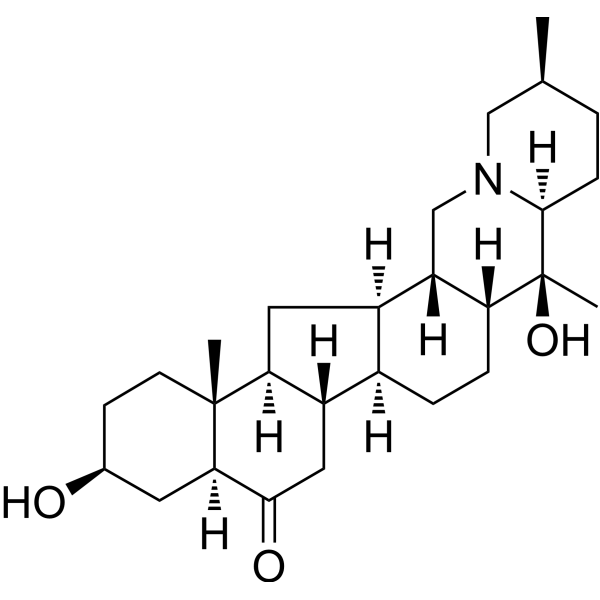
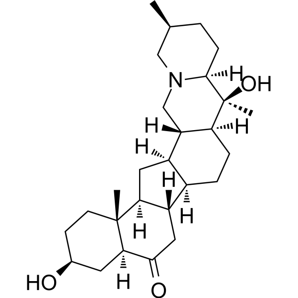
Sipeimine (Imperialine) is an inhibitor targeting the PI3K/AKT/NF-κB pathway and NLRP3 inflammasome, which can competitively bind to PI3K and p65. Sipeimine inhibits PI3K/AKT phosphorylation, blocks NF-κB nuclear translocation and NLRP3 inflammasome activation. Sipeimine exerts anti-inflammatory activities, inhibits pyroptosis and ferroptosis, and protects the extracellular matrix. Sipeimine can reduce cartilage degradation and synovial inflammation in osteoarthritis and improve PM2.5-induced lung injury. Sipeimine is mainly used in the study of anti-inflammatory and degenerative diseases.
Nur für Forschungszwecke. Wir verkaufen nicht an Patienten.
- Reinheit : 99.51%
- CAS. Nr.: 61825-98-7
- Formel: C27H43NO3
- Molecular Weight:429.64
-
Speicherung:Powder -20°C, 3 years , 4°C, 2 years ; In solvent -80°C, 2 years , -20°C, 1 year

To place orders, for customer services and technical support, please contact: MedChemExpress USA
Tel: 609-228-6898 E-mail: [email protected] [email protected]


-
 Biologische Aktivität
-
Chemical Information
-
Lösungsmittel & Löslichkeit
- Protokoll
- Reinheit & Dokumentation
- Verweise
-
 Help & FAQs



Publications Citing Use of MedChemExpress (MCE) Sipeimine
More

Biologische Aktivität
Beschreibung
In Vitro
Sipeimine (100 μM, 200 μM; 24-48 h) significantly reduces cell viability in mouse chondrocytes[1].
Sipeimine (10-50 μM; 24 h) dose-dependently inhibits lipopolysaccharide (LPS)-induced cytotoxicity in the LDH release assay[1].
Sipeimine (25 μM, 50 μM; 24 h) inhibits the mRNA and protein expression of proinflammatory factors (COX-2, iNOS, IL-1β, IL-18) in 1 μg/mL LPS-induced mouse chondrocytes and reduces PGE2 and nitrite production[1].
Sipeimine (25 μM, 50 μM; 4 h) increases the extracellular matrix components (collagen-II, aggrecan) and reduces the protein expression of degradative enzymes (MMP-13, ADAMTS-5) in mouse chondrocytes in WB experiments[1].
Sipeimine (50 μM; 2-24 h) inhibits LPS-induced NF-κB pathway activation (reduced p-IκBα, p-p65 phosphorylation and p65 nuclear translocation) and NLRP3 inflammasome-mediated cell pyroptosis (reduced NLRP3, ASC, cleaved-caspase-1, cleaved-GSDMD protein expression) in mouse chondrocytes[1].
MedChemExpress (MCE) has not independently confirmed the accuracy of these methods. They are for reference only.
-
Cell Line:Mouse chondrocytes
-
Concentration:0, 25, 50 μM
-
Incubation Time:24 h (after LPS stimulation)
-
Result:Significantly reduced the mRNA levels of pro-inflammatory factors (IL-1β, IL-18, COX-2, iNOS) induced by LPS.
-
Cell Line:Mouse chondrocytes
-
Concentration:0, 25, 50 μM
-
Incubation Time:24 h (after LPS stimulation)
-
Result:Decreased the protein expressions of COX-2, iNOS, MMP-13, ADAMTS-5, NLRP3, ASC, cleaved-caspase-1, cleaved-GSDMD, p-IκBα, and p-p65, while increased the expressions of collagen-II, aggrecan, and total IκBα, AKT, PI3K.
In Vivo
Sipeimine (15, 30 mg/kg; intraperitoneal injection; once daily; 3 days) can reduce lung tissue damage, pulmonary edema, inflammatory response, inhibit ferroptosis-related indicators (4-HNE, MDA, tissue iron) and upregulate antioxidant factors (GSH, Nrf2, GPX4, HO-1) in the PM2.5-induced lung injury Sprague-Dawley rat model[1].
MedChemExpress (MCE) has not independently confirmed the accuracy of these methods. They are for reference only.
-
Animal Model:C57BL/6 mice (8-10 weeks old), medial meniscus destabilization (DMM)-induced OA model[1]
-
Dosage:30 mg/kg (dissolved in saline with 0.1% DMSO).
-
Administration:Intra-articular injection, once daily for 14 days, followed by an 8-week observation period.
-
Result:Significantly reduced subchondral bone volume/tissue volume (BV/TV), trabecular thickness (Tb.Th), and trabecular number (Tb.N), and increased trabecular separation (Tb.Sp) compared to the DMM group.
Histological analysis using Safranin O-fast green and H&E staining showed decreased cartilage degeneration (lower OARSI scores) and synovitis.
Immunohistochemistry revealed reduced expressions of MMP-13, NLRP3, and p-PI3K, and increased collagen-II expression in cartilage tissues.
-
Animal Model:Male Sprague-Dawley rats (8 weeks old, 110-150 g), PM2.5-induced lung injury model via intratracheal instillation of PM2.5 (7.5 mg/kg/day for 2 days)[2]
-
Dosage:15 mg/kg and 30 mg/kg (dissolved in saline with 0.1% DMSO).
-
Administration:Intraperitoneal injection, once daily for 3 days before PM2.5 exposure.
-
Result:Dose-dependently alleviated PM2.5-induced lung injury, as indicated by reduced histological scores (thickened alveolar walls, inflammatory cell infiltration, hemorrhage), lower lung wet/dry (W/D) ratio, and decreased levels of TNF-α and IL-1β in bronchoalveolar lavage fluid (BALF).
Reversed PM2.5-induced increases in tissue iron, 4-HNE, and MDA, and restored GSH levels.
Transmission electron microscopy showed attenuated mitochondrial morphological changes (reduced cristae loss and electron density) in type II alveolar epithelial cells.
Western blot revealed upregulated expressions of Nrf2, GPX4, HO-1, SLC7A11, and FTH1, and increased phosphorylation of PI3K and Akt.
- Western Blot
- Collagen: Sirius Red Staining
- ECM-Embedded Organoid (Matrigel/Dome) Culture
- Research Protocol for Inflammation-related Diseases
- Matrigel Transwell/Boyden Chamber Invasion Assay
- Kinase activity and phosphorylation assays
- Fibrosis/Collagen Morphometry
- Cytoplasmic-Nuclear Fractionated Protein Extraction
- Matrigel/ECM Transwell Invasion Assay
- Protocol for Kinase activity and phosphorylation assays
- Ferroptosis Solutions
- Inhalation Toxicity Study
- Pyroptosis Solutions


Chemical Information
-
CAS. Nr. 61825-98-7
-
Appearance Solid
-
Molecular Weight 429.64
-
Formel C27H43NO3
-
Color White to off-white
-
SMILES
C[C@@]([C@@]1([H])C2=O)(CC[C@H](O)C1)[C@]3([H])[C@](C2)([H])[C@@](CC[C@]4([H])[C@@]5([H])CN(C[C@@H](C)CC6)[C@]6([H])[C@]4(O)C)([H])[C@@]5([H])C3
-
Synonyms
Imperialine
-
Structure Classification
-
Initial Source
-
Versand
Room temperature in continental US; may vary elsewhere.
-
Speicherung
Powder -20°C 3 years 4°C 2 years In solvent -80°C 2 years -20°C 1 year


Publications (4)
-
Journal Impact Factor
-
Most Recent
-
Phytomedicine
Total alkaloids of Fritillaria unibracteata var. wabuensis bulbus ameliorate chronic asthma via the TRPV1/Ca2+/NFAT pathway. [Abstract]2023 Sep:118:154946. PMID: 37421766 -
Molecules
hERG Channel Blockade and Antagonistic Interactions of Three Steroidal Alkaloids from Fritillaria Species. [Abstract]2025 Sep 25;30(19):3882. PMID: 41097303 -
Molecules
Metabolite Profiling and Anti-Inflammatory Activities of Fritillaria cirrhosa D. Don Bulbs Derived from Tissue Culture. [Abstract]2025 Jan 31;30(3):623. PMID: 39942727 -
Sci Rep
Sipeimine reduces ethanol-induced gastric ulcer in mice by suppressing Jak-Stat activation and restoring gut microbiota balance. [Abstract]2025 Aug 6;15(1):28683. PMID: 40770008


Lösungsmittel & Löslichkeit
In Vitro:
DMSO : 8.33 mg/mL (19.39 mM; ultrasonic and warming and heat to 60°C; Hygroscopic DMSO has a significant impact on the solubility of product, please use newly opened DMSO)
Please refer to the solubility information to select the appropriate solvent. Once prepared, please aliquot and store the solution to prevent product inactivation from repeated freeze-thaw cycles.
Storage method and period of stock solution: -80°C, 2 years; -20°C, 1 year. When stored at -80°C, please use it within 2 years. When stored at -20°C, please use it within 1 year.
Please refer to the solubility information to select the appropriate solvent. Once prepared, please aliquot and store the solution to prevent product inactivation from repeated freeze-thaw cycles.
Storage method and period of stock solution: -80°C, 2 years; -20°C, 1 year. When stored at -80°C, please use it within 2 years. When stored at -20°C, please use it within 1 year.
Konzentration (Stammlösung) × Volumen (Stammlösung) = Konzentration (Ziellösung) × Volumen (Ziellösung)
In Vivo:
Select the appropriate dissolution method based on your experimental animal and administration route.
- For the following dissolution methods, please ensure to first prepare a clear stock solution using an In Vitro approach and then sequentially add co-solvents:
- To ensure reliable experimental results, the clarified stock solution can be appropriately stored based on storage conditions. As for the working solution for In Vivo experiments, it is recommended to prepare freshly and use it on the same day.
- The percentages shown for the solvents indicate their volumetric ratio in the final prepared solution. If precipitation or phase separation occurs during preparation, heat and/or sonication can be used to aid dissolution.
Add each solvent one by one: 10% DMSO 40% PEG300 5% Tween-80 45% Saline
Solubility: ≥ 0.77 mg/mL (1.79 mM); Clear solution
This protocol yields a clear solution of ≥ 0.77 mg/mL (saturation unknown).
Taking 1 mL working solution as an example, add 100 μL DMSO stock solution (7.7 mg/mL) to 400 μL PEG300, and mix evenly; then add 50 μL Tween-80 and mix evenly; then add 450 μL Saline to adjust the volume to 1 mL.
Preparation of Saline: Dissolve 0.9 g sodium chloride in ddH₂O and dilute to 100 mL to obtain a clear Saline solution.
Add each solvent one by one: 10% DMSO 90% (20% SBE-β-CD in Saline)
Solubility: ≥ 0.77 mg/mL (1.79 mM); Clear solution
This protocol yields a clear solution of ≥ 0.77 mg/mL (saturation unknown).
Taking 1 mL working solution as an example, add 100 μL DMSO stock solution (7.7 mg/mL) to 900 μL 20% SBE-β-CD in Saline, and mix evenly.
Preparation of 20% SBE-β-CD in Saline (4°C, storage for one week): 2 g SBE-β-CD powder is dissolved in 10 mL Saline, completely dissolve until clear.
In Vivo Dissolution Calculator
Please enter the basic information of animal experiments:
-
-
-
-
Recommended: Prepare an additional quantity of animals to account for potential losses during experiments.
Please enter your animal formula composition:
-
%DMSO +
Recommended: Keep the proportion of DMSO in working solution below 2% if your animal is weak.
-
%+
-
+%Tween-80 + +
-
%Saline +
The co-solvents required include: DMSO, . All of co-solvents are available by MedChemExpress (MCE). , Tween 80. All of co-solvents are available by MedChemExpress (MCE).
Working solution concentration: 0.22 mg/mL
Method for preparing stock solution: mg drug dissolved in μL DMSO. Stock solution concentration: mg/mL.
1. Take μL DMSO stock solution;
2. Add μL .
μL , mix evenly;
3. Then add μL Tween 80, mix evenly;
4. Then add μL
Please ensure that the stock solution in the first step is dissolved to a clear state, and add co-solvents in sequence. You can use ultrasonic heating (ultrasonic cleaner, recommended frequency 20-40 kHz), vortexing, etc. to assist dissolution.


Reinheit & Dokumentation
-
Data Sheet (286 KB)
-
SDS (394 KB)
- English - EN (394 KB)
- Français - FR (394 KB)
- Deutsch - DE (394 KB)
- Norwegian - NO (394 KB)
- Español - ES (394 KB)
- Swedish - SV (394 KB)
- Italian - IT (394 KB)
- Korean - KR (394 KB)
- Portuguese - PT (394 KB)
-
Handling Instructions (2659 KB)
Verweise
[1]. Fang Y, et al. Sipeimine ameliorates osteoarthritis progression by suppression of NLRP3 inflammasome-mediated pyroptosis through inhibition of PI3K/AKT/NF-κB pathway: An in vitro and in vivo study. J Orthop Translat. 2024 May 9;46:1-17. [Content Brief]
[2]. Wang Y, et al. Sipeimine ameliorates PM2.5-induced lung injury by inhibiting ferroptosis via the PI3K/Akt/Nrf2 pathway: A network pharmacology approach. Ecotoxicol Environ Saf. 2022 Jul 1;239:113615. [Content Brief]


Complete Stock Solution Preparation Table
Please refer to the solubility information to select the appropriate solvent. Once prepared, please aliquot and store the solution to prevent product inactivation from repeated freeze-thaw cycles.
Storage method and period of stock solution: -80°C, 2 years; -20°C, 1 year. When stored at -80°C, please use it within 2 years. When stored at -20°C, please use it within 1 year.
| Optional Solvent | Concentration Solvent Mass | 1 mg | 5 mg | 10 mg | 25 mg |
|---|---|---|---|---|---|
| DMSO | 1 mM | 2.3275 mL | 11.6376 mL | 23.2753 mL | 58.1882 mL |
| 5 mM | 0.4655 mL | 2.3275 mL | 4.6551 mL | 11.6376 mL | |
| 10 mM | 0.2328 mL | 1.1638 mL | 2.3275 mL | 5.8188 mL | |
| 15 mM | 0.1552 mL | 0.7758 mL | 1.5517 mL | 3.8792 mL |


Sipeimine Related Classifications
HY-N0696 Related Classifications
 Powered by Bioz