



MS177
Based on 3 publication(s) in Google Scholar
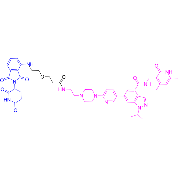
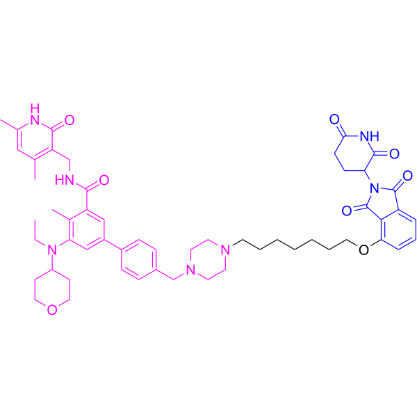
MS177 is an effective and fast-acting EZH2 degrader. MS177 is a PROTAC that consists of a CRBN ligand, linker, and a potent enzymatic EZH2 inhibitor C24 (C24 IC50): 12 nM). MS177 effectively depletes both canonical EZH2-PRC2 and noncanonical EZH2-cMyc complexes. MS177 induces leukaemia cell growth inhibition, apoptosis and cell cycle progression arrest.
(Pink: EZH2 ligand (HY-W1063360); Blue: Cereblon ligand (HY-41547); Black: linker (HY-140002)).
Nur für Forschungszwecke. Wir verkaufen nicht an Patienten.
- Reinheit : 99.55%
- CAS. Nr.: 2225938-86-1
- Formel: C48H55N11O8
- Molecular Weight:914.02
-
Speicherung:Powder -20°C, 3 years , 4°C, 2 years ; In solvent -80°C, 6 months , -20°C, 1 month

To place orders, for customer services and technical support, please contact: MedChemExpress USA
Tel: 609-228-6898 E-mail: [email protected] [email protected]


-
 Biologische Aktivität
-
Chemical Information
-
Lösungsmittel & Löslichkeit
- Protokoll
- Reinheit & Dokumentation
- Verweise
-
 Help & FAQs


-
HY-148334
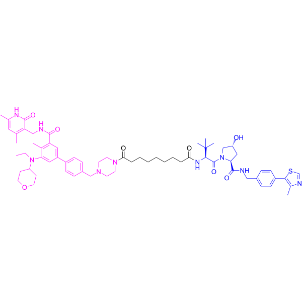
MS8815PROTAC EZH2 Degrader -
HY-13803
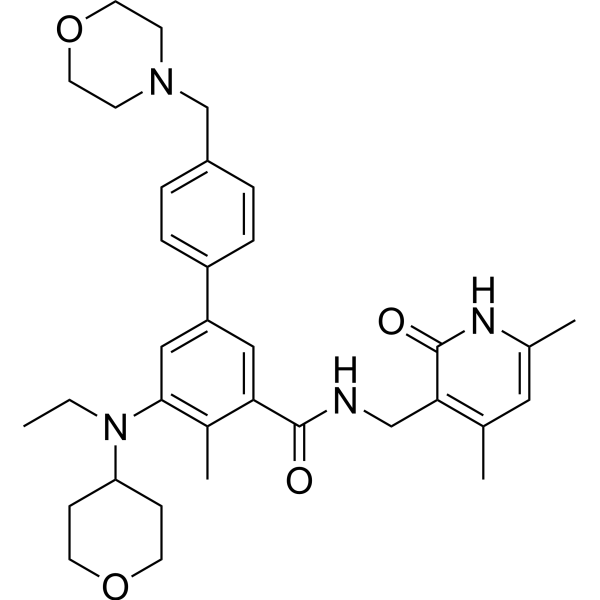
TazemetostatEZH2 Inhibitor -
HY-133129
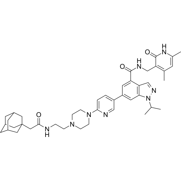
MS1943EZH2 HyT Degrader -
HY-147525
PROTAC EZH2 Degrader-1PROTAC EZH2 Degrader -
HY-13470
GSK126EZH2 Inhibitor -
HY-13030
(+)-JQ-1BET Inhibitor -
HY-10492
DinaciclibCDK2/5/9 Inhibitor -

HY-10431
SB-431542TGF-β Receptor Kinase Inhibitor -

HY-100579
Ferrostatin-1Ferroptosis Inhibitor -
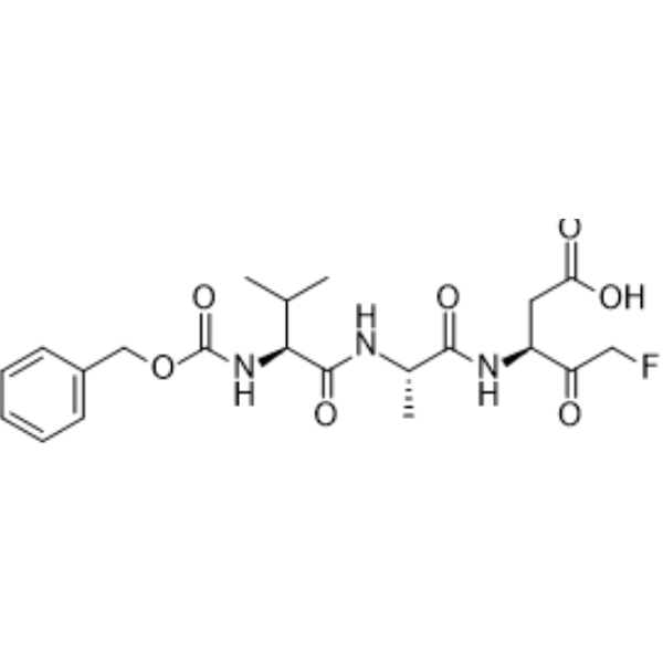
HY-16658B
Z-VAD-FMKPan-Caspase Inhibitor -
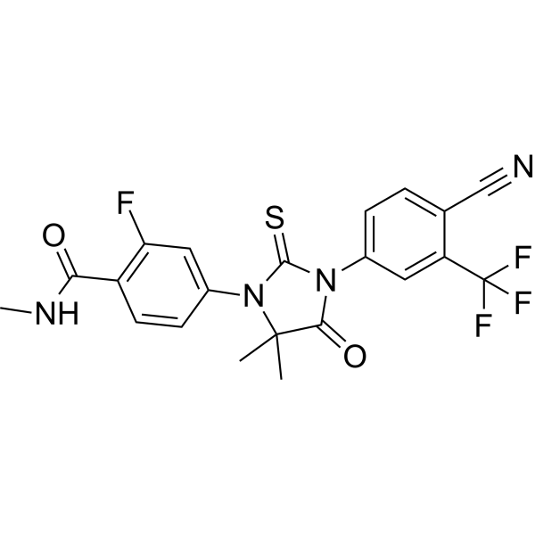
HY-70002
EnzalutamideAndrogen Receptor Antagonist -
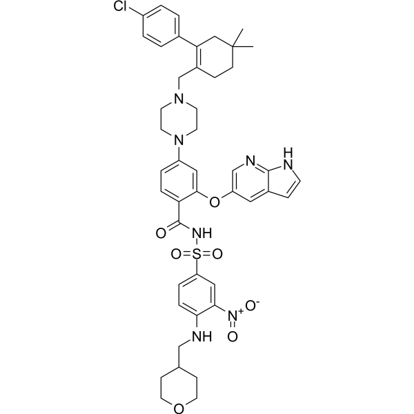
HY-15531
VenetoclaxBCL-2 Inhibitor
Publications Citing Use of MedChemExpress (MCE) MS177
More Customer Validation & Images

-
WB

-
WB

-
IF

-
RT-PCR

-
WB

Alle PROTACs Isoform-spezifische Produkte anzeigen
MoreAlle Histone Methyltransferase Isoform-spezifische Produkte anzeigen
More

Biologische Aktivität
Beschreibung
IC50 & Target
[1]|
EZH2 |
In Vitro
MS177 inhibits the enzymatic activities of EZH2-PRC2 (IC50: 7 nM)[1].
MS177 (5 μM, 24 h) decreases H3K27me3 and increases H3K27activity in HeLa cells[1].
MS177 (0.1-5 μM, 16 h) effectively degrades cellular EZH2-PRC2 and suppresses global H3K27me3 in EOL-1 cells[1].
MS177 (0.1-5 μM, 16 h) induces Myc degradation in EOL-1 and MV4 cells[1].
MS177 (4 days) shows antiproliferation effects in a panel of MLL-r leukaemia cells and samples from patients with AML, with IC50s below 2 μM[1].
MS177 (0.5-2.5 μM, 24 h) decreases colony-forming capabilities in MV4;11 cells[1].
MS177 (0.5-2.5 μM, 24 h) slows cell cycle progression and induces MOLM-13 cell apoptosis[1].
MedChemExpress (MCE) has not independently confirmed the accuracy of these methods. They are for reference only.
-
Cell Line:AML cell line: MV4;11, MOML-13, RS4;11, KOPN-8 THP-1, EOL-1 (MLL-r cells)
Control cell line: K562 (CML cells)
Patient sample: AML cells -
Concentration:0-100 μM approximately
-
Incubation Time:4 days
-
Result:Inhibited cell proliferation with IC50s of 0.1-0.57 μM for MLL-r cells, 0.09-1.35 μM for Patient sample, >100 μM for K562 cell.
-
Cell Line:EOL-1 cell
-
Concentration:0.1, 0.5, 1, 2.5, 5 μM
-
Incubation Time:16 h
-
Result:Depleted EZH2, EED and SUZ12 in a concentration-dependent manner and suppressed global H3K27me3.
In Vivo
MS177 (50 mg/kg, i.p.) achieves intraplasma concentrations about 1 μM in male Swiss Albino mice[1].
MS177 (100 mg/kg, i.p., BID for 6 days per week; and 200 mg/ kg, i.p. BID 3 days per week) is well tolerated and lacks apparent toxicity in mice[1].
MedChemExpress (MCE) has not independently confirmed the accuracy of these methods. They are for reference only.
-
Animal Model:PDX animal model of MLL-r AML[1]
-
Dosage:100 mg/kg
-
Administration:Intraperitoneal injection (i.p.), BID for 6 days.
-
Result:Inhibited tumor growth and prolonged survival.
- Annexin V plus membrane-impermeant dye apoptosis staining
- BrdU Incorporation Assay
- Somatic Cell Culture
- CCK-8/WST-8 Cell Proliferation Assay
- Protocol for Cell Cycle
- Cell Counting-Based Growth Curve Assay
- Cell Viability Determination by MTT Colorimetric Assay
- Detection of Apoptosis
- TUNEL staining for apoptotic DNA fragmentation
- Flow cytometric DNA-content cell-cycle staining
- Protocol for Cell Counting and Cell Density Analysis
- Apoptosis Solutions
- MTT Cell Proliferation Assay


Chemical Information
-
CAS. Nr. 2225938-86-1
-
Appearance Solid
-
Molecular Weight 914.02
-
Formel C48H55N11O8
-
Color Light yellow to yellow
-
SMILES
O=C(C1=CC(C2=CC=C(N3CCN(CCNC(CCOCCNC4=CC=CC(C(N5C(CC6)C(NC6=O)=O)=O)=C4C5=O)=O)CC3)N=C2)=CC7=C1C=NN7C(C)C)NCC8=C(C)C=C(C)NC8=O
-
Versand
Room temperature in continental US; may vary elsewhere.
-
Speicherung
Powder -20°C 3 years 4°C 2 years In solvent -80°C 6 months -20°C 1 month


Publications (3)
-
Journal Impact Factor
-
Most Recent
-
Cell Mol Gastroenterol Hepatol
2025;19(1):101404. PMID: 39278404
MS177 purchased from MedChemExpress. Usage Cited in: Cell Mol Gastroenterol Hepatol. 2025;19(1):101404. [Abstract]
MS177 (10 μM; 48 h) decreased the nuclear localization of β-catenin in HuCCT1 and CCLP1 cells.
MS177 purchased from MedChemExpress. Usage Cited in: Cell Mol Gastroenterol Hepatol. 2025;19(1):101404. [Abstract]
MS177 (10 μM; 48 h) decreased the level of active β-catenin in HuCCT1 and CCLP1 cells.
MS177 purchased from MedChemExpress. Usage Cited in: Cell Mol Gastroenterol Hepatol. 2025;19(1):101404. [Abstract]
MS177 (10 μM; 48 h) revealed a reduction in the nuclear localization of β-catenin in HuCCT1 and CCLP1 cells.
MS177 purchased from MedChemExpress. Usage Cited in: Cell Mol Gastroenterol Hepatol. 2025;19(1):101404. [Abstract]
MS177 (10 μM; 48 h) showed that the mRNA of β-catenin targets (c-Myc, BIRC5, and AXIN2) was markedly downregulated in HuCCT1 and CCLP1 cells.
-
Biochim Biophys Acta Mol Basis Dis
EZH2 contributes to sepsis-induced acute lung injury through regulating macrophage polarization. [Abstract]2025 Jan;1871(1):167554. PMID: 39471914 -
Mol Immunol
Sigmoidin B attenuates sepsis-associated encephalopathy through EZH2-AKT2-mediated regulation of microglial polarization. [Abstract]2025 Oct:186:95-113. PMID: 40834508
MS177 purchased from MedChemExpress. Usage Cited in: Mol Immunol. 2025 Oct:186:95-113. [Abstract]
MS177 (4 mg/kg) led to a significant reduction in p-AKT2 expression in microglia of SAE mice.


Lösungsmittel & Löslichkeit
In Vitro:
DMSO : 100 mg/mL (109.41 mM; Need ultrasonic; Hygroscopic DMSO has a significant impact on the solubility of product, please use newly opened DMSO)
Please refer to the solubility information to select the appropriate solvent. Once prepared, please aliquot and store the solution to prevent product inactivation from repeated freeze-thaw cycles.
Storage method and period of stock solution: -80°C, 6 months; -20°C, 1 month. When stored at -80°C, please use it within 6 months. When stored at -20°C, please use it within 1 month.
Please refer to the solubility information to select the appropriate solvent. Once prepared, please aliquot and store the solution to prevent product inactivation from repeated freeze-thaw cycles.
Storage method and period of stock solution: -80°C, 6 months; -20°C, 1 month. When stored at -80°C, please use it within 6 months. When stored at -20°C, please use it within 1 month.
Konzentration (Stammlösung) × Volumen (Stammlösung) = Konzentration (Ziellösung) × Volumen (Ziellösung)
In Vivo:
Select the appropriate dissolution method based on your experimental animal and administration route.
- For the following dissolution methods, please ensure to first prepare a clear stock solution using an In Vitro approach and then sequentially add co-solvents:
- To ensure reliable experimental results, the clarified stock solution can be appropriately stored based on storage conditions. As for the working solution for In Vivo experiments, it is recommended to prepare freshly and use it on the same day.
- The percentages shown for the solvents indicate their volumetric ratio in the final prepared solution. If precipitation or phase separation occurs during preparation, heat and/or sonication can be used to aid dissolution.
Add each solvent one by one: 10% DMSO 40% PEG300 5% Tween-80 45% Saline
Solubility: 2.08 mg/mL (2.28 mM); Suspended solution; Need ultrasonic
This protocol yields a suspended solution of 2.08 mg/mL. Suspended solution can be used for oral and intraperitoneal injection.
Taking 1 mL working solution as an example, add 100 μL DMSO stock solution (20.8 mg/mL) to 400 μL PEG300, and mix evenly; then add 50 μL Tween-80 and mix evenly; then add 450 μL Saline to adjust the volume to 1 mL.
Preparation of Saline: Dissolve 0.9 g sodium chloride in ddH₂O and dilute to 100 mL to obtain a clear Saline solution.
In Vivo Dissolution Calculator
Please enter the basic information of animal experiments:
-
-
-
-
Recommended: Prepare an additional quantity of animals to account for potential losses during experiments.
Please enter your animal formula composition:
-
%DMSO +
Recommended: Keep the proportion of DMSO in working solution below 2% if your animal is weak.
-
%+
-
+%Tween-80 + +
-
%Saline +
The co-solvents required include: DMSO, . All of co-solvents are available by MedChemExpress (MCE). , Tween 80. All of co-solvents are available by MedChemExpress (MCE).
Working solution concentration: 0.22 mg/mL
Method for preparing stock solution: mg drug dissolved in μL DMSO. Stock solution concentration: mg/mL.
1. Take μL DMSO stock solution;
2. Add μL .
μL , mix evenly;
3. Then add μL Tween 80, mix evenly;
4. Then add μL
Please ensure that the stock solution in the first step is dissolved to a clear state, and add co-solvents in sequence. You can use ultrasonic heating (ultrasonic cleaner, recommended frequency 20-40 kHz), vortexing, etc. to assist dissolution.


Reinheit & Dokumentation
-
Data Sheet (283 KB)
-
SDS (254 KB)
- English - EN (254 KB)
- Français - FR (254 KB)
- Deutsch - DE (254 KB)
- Norwegian - NO (254 KB)
- Español - ES (254 KB)
- Swedish - SV (254 KB)
- Italian - IT (254 KB)
- Korean - KR (254 KB)
- Portuguese - PT (254 KB)
-
Handling Instructions (2659 KB)
Verweise


Complete Stock Solution Preparation Table
Please refer to the solubility information to select the appropriate solvent. Once prepared, please aliquot and store the solution to prevent product inactivation from repeated freeze-thaw cycles.
Storage method and period of stock solution: -80°C, 6 months; -20°C, 1 month. When stored at -80°C, please use it within 6 months. When stored at -20°C, please use it within 1 month.
| Optional Solvent | Concentration Solvent Mass | 1 mg | 5 mg | 10 mg | 25 mg |
|---|---|---|---|---|---|
| DMSO | 1 mM | 1.0941 mL | 5.4703 mL | 10.9407 mL | 27.3517 mL |
| 5 mM | 0.2188 mL | 1.0941 mL | 2.1881 mL | 5.4703 mL | |
| 10 mM | 0.1094 mL | 0.5470 mL | 1.0941 mL | 2.7352 mL | |
| 15 mM | 0.0729 mL | 0.3647 mL | 0.7294 mL | 1.8234 mL | |
| 20 mM | 0.0547 mL | 0.2735 mL | 0.5470 mL | 1.3676 mL | |
| 25 mM | 0.0438 mL | 0.2188 mL | 0.4376 mL | 1.0941 mL | |
| 30 mM | 0.0365 mL | 0.1823 mL | 0.3647 mL | 0.9117 mL | |
| 40 mM | 0.0274 mL | 0.1368 mL | 0.2735 mL | 0.6838 mL | |
| 50 mM | 0.0219 mL | 0.1094 mL | 0.2188 mL | 0.5470 mL | |
| 60 mM | 0.0182 mL | 0.0912 mL | 0.1823 mL | 0.4559 mL | |
| 80 mM | 0.0137 mL | 0.0684 mL | 0.1368 mL | 0.3419 mL | |
| 100 mM | 0.0109 mL | 0.0547 mL | 0.1094 mL | 0.2735 mL |


MS177 Related Classifications
HY-148333 Related Classifications
 Powered by Bioz