



Murrayafoline A

Based on 1 Customer Validation
Murrayafoline A is a carbazole alkaloid that can be extracted from Murraya tetramera. Murrayafoline A directly targets Specificity protein 1 (Sp1), thereby inhibiting NF-κB and MAPK signaling pathways. Murrayafoline a induces a G0/G1-phase arrest in platelet-derived growth factor (PDGF)-stimulated vascular smooth muscle cells. Murrayafoline A attenuates the Wnt/β-catenin pathway by promoting the degradation of intracellular β-catenin proteins. Murrayafoline A enhances the contraction of rat ventricular myocytes and L-type calcium current by activating protein kinase C. Murrayafoline A inhibits LPS (HY-D1056)-induced neuroinflammation in vivo. Murrayafoline A can be used for the study of inflammation, vascular complications and colon cancer.
Nos produits utilisent uniquement pour la recherche. Nous ne vendons pas aux patients.
- Pureté : 99.73%
- CAS No.: 4532-33-6
- Formule: C14H13NO
- Masse moléculaire:211.26
-
Stockage:Powder -20°C, 3 years , 4°C, 2 years ; In solvent -80°C, 6 months , -20°C, 1 month


To place orders, for customer services and technical support, please contact: MedChemExpress USA
Tel: 609-228-6898 E-mail: [email protected] [email protected]


-
 Activité biologique
-
Chemical Information
-
Solvant et solubilité
- Protocole
- Pureté et documentation
- Références
-
 Help & FAQs


-
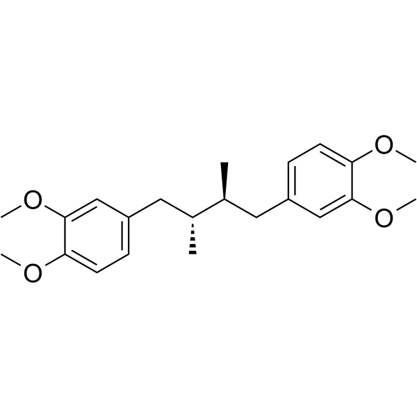
HY-10447
TerameprocolSp1 Inhibitor -
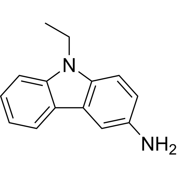
HY-W014386
3-Amino-9-ethylcarbazoleBiochemical Assay Reagent -
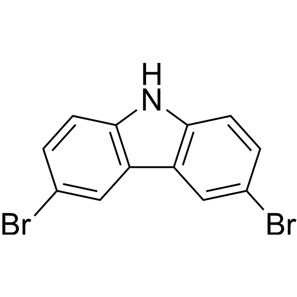
HY-W009154
3,6-DibromocarbazoleGlucocorticoid Receptor Inhibitor -
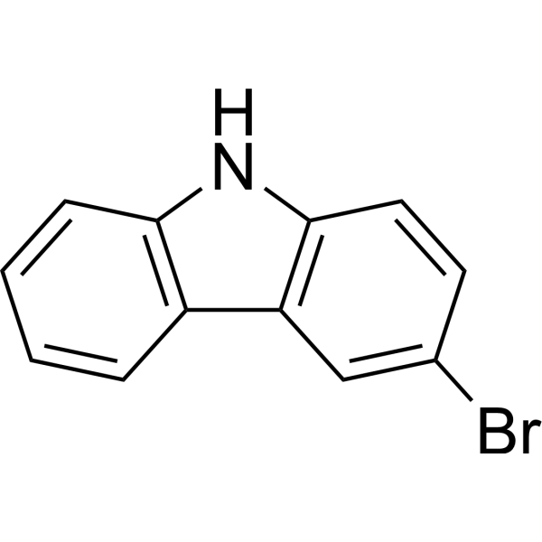
HY-33798
3-BromocarbazoleAHR Inhibitor -

HY-W016331
9H-Carbazol-4-olAromatic Heterocycle -

HY-W013429
4-Glycidyloxycarbazole -

HY-33672R
9-Phenylcarbazole (Standard) -

HY-W100287R
Murrayafoline A (Standard)Anti-neuroinflammatory Agent


Activité biologique
Description
Cellular Effect
|
Cell Line

|
Type | Value | Description | References |
|---|---|---|---|---|
| PBMC | IC50 |
7 μM
Compound: 58
|
Inhibition of TNF-alpha production in LPS-stimulated human PBMC cells
Inhibition of TNF-alpha production in LPS-stimulated human PBMC cells
|
[PMID: 33454546] |
| PBMC | IC50 |
8.4 μM
Compound: 58
|
Inhibition of IL6 production in LPS-stimulated human PBMC cells
Inhibition of IL6 production in LPS-stimulated human PBMC cells
|
[PMID: 33454546] |
In Vitro
Murrayafoline A (5-20 μM, 24 h) potently inhibits the production of neuroinflammation mediators, such as nitric oxide (NO), TNF-α, IL-6 and IL-1β in LPS (HY-D1056)-induced BV-2 microglial cells, an effect that can be significantly antagonized by Sp1 knockdown[1].
Murrayafoline A (20 μM, 2 h) directly interacts with Sp1, as confirmed by surface plasmon resonance (SPR) with a dissociation constant (KD) of 11.34 pM in BV-2 cells[1].
Murrayafoline A (20 μM) inhibits IKKβ/NF-κB and p38/JNK MAPKs pathways that can be markedly reversed by Sp1 knockdown in LPS-induced BV-2 cells[1].
Murrayafoline A (1-5 μM, 24 h) inhibits PDGF-BB-stimulated proliferation of vascular smooth muscle cells (VSMCs), DNA synthesis and induces a G0/G1-phase arrest[2].
Murrayafoline A (1-5 μM) down-regulates the expression of cyclin D1, cyclin E, CDK2, CDK4, and PCNA, and inhibits the phosphorylation of pRb in PDGF-BB-stimulated VSMCs[2].
Murrayafoline A (5-40 μM, 15 h) dose-dependently reduces LiCl- and Wnt3a-CM-induced TOPFlash reporter activity and SEAP activity in HEK293 reporter cells without affecting FOPFlash activity[3].
Murrayafoline A (20-40 μM, 15 h) decreases cytosolic β-catenin level in LiCl- and Wnt3a-CM-treated HEK293 reporter cells[3].
Murrayafoline A (10-80 μM, 15-48 h) reduces cytosolic β-catenin level, cyclin D1 and c-myc levels in SW480 and HCT-116 colon cancer cells and dose-dependently inhibits the proliferation of SW480, DLD-1, HCT-116 and LS174T colon cancer cells[3].
Murrayafoline A (25 μM, 2 min) increases cell shortening, increases L-type Ca2+ current, enhances PKC phosphorylation and enhances the rate of contraction in rat ventricular myocytes[4].
MedChemExpress (MCE) has not independently confirmed the accuracy of these methods. They are for reference only.
-
Cell Line:Vascular smooth muscle cells (VSMCs)
-
Concentration:1, 3, 5 μM
-
Incubation Time:24 h
-
Result:Increased the cell population in G0/G1phase.
Decreased the S, G2, and M phases.
-
Cell Line:SW480, DLD-1, HCT-116 and LS174T colon cancer cells
-
Concentration:5, 10, 20, 40, 80 μM
-
Incubation Time:48 h
-
Result:Inhibited the proliferation of SW480, DLD-1, HCT-116 and LS174T colon cancer cells.
In Vivo
MedChemExpress (MCE) has not independently confirmed the accuracy of these methods. They are for reference only.
-
Animal Model:LPS (1.5 mg/kg) was used to induce neuroinflammation in 6-7-week-old male Balb/c mice[1]
-
Dosage:10, 50 mg/kg
-
Administration:1h pretreatment for a single dose
-
Result:Reduced the expression of Iba-1, TNF-α, and IL-6 in the brains of LPS-induced male Balb/c mice.
Increased the number of Nissl bodies in the brains of LPS-induced male Balb/c mice.


Chemical Information
-
CAS No. 4532-33-6
-
Appearance Solid
-
Masse moléculaire 211.26
-
Formule C14H13NO
-
Color White to light yellow
-
SMILES
CC1=CC2=C(C(=C1)OC)NC3=CC=CC=C23
-
Structure Classification
-
Initial Source
-
Livraison
Room temperature in continental US; may vary elsewhere.
-
Stockage
Powder -20°C 3 years 4°C 2 years In solvent -80°C 6 months -20°C 1 month


Solvant et solubilité
In Vitro:
DMSO : 100 mg/mL (473.35 mM; Need ultrasonic; Hygroscopic DMSO has a significant impact on the solubility of product, please use newly opened DMSO)
H2O : < 0.1 mg/mL (insoluble)
Please refer to the solubility information to select the appropriate solvent. Once prepared, please aliquot and store the solution to prevent product inactivation from repeated freeze-thaw cycles.
Storage method and period of stock solution: -80°C, 6 months; -20°C, 1 month. When stored at -80°C, please use it within 6 months. When stored at -20°C, please use it within 1 month.
Please refer to the solubility information to select the appropriate solvent. Once prepared, please aliquot and store the solution to prevent product inactivation from repeated freeze-thaw cycles.
Storage method and period of stock solution: -80°C, 6 months; -20°C, 1 month. When stored at -80°C, please use it within 6 months. When stored at -20°C, please use it within 1 month.
Concentration (start) × Volume (start) = Concentration (final) × Volume (final)
In Vivo:
Select the appropriate dissolution method based on your experimental animal and administration route.
- For the following dissolution methods, please ensure to first prepare a clear stock solution using an In Vitro approach and then sequentially add co-solvents:
- To ensure reliable experimental results, the clarified stock solution can be appropriately stored based on storage conditions. As for the working solution for In Vivo experiments, it is recommended to prepare freshly and use it on the same day.
- The percentages shown for the solvents indicate their volumetric ratio in the final prepared solution. If precipitation or phase separation occurs during preparation, heat and/or sonication can be used to aid dissolution.
Add each solvent one by one: 10% DMSO 40% PEG300 5% Tween-80 45% Saline
Solubility: ≥ 2.5 mg/mL (11.83 mM); Clear solution
This protocol yields a clear solution of ≥ 2.5 mg/mL (saturation unknown).
Taking 1 mL working solution as an example, add 100 μL DMSO stock solution (25.0 mg/mL) to 400 μL PEG300, and mix evenly; then add 50 μL Tween-80 and mix evenly; then add 450 μL Saline to adjust the volume to 1 mL.
Preparation of Saline: Dissolve 0.9 g sodium chloride in ddH₂O and dilute to 100 mL to obtain a clear Saline solution.
In Vivo Dissolution Calculator
Please enter the basic information of animal experiments:
-
-
-
-
Recommended: Prepare an additional quantity of animals to account for potential losses during experiments.
Please enter your animal formula composition:
-
%DMSO +
Recommended: Keep the proportion of DMSO in working solution below 2% if your animal is weak.
-
%+
-
+%Tween-80 + +
-
%Saline +
The co-solvents required include: DMSO, . All of co-solvents are available by MedChemExpress (MCE). , Tween 80. All of co-solvents are available by MedChemExpress (MCE).
Working solution concentration: 0.22 mg/mL
Method for preparing stock solution: mg drug dissolved in μL DMSO. Stock solution concentration: mg/mL.
1. Take μL DMSO stock solution;
2. Add μL .
μL , mix evenly;
3. Then add μL Tween 80, mix evenly;
4. Then add μL
Please ensure that the stock solution in the first step is dissolved to a clear state, and add co-solvents in sequence. You can use ultrasonic heating (ultrasonic cleaner, recommended frequency 20-40 kHz), vortexing, etc. to assist dissolution.


Pureté et documentation
-
Fiche technique (287 KB)
-
SDS (418 KB)
- English - EN (418 KB)
- Français - FR (418 KB)
- Deutsch - DE (418 KB)
- Norwegian - NO (418 KB)
- Español - ES (418 KB)
- Swedish - SV (418 KB)
- Italian - IT (418 KB)
- Korean - KR (418 KB)
- Portuguese - PT (418 KB)
-
Instruction de manipulation (2659 KB)
Références
[1]. Li CH, et al. Natural carbazole alkaloid murrayafoline A displays potent anti-neuroinflammatory effect by directly targeting transcription factor Sp1 in LPS-induced microglial cells. Bioorg Chem. 2022 Dec;129:106178. [Content Brief]
[2]. Han JH, et al. Murrayafoline A Induces a G0/G1-Phase Arrest in Platelet-Derived Growth Factor-Stimulated Vascular Smooth Muscle Cells. Korean J Physiol Pharmacol. 2015 Sep;19(5):421-6. [Content Brief]
[3]. Choi H, et al. Murrayafoline A attenuates the Wnt/beta-catenin pathway by promoting the degradation of intracellular beta-catenin proteins. Biochem Biophys Res Commun. 2010 Jan 1;391(1):915-20. [Content Brief]
[4]. Chidipi B, et al. Enhancement of contraction and L-type Ca(2+) current by murrayafoline-A via protein kinase C in rat ventricular myocytes. Eur J Pharmacol. 2016 Aug 5;784:33-41. [Content Brief]


Complete Stock Solution Preparation Table
Please refer to the solubility information to select the appropriate solvent. Once prepared, please aliquot and store the solution to prevent product inactivation from repeated freeze-thaw cycles.
Storage method and period of stock solution: -80°C, 6 months; -20°C, 1 month. When stored at -80°C, please use it within 6 months. When stored at -20°C, please use it within 1 month.
| Optional Solvent | Concentration Solvent Mass | 1 mg | 5 mg | 10 mg | 25 mg |
|---|---|---|---|---|---|
| DMSO | 1 mM | 4.7335 mL | 23.6675 mL | 47.3350 mL | 118.3376 mL |
| 5 mM | 0.9467 mL | 4.7335 mL | 9.4670 mL | 23.6675 mL | |
| 10 mM | 0.4734 mL | 2.3668 mL | 4.7335 mL | 11.8338 mL | |
| 15 mM | 0.3156 mL | 1.5778 mL | 3.1557 mL | 7.8892 mL | |
| 20 mM | 0.2367 mL | 1.1834 mL | 2.3668 mL | 5.9169 mL | |
| 25 mM | 0.1893 mL | 0.9467 mL | 1.8934 mL | 4.7335 mL | |
| 30 mM | 0.1578 mL | 0.7889 mL | 1.5778 mL | 3.9446 mL | |
| 40 mM | 0.1183 mL | 0.5917 mL | 1.1834 mL | 2.9584 mL | |
| 50 mM | 0.0947 mL | 0.4734 mL | 0.9467 mL | 2.3668 mL | |
| 60 mM | 0.0789 mL | 0.3945 mL | 0.7889 mL | 1.9723 mL | |
| 80 mM | 0.0592 mL | 0.2958 mL | 0.5917 mL | 1.4792 mL | |
| 100 mM | 0.0473 mL | 0.2367 mL | 0.4734 mL | 1.1834 mL |


Murrayafoline A Related Classifications
HY-W100287 Related Classifications
 Powered by Bioz