



SKI II
Based on 8 publication(s) in Google Scholar
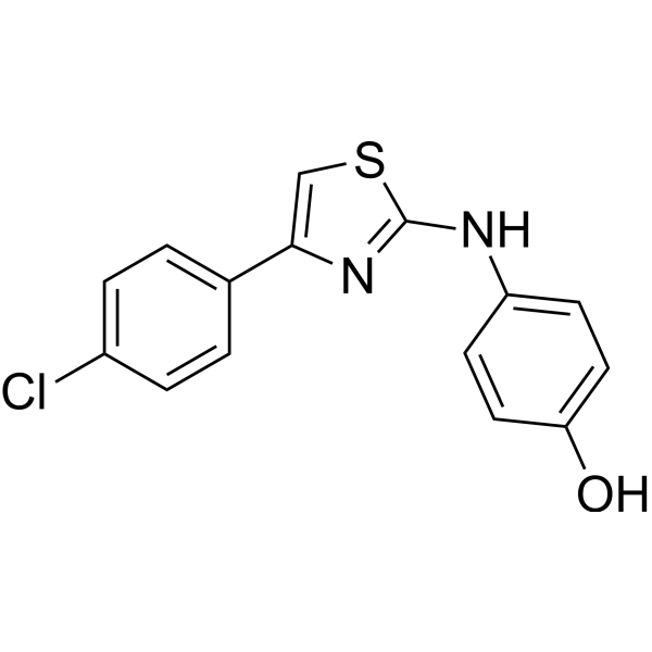
SKI-II is an orally active dual-target inhibitor of SphK1/2 and DES1, with Ki values of 16 µM and 0.3 µM against SphK1 and DES1, respectively. SKI-II activates the PERK-Nrf2 antioxidant pathway by stabilizing Nrf2, and synergizes with Temozolomide (HY-17364) to induce oxidative/endoplasmic reticulum stress and cancer cell death under hypoxic conditions. In vivo, SKI-II inhibits SphK activity, promotes ceramide accumulation and apoptosis, and synergizes with Cisplatin (HY-17394) to reverse drug resistance via the ras/MAPK pathway. SKI-II reduces measles virus replication by inhibiting mTORC1 activity. SKI-II binds to VCP to induce M1 polarization, enhances host tolerance to Staphylococcus aureus infection, and exerts radioprotective effects through Nrf2 activation and accelerated DNA repair. SKI-II can be used in research related to glioblastoma, acute myeloid leukemia, gastric cancer, measles virus infection, Staphylococcus aureus infection, and acute radiation syndrome.
For research use only. We do not sell to patients.
- Purity: 99.25%
- CAS No.: 312636-16-1
- Formula: C15H11ClN2OS
- Molecular Weight:302.78
-
Storage:Powder -20°C, 3 years , 4°C, 2 years
* The compound is unstable in solutions, freshly prepared is recommended.

To place orders, for customer services and technical support, please contact: MedChemExpress USA
Tel: 609-228-6898 E-mail: [email protected] [email protected]


-
 Biological Activity
-
Chemical Information
-
Solvent & Solubility
- Purity & Documentation
- References
-
 Help & FAQs


Publications Citing Use of MedChemExpress (MCE) SKI II
More- Cancer Commun (Lond). 2025 Jul 16. [Abstract]
- Autophagy. 2025 Apr 3. [Abstract]
- Cell Commun Signal. 2024 Aug 7;22(1):391. [Abstract]
- Mol Med Rep. 2024 Jan;29(1):16. [Abstract]
- J Virol. 2025 Feb 25;99(2):e0122024. [Abstract]
- Cell Biol Int. 2021 Apr;45(4):775-784. [Abstract]
- J Leukoc Biol. 2025 Aug 5;117(8):qiaf111. [Abstract]
- bioRxiv. 2024 July 15.
 Customer Validation & Images

-
IF

-
Others

-
WB

-
WB
-
Flow Cytometry


Biological Activity
Description
|
SphK1 16 μM (Ki) |
SphK2 45 μM (IC50) |
DES1 0.3 μM (Ki) |
Cellular Effect
|
Cell Line

|
Type | Value | Description | References |
|---|---|---|---|---|
| A549 | IC50 |
7.8 μM
Compound: 13, SKI-II
|
Cytotoxicity against human A549 cells after 72 hrs by SRB assay
Cytotoxicity against human A549 cells after 72 hrs by SRB assay
|
[PMID: 20598402] |
| HeLa | EC50 |
<1.2 μM
Compound: 12
|
Inhibition of VCP in human HeLa cells assessed as stabilization of ubiquitin-tagged luciferase after 24 hrs by reporter gene assay
Inhibition of VCP in human HeLa cells assessed as stabilization of ubiquitin-tagged luciferase after 24 hrs by reporter gene assay
|
[PMID: 20137940] |
| HeLa | IC50 |
9.1 μM
Compound: 13, SKI-II
|
Cytotoxicity against human HeLa cells after 72 hrs by SRB assay
Cytotoxicity against human HeLa cells after 72 hrs by SRB assay
|
[PMID: 20598402] |
| MCF-10A | IC50 |
14.5 μM
Compound: 13, SKI-II
|
Cytotoxicity against human MCF10A cells after 72 hrs by SRB assay
Cytotoxicity against human MCF10A cells after 72 hrs by SRB assay
|
[PMID: 20598402] |
| MDA-MB-231 | IC50 |
8.2 μM
Compound: 13, SKI-II
|
Cytotoxicity against human MDA-MB-231 cells after 72 hrs by SRB assay
Cytotoxicity against human MDA-MB-231 cells after 72 hrs by SRB assay
|
[PMID: 20598402] |
| MEF | IC50 |
8.9 μM
Compound: 13, SKI-II
|
Cytotoxicity against MEF after 72 hrs by SRB assay
Cytotoxicity against MEF after 72 hrs by SRB assay
|
[PMID: 20598402] |
| MIA PaCa-2 | IC50 |
7.7 μM
Compound: 13, SKI-II
|
Cytotoxicity against human MIAPaCa2 cells after 72 hrs by SRB assay
Cytotoxicity against human MIAPaCa2 cells after 72 hrs by SRB assay
|
[PMID: 20598402] |
| NCI-H226 | IC50 |
8.6 μM
Compound: 13, SKI-II
|
Cytotoxicity against human NCI-H226 cells after 72 hrs by SRB assay
Cytotoxicity against human NCI-H226 cells after 72 hrs by SRB assay
|
[PMID: 20598402] |
| NCI-H441 | IC50 |
8 μM
Compound: 13, SKI-II
|
Cytotoxicity against human H441 cells after 72 hrs by SRB assay
Cytotoxicity against human H441 cells after 72 hrs by SRB assay
|
[PMID: 20598402] |
| NCI-H460 | IC50 |
9.2 μM
Compound: 13, SKI-II
|
Cytotoxicity against human H460 cells after 72 hrs by SRB assay
Cytotoxicity against human H460 cells after 72 hrs by SRB assay
|
[PMID: 20598402] |
| PBL | CC50 |
>5 μM
Compound: 99; SKI II
|
Cytotoxicity in human PBL cells
Cytotoxicity in human PBL cells
|
[PMID: 33539089] |
| PC-3 | GI50 |
11 μM
Compound: 1; SKI-II
|
Antiproliferative activity against human PC3 cells after 72 hrs by MTS assay
Antiproliferative activity against human PC3 cells after 72 hrs by MTS assay
|
[PMID: 26780304] |
| U-87MG ATCC | IC50 |
10.7 μM
Compound: 13, SKI-II
|
Cytotoxicity against human U87MG cells after 72 hrs by SRB assay
Cytotoxicity against human U87MG cells after 72 hrs by SRB assay
|
[PMID: 20598402] |
In Vitro
SKI II (1.25 μM; 48 h) reverses the resistance of human gastric cancer SGC7901/DDP cells to Cisplatin (HY-17394)[1].
SKI II (5-10 μM; 48 h) downregulates the expressions of P-gp, MRP1, p-ERK and p-JNK in human gastric cancer cell line SGC7901/DDP, and the effect is more significant when used in combination with Cisplatin[1].
SKI II (5-10 μM; 48 h) reduces the protein expression levels of P-gp, MRP1, p-ERK and p-JNK in human gastric cancer cell line SGC7901/DDP, and the inhibitory effect is enhanced when used in combination with Cisplatin[1].
SKI II (5-10 μM; 48 h) downregulates the mRNA expression of MRP1 and GST in human gastric cancer cell line SGC7901/DDP, and the inhibitory effect is enhanced when used in combination with Cisplatin[1].
SKI II (5-10 μM; 48 h) reduces the intracellular GSH level in human gastric cancer cell line SGC7901/DDP, and its combination with cisplatin further decreases GSH level and inhibits GST activity[1].
SKI II (0.33-5.32 μM; 5 days) inhibits the growth of human glioblastoma NCH82 cells, with an ED50 of approximately 1.3 μM under both normoxic and hypoxic conditions; it also exerts a synergistic effect with Temozolomide (HY-17364)[3].
SKI II (2.66 μM; 4 weeks) impairs the self-renewal capacity of human DMSO-1080 and temozolomide (TMZ)-resistant TMZ-1080 glioblastoma stem-like cells under both normoxic and hypoxic conditions, with an efficacy comparable to that of combined treatment with temozolomide, and its effect on TMZ-resistant GSCs is particularly pronounced under hypoxic conditions[3].
SKI II (2.66 μM; 5 days) impairs the invasive capacity of human mesenchymal glioblastoma U3054MG spheroids under both normoxic and hypoxic conditions[3].
SKI II (100 nM) exerts radioprotective effects by promoting the proliferation of Raw 264.7 mouse macrophages and INT-407 human intestinal epithelial cells[6].
SKI II (100 nM; 2 h pretreatment before IR, 7-10 days) enhances the clonogenic radioresistance of Raw 264.7 mouse macrophages and INT-407 human intestinal epithelial cells[6].
SKI II (100 nM; 2 h pretreatment prior to 2 Gy IR, 60 min-72 h post-IR) reduces ionizing radiation (IR)-induced DNA double-strand breaks and cytogenetic damage in Raw 264.7 mouse macrophages, as evidenced by decreased γ-H2AX foci and reduced micronucleus formation rate[6].
SKI II (5 μM; 6-24 h) reduces the phosphorylation level of eIF4E in uninfected primary human peripheral blood lymphocytes (PBL), inhibits the increase in total MNK1 expression, and shows a tendency to decrease the phosphorylation level of rpS6[2].
SKI II (100 nM-5000 nM; 48-72 h) shows no toxicity in Raw 264.7 mouse macrophages and INT-407 human intestinal epithelial cells; concentration-dependent cytotoxicity is observed only in Raw 264.7 cells at concentrations of 700 nM and higher[6].
SKI II (5 μM; 3-24 h) reduces the expression levels of total rpS6, phosphorylated rpS6, phosphorylated eIF4E, and total IKK in primary human peripheral blood lymphocytes (PBL) infected with measles virus (MV)[2].
SKI II (1-5 μM; 2-3 days) reduces the replication level of MV in primary human peripheral blood lymphocytes (PBLs) by approximately one log order upon infection[2].
SKI II (5 μM; 24 h) reduces the expression of GFP and MV-H proteins in primary human peripheral blood lymphocytes (PBL) infected with measles virus (MV), while the expression of MV-N protein remains unchanged[2].
SKI II (2.66 μM; 3-5 days) induces approximately 30% cell death in human glioblastoma NCH82 cells under normoxic conditions; when combined with Temozolomide, it further increases the proportion of cell death by approximately 30%, and its cell death-inducing effect under hypoxic conditions is significantly stronger than that of Temozolomide used alone[3].
SKI II (2.66 μM; 48 h) hydrochloride, either used alone or in combination with Temozolomide, acts on human glioblastoma NCH82 cells under both normoxic and hypoxic conditions, reduces the levels of ceramide and its metabolites, and downregulates the ceramide/dihydrosphingosine ratio[3].
SKI II (10 nM) binds to the ATPase domain of human VCP, as well as Caenorhabditis elegans CDC-48.1 and CDC-48.2, with nanomolar-level binding affinity, and acts as a competitive inhibitor of ATP binding[5].
SKI II (4 μg/mL; 2-24 h) activates Nrf2-mediated oxidative stress responses in mouse RAW264.7 macrophages via a PERK-dependent pathway, reduces infection-induced reactive oxygen species levels, and decreases intracellular Staphylococcus aureus loads[5].
SKI II (4 μg/mL; 2-24 h) promotes M1 polarization of mouse RAW264.7 macrophages, enhances their sensitivity to subsequent LPS stimulation, and induces a metabolic shift from mitochondrial oxidative phosphorylation to glycolysis[5].
SKI II (8-32 μg/mL; 8 h-5 days) activates the SKN-1/Nrf2 pathway in Caenorhabditis elegans in a CDC-48-dependent manner, reduces infection-induced reactive oxygen species levels, and improves host survival during Staphylococcus aureus infection via a host-directed mechanism[5].
SKI II (1 nM-1000 nM; 1 h-24 h) upregulates Nrf2 protein expression in a concentration- and time-dependent manner in Raw 264.7 mouse macrophages and INT-407 human intestinal epithelial cells[6].
SKI II (100 nM; 2 h pretreatment prior to 2 Gy IR, 4 h-36 h post-IR) alleviates IR-induced oxidative stress in Raw 264.7 mouse macrophages by reducing ROS levels, restoring MMP, enhancing antioxidant enzyme activity and GSH levels, and upregulating the Nrf2 signaling pathway[6].
SKI II (100 nM; pretreated for 2 h before 2 Gy irradiation and treated for 24 h after irradiation) reduces irradiation-induced apoptosis, decreases caspase-3/7 activity, and regulates the expression of pro-apoptotic and anti-apoptotic proteins in Raw 264.7 mouse macrophages[6].
MedChemExpress (MCE) has not independently confirmed the accuracy of these methods. They are for reference only.
-
Cell Line:human gastric carcinoma SGC7901/DDP cells
-
Concentration:1.25 μM (alone or in combination with DDP)
-
Incubation Time:48 h
-
Result:Showed no cytotoxicity against SGC7901/DDP cells when used alone.
Reduced the IC50 of DDP against SGC7901/DDP cells from 3.33 μM to 2.062 μM when used in combination with DDP.
Resulted in a 1.615-fold reversal of DDP resistance when used in combination with DDP.
-
Cell Line:human gastric carcinoma SGC7901/DDP cells
-
Concentration:5 and 10 μM (alone or in combination with 2.5 mg/l DDP)
-
Incubation Time:48 h
-
Result:Significantly reduced the OD values (indicating decreased protein expression) of P-gp, MRP1, p-ERK, and p-JNK in SGC7901/DDP cells when used alone at 5 μM and 10 μM compared to control or DDP-only groups.
Caused a greater reduction in OD values when combined with 2.5 mg/l DDP compared to SKI‑Ⅱ alone.
-
Cell Line:human gastric carcinoma SGC7901/DDP cells
-
Concentration:5 and 10 μM (alone or in combination with 2.5 mg/l DDP)
-
Incubation Time:48 h
-
Result:Significantly decreased MRP1 and GST mRNA expression in SGC7901/DDP cells when used alone at 5 μM and 10 μM.
Caused a greater reduction in MRP1 and GST mRNA expression when combined with 2.5 mg/l DDP compared to SKI‑Ⅱ alone.
-
Cell Line:human glioblastoma NCH82 cells
-
Concentration:0.33, 0.66, 1.33, 2.66 and 5.32 μM; 2.66 μM (in combination with 48 μM Temozolomide)
-
Incubation Time:5 days
-
Result:Inhibited NCH82 cell growth with an ED50 of approximately 1.3 μM under both normoxia and hypoxia.
Synergistically inhibited cell growth by almost 80% under both oxygen conditions when used at 2.66 μM in combination with 48 μM temozolomide, with a combination index of 0.45 under normoxia and 0.51 under hypoxia.
-
Cell Line:human glioblastoma NCH82 cells
-
Concentration:2.66 μM; 2.66 μM (in combination with 48 μM Temozolomide)
-
Incubation Time:3 days; 5 days
-
Result:Induced approximately 30% cell death (Annexin V-FITC+/PI− and Annexin V-FITC+/PI+ cells) after 3 and 5 days under normoxia.
Enhanced cell death by about 30% after 5 days under normoxia when combined with 48 μM temozolomide compared to single treatments.
Induced similar cytotoxicity to temozolomide alone after 3 days under hypoxia; significantly potentiated cell death compared to temozolomide alone after 5 days under hypoxia when combined with 48 μM temozolomide.
-
Cell Line:human mesenchymal glioblastoma U3054MG cells
-
Concentration:2.66 μM; 2.66 μM (in combination with 48 μM Temozolomide)
-
Incubation Time:5 days
-
Result:Reduced U3054MG spheroid invasion by about 30-40% after 5 days under both normoxia and hypoxia.
Yielded similar invasion reduction to SKI II (hydrochloride) alone when combined with 48 μM temozolomide.
-
Cell Line:Raw 264.7 mouse macrophage cells, INT-407 human intestinal epithelial cells
-
Concentration:100, 200, 500, 700, 1000, 2000 and 5000 nM
-
Incubation Time:48 h (Raw 264.7); 72 h (INT-407)
-
Result:Showed no cytotoxicity at concentrations up to 500 nM in Raw 264.7 cells.
Induced concentration-dependent cytotoxicity at 700 nM to 5000 nM in Raw 264.7 cells.
Exhibited no toxicity at all tested concentrations in INT-407 cells.
-
Cell Line:Raw 264.7 mouse macrophage cells, INT-407 human intestinal epithelial cells
-
Concentration:1, 10, 100, 200, 500 and 1000 nM (2 h incubation); 100 nM (1 h, 2 h, 4 h, 6 h, 8 h, 24 h incubation)
-
Incubation Time:1 h, 2 h, 4 h, 6 h, 8 h, 24 h
-
Result:Significantly upregulated Nrf2 expression at all tested concentrations in Raw 264.7 cells, with maximum ~1.8-fold relative to control at 100 nM.
Significantly upregulated Nrf2 expression first detected at 2 h and persisted up to 24 h with 100 nM treatment in Raw 264.7 cells.
Significantly upregulated Nrf2 expression at all tested concentrations in INT-407 cells, with maximum ~2.8-fold relative to control at 200 nM.
Significantly upregulated Nrf2 expression first detected at 1 h and persisted up to 24 h with 100 nM treatment in INT-407 cells.
-
Cell Line:Raw 264.7 mouse macrophage cells, INT-407 human intestinal epithelial cells, IEC-6 rat intestinal epithelial cells, NIH/3T3 mouse fibroblast cells
-
Concentration:100 nM
-
Incubation Time:2 h pretreatment before IR; 24 h, 48 h post-2 Gy IR (Raw 264.7); 24 h, 48 h, 72 h post-6 Gy IR (INT-407, IEC-6); 24 h, 48 h, 72 h post-4 Gy IR (NIH/3T3)
-
Result:Significantly increased cell proliferation relative to IR-only treatment in all cell lines.
Increased proliferation index by 43% at 48 h post-2 Gy IR in Raw 264.7 cells compared to IR-only cells.
Increased proliferation index by ~39% at 72 h post-IR in INT-407, IEC-6, and NIH/3T3 cells compared to IR-only cells.
-
Cell Line:Raw 264.7 mouse macrophage cells
-
Concentration:100 nM
-
Incubation Time:2 h pretreatment before 2 Gy IR; 24 h post-IR analysis
-
Result:Reduced percentage of dead cells relative to IR-only treatment.
Decreased total apoptotic cells (early + late) from 53.3% (IR-only) to 24.7% (SKI-II + IR).
Significantly lowered caspase-3/7 activity compared to IR-only cells.
Reduced cleaved PARP and p53 expression (1.4-fold vs. 4-fold relative to control in IR-only).
Reduced pro-apoptotic Bax expression relative to IR-only cells.
Increased anti-apoptotic Bcl-xL expression relative to IR-only cells.
In Vivo
Hydrochloride of SKI II (10-50 mg/kg; intraperitoneal injection; once daily; for 7 consecutive days) dose-dependently inhibits the growth of U937 acute myeloid leukemia xenografts in SCID/beige mice without inducing obvious toxicity[4].
Treatment with SKI II (50.0 mg/kg, i.p.; 100 mg/kg, p.o.) reduces tumor growth in mice bearing solid tumor models[7].
SKI II (4-32 μg/mL; liquid incubation; 8-24 hours; liquid infection assay; 5 days) activates the SKN-1/Nrf2 pathway in Caenorhabditis elegans, thereby reducing pathogenic ROS, restoring physiological functions, and improving survival rate during Staphylococcus aureus infection[5].
Hydrochloride form of SKI II (0.1 mg/kg; i.p.; administered once before irradiation and once every 24 h after irradiation, for a total of 3 doses) exerts radioprotective effects on C57BL/6 mice exposed to lethal ionizing radiation[6].
Hydrochloride form of SKI II (0.1 mg/kg; i.p.; administered once before irradiation, followed by once every 24 h post-irradiation until 72 h post-irradiation) protects C57BL/6 mice against radiation-induced cytopenia by reducing initial cytopenia and promoting hematopoietic recovery[6].
MedChemExpress (MCE) has not independently confirmed the accuracy of these methods. They are for reference only.
-
Animal Model:BALB/C nude (female, 4 weeks old, 15-19 g, subcutaneous implantation of human gastric cancer SGC7901/DDP cells)[1]
-
Dosage:0.3 mg/kg (single agent); 0.3 mg/kg + 2.5 mg/kg (combination with Cisplatin)
-
Administration:i.p.; single dose; 48-hour exposure
-
Result:Reduced protein expression rates of P-gp (51.503%), MRP1 (50.624%), p-ERK (52.500%), and p-JNK (52.173%) via immunohistochemistry.
Reduced optical density values of P-gp (1.047), MRP1 (0.997), p-ERK (0.813), and p-JNK (0.817) via western blotting.
Reduced tumor GSH content.
Further reduced protein expression rates of P-gp (45.637%), MRP1 (42.321%), p-ERK (44.534%), and p-JNK (46.040%) via immunohistochemistry when combined with Cisplatin.
Further reduced optical density values of P-gp (0.870), MRP1 (0.810), p-ERK (0.713), and p-JNK (0.680) via western blotting when combined with Cisplatin.
Further reduced tumor GSH content and reduced tumor GST activity when combined with Cisplatin.
-
Animal Model:CB17 severe combined immunodeficient (SCID)/beige (6-week-old)[4]
-
Dosage:10 mg/kg; 50 mg/kg
-
Administration:i.p.; daily; 7 consecutive days
-
Result:Reduced mean U937 tumor volume by 50% relative to vehicle controls at day 10.
Reduced mean U937 tumor volume by over 75% relative to vehicle controls at day 10.
Lowered tumor weights relative to vehicle controls at euthanasia.
Showed no signs of wasting, with body weights not different from vehicle controls.
-
Animal Model:AU37 [glp-4(bn2);sek-1(km4)][5]
-
Dosage:4 μg/mL (gst-4 activation assay, 8 hours); 8 μg/mL (gst-4 activation assay, 16/24 hours; infection survival assay, 5 days); 16 μg/mL (gst-4 activation assay, 16/24 hours; infection survival assay, 5 days; ROS measurement, 3 days; SKN-1 expression analysis, 24 hours; pharyngeal pumping measurement, 24 hours); 32 μg/mL (gst-4 activation assay, 16/24 hours)
-
Administration:liquid incubation (8, 16, 24 hours); liquid infection assay (5 days)
-
Result:Increased survival of Caenorhabditis elegans during S. aureus infection at 8, 16, or 32 μg/mL.
Induced a modest increase in gst-4p::gfp expression after 8 hours at 4 μg/mL.
Sustained increased gst-4p::gfp expression after 16 and 24 hours at 8, 16, and 32 μg/mL.
Reduced ROS levels in S. aureus-infected Caenorhabditis elegans to levels comparable to vancomycin treatment at 16 μg/mL.
Increased the percentage of skn-1B/C::gfp Caenorhabditis elegans with high SKN-1 expression from 3.7% to 44.4% at 16 μg/mL.
Abolished gst-4p::gfp expression and protective effects during infection when SKN-1 was knocked down.
Restored near-normal pharyngeal pumping in S. aureus-infected *Caenorhabditis elegans* and increased intestinal S. aureus load while improving survival at 16 μg/mL.
-
Animal Model:C57BL/6 (adult female, 6-8 weeks old, average weight 22-25 g, whole-body exposure to single lethal 7.5 Gy ionizing radiation)[6]
-
Dosage:0.1 mg/kg
-
Administration:i.p.; 1 dose 2 hours pre-irradiation, plus 3 doses at 24-hour intervals post-irradiation
-
Result:Achieved 75% survival rate at 30 days post-irradiation.
-
Animal Model:C57BL/6 (adult female, 6-8 weeks old, average weight 22-25 g, whole-body exposure to single 2 Gy ionizing radiation)[6]
-
Dosage:0.1 mg/kg
-
Administration:i.p.; 1 dose 2 hours pre-irradiation, plus additional doses at 24-hour intervals up to 72 hours post-irradiation
-
Result:Reduced initial dip in white blood cell counts to 3.145 × 10⁹/L at 24 hours post-irradiation.
Accelerated white blood cell recovery starting at day 14, with nearly restored baseline levels by day 28.
Reduced initial dip in lymphocyte counts, with complete recovery by day 21.
Initiated granulocyte recovery at day 7.
Induced earlier platelet count incline at day 7 compared to irradiated-only mice.
Caused no changes in red blood cell counts across groups.
-
Animal Model:BALB/c mouse solid tumor model that uses JC mammary adenocarcinoma cells[7]
-
Dosage:50.0 mg/kg
-
Administration:IP injection daily, 3 days a week for 16 weeks.
-
Result:Had strong inhibition of tumor growth from the start of treatment of 65%, with no toxicity or weight loss.
-
Animal Model:BALB/c JC tumor model[7]
-
Dosage:100 mg/kg
-
Administration:PO every other day
-
Result:Caused significant antitumor activity in well-established tumors as early as day 5, with maximal response seen at the end of the study. Showed 79% inhibition of tumor growth from the start of treatment.


Chemical Information
-
CAS No. 312636-16-1
-
Appearance Solid
-
Molecular Weight 302.78
-
Formula C15H11ClN2OS
-
Color White to off-white
-
SMILES
OC1=CC=C(NC2=NC(C3=CC=C(Cl)C=C3)=CS2)C=C1
-
Shipping
Room temperature in continental US; may vary elsewhere.
-
Storage
Powder -20°C 3 years 4°C 2 years * The compound is unstable in solutions, freshly prepared is recommended.


Publications (8)
-
Journal Impact Factor
-
Most Recent
-
Cancer Commun (Lond)
Targeting SPHK1 in macrophages remodels the tumor microenvironment and enhances anti-PD-1 immunotherapy efficacy in colorectal cancer liver metastasis. [Abstract]2025 Jul 16. PMID: 40665874
SKI II purchased from MedChemExpress. Usage Cited in: Cancer Commun (Lond). 2025 Jul 16. [Abstract]
IF staining showed that the liver tumors with PF543 or SKI II (50 mg/kg) treatment exhibited a decreased number of phosphorylated SPHK1+ TAMs (activated SPHK1) in the TME.
-
Autophagy
Mitophagy-mediated S1P facilitates muscle adaptive responses to endurance exercise through SPHK1-S1PR1/S1PR2 in slow-twitch myofibers. [Abstract]2025 Apr 3. PMID: 40181214
SKI II purchased from MedChemExpress. Usage Cited in: Autophagy. 2025 Apr 3. [Abstract]
Quantification of S1P concentrations in C2C12 myotubes treated with 10 μM FCCP for 4 h with/without 50 μM NOE or 5 μM SKI-II pre-treatment (n = 4).
SKI II purchased from MedChemExpress. Usage Cited in: Autophagy. 2025 Apr 3. [Abstract]
PPARGC1A protein levels in the C2C12 myotubes 48 h after the stimulation of 10 μM FCCP with/without 5 μM SKI-II in the absence or presence of 5 μM S1P (n = 4).
-
Cell Commun Signal
Concomitant targeting of FLT3 and SPHK1 exerts synergistic cytotoxicity in FLT3-ITD+ acute myeloid leukemia by inhibiting β-catenin activity via the PP2A-GSK3β axis. [Abstract]2024 Aug 7;22(1):391. PMID: 39113090
SKI II purchased from MedChemExpress. Usage Cited in: Cell Commun Signal. 2024 Aug 7;22(1):391. [Abstract]
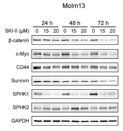
Western blot analysis of Molm13 cells after treatment with the indicated concentrations of SKI-II for 24, 48, and 72 h.
SKI II purchased from MedChemExpress. Usage Cited in: Cell Commun Signal. 2024 Aug 7;22(1):391. [Abstract]
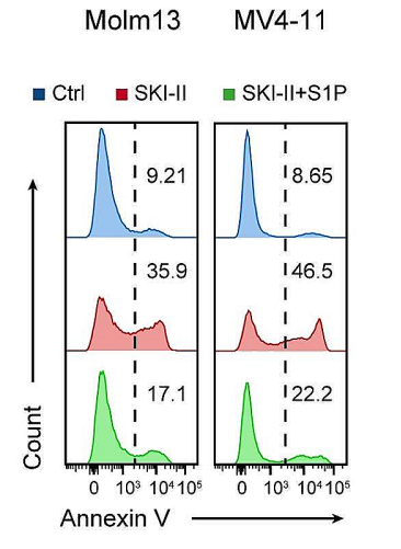
Apoptosis in Molm13 and MV4-11 cells pretreated with or without exogenous BSA-conjugated S1P (1 µM, added once every 3 h for 24 h) for 2 h, followed by treatment with SKI-II (25 µM) for 24 h.
-
Mol Med Rep
Notoginsenoside R1 ameliorates the inflammation induced by amyloid‑β by suppressing SphK1‑mediated NF‑κB activation in PC12 cells. [Abstract]2024 Jan;29(1):16. PMID: 38063180 -
J Virol
Epithelial-to-mesenchymal transition and live cell extrusion contribute to measles virus release from human airway epithelia. [Abstract]2025 Feb 25;99(2):e0122024. PMID: 39791903 -
Cell Biol Int
Bone marrow mesenchymal stem cells-derived exosomes reduce Aβ deposition and improve cognitive function recovery in mice with Alzheimer's disease by activating sphingosine kinase/sphingosine-1-phosphate signaling pathway. [Abstract]2021 Apr;45(4):775-784. PMID: 33300254 -
J Leukoc Biol
Microvesicles derived from activated T cells promote human mast cell migration via the S1P1 receptor. [Abstract]2025 Aug 5;117(8):qiaf111. PMID: 40743264 -


Solvent & Solubility
In Vitro:
DMSO : ≥ 100 mg/mL (330.27 mM; Hygroscopic DMSO has a significant impact on the solubility of product, please use newly opened DMSO)
* "≥" means soluble, but saturation unknown.
Please refer to the solubility information to select the appropriate solvent. The compound is unstable in solutions, freshly prepared is recommended.
Please refer to the solubility information to select the appropriate solvent. The compound is unstable in solutions, freshly prepared is recommended.
Concentration (start) × Volume (start) = Concentration (final) × Volume (final)
In Vivo:
Select the appropriate dissolution method based on your experimental animal and administration route.
- For the following dissolution methods, please ensure to first prepare a clear stock solution using an In Vitro approach and then sequentially add co-solvents:
- To ensure reliable experimental results, the clarified stock solution can be appropriately stored based on storage conditions. As for the working solution for In Vivo experiments, it is recommended to prepare freshly and use it on the same day.
- The percentages shown for the solvents indicate their volumetric ratio in the final prepared solution. If precipitation or phase separation occurs during preparation, heat and/or sonication can be used to aid dissolution.
Add each solvent one by one: 10% DMSO 40% PEG300 5% Tween-80 45% Saline
Solubility: ≥ 2.75 mg/mL (9.08 mM); Clear solution
This protocol yields a clear solution of ≥ 2.75 mg/mL (saturation unknown).
Taking 1 mL working solution as an example, add 100 μL DMSO stock solution (27.5 mg/mL) to 400 μL PEG300, and mix evenly; then add 50 μL Tween-80 and mix evenly; then add 450 μL Saline to adjust the volume to 1 mL.
Preparation of Saline: Dissolve 0.9 g sodium chloride in ddH₂O and dilute to 100 mL to obtain a clear Saline solution.
Add each solvent one by one: 10% DMSO 90% (20% SBE-β-CD in Saline)
Solubility: ≥ 2.75 mg/mL (9.08 mM); Clear solution
This protocol yields a clear solution of ≥ 2.75 mg/mL (saturation unknown).
Taking 1 mL working solution as an example, add 100 μL DMSO stock solution (27.5 mg/mL) to 900 μL 20% SBE-β-CD in Saline, and mix evenly.
Preparation of 20% SBE-β-CD in Saline (4°C, storage for one week): 2 g SBE-β-CD powder is dissolved in 10 mL Saline, completely dissolve until clear.
In Vivo Dissolution Calculator
Please enter the basic information of animal experiments:
-
-
-
-
Recommended: Prepare an additional quantity of animals to account for potential losses during experiments.
Please enter your animal formula composition:
-
%DMSO +
Recommended: Keep the proportion of DMSO in working solution below 2% if your animal is weak.
-
%+
-
+%Tween-80 + +
-
%Saline +
The co-solvents required include: DMSO, . All of co-solvents are available by MedChemExpress (MCE). , Tween 80. All of co-solvents are available by MedChemExpress (MCE).
Working solution concentration: 0.22 mg/mL
Method for preparing stock solution: mg drug dissolved in μL DMSO. Stock solution concentration: mg/mL. * The compound is unstable in solutions, freshly prepared is recommended.
1. Take μL DMSO stock solution;
2. Add μL .
μL , mix evenly;
3. Then add μL Tween 80, mix evenly;
4. Then add μL
Please ensure that the stock solution in the first step is dissolved to a clear state, and add co-solvents in sequence. You can use ultrasonic heating (ultrasonic cleaner, recommended frequency 20-40 kHz), vortexing, etc. to assist dissolution.


Purity & Documentation
-
Data Sheet (318 KB)
-
SDS (392 KB)
- English - EN (392 KB)
- Français - FR (392 KB)
- Deutsch - DE (392 KB)
- Norwegian - NO (392 KB)
- Español - ES (392 KB)
- Swedish - SV (392 KB)
- Italian - IT (392 KB)
- Korean - KR (392 KB)
- Portuguese - PT (392 KB)
-
Handling Instructions (2659 KB)
References
[1]. Liu Y, et al. SKI-II reverses the chemoresistance of SGC7901/DDP gastric cancer cells. Oncology letters. 2014 Jul;8(1):367-373. [Content Brief]
[8]. Potì F, et al. SKI-II--a sphingosine kinase 1 inhibitor--exacerbates atherosclerosis in low-density lipoprotein receptor-deficient (LDL-R-/-) mice on high cholesterol diet. Atherosclerosis. 2015 May;240(1):212-5. [Content Brief]


Complete Stock Solution Preparation Table
Please refer to the solubility information to select the appropriate solvent. The compound is unstable in solutions, freshly prepared is recommended.
| Optional Solvent | Concentration Solvent Mass | 1 mg | 5 mg | 10 mg | 25 mg |
|---|---|---|---|---|---|
| DMSO | 1 mM | 3.3027 mL | 16.5136 mL | 33.0273 mL | 82.5682 mL |
| 5 mM | 0.6605 mL | 3.3027 mL | 6.6055 mL | 16.5136 mL | |
| 10 mM | 0.3303 mL | 1.6514 mL | 3.3027 mL | 8.2568 mL | |
| 15 mM | 0.2202 mL | 1.1009 mL | 2.2018 mL | 5.5045 mL | |
| 20 mM | 0.1651 mL | 0.8257 mL | 1.6514 mL | 4.1284 mL | |
| 25 mM | 0.1321 mL | 0.6605 mL | 1.3211 mL | 3.3027 mL | |
| 30 mM | 0.1101 mL | 0.5505 mL | 1.1009 mL | 2.7523 mL | |
| 40 mM | 0.0826 mL | 0.4128 mL | 0.8257 mL | 2.0642 mL | |
| 50 mM | 0.0661 mL | 0.3303 mL | 0.6605 mL | 1.6514 mL | |
| 60 mM | 0.0550 mL | 0.2752 mL | 0.5505 mL | 1.3761 mL | |
| 80 mM | 0.0413 mL | 0.2064 mL | 0.4128 mL | 1.0321 mL | |
| 100 mM | 0.0330 mL | 0.1651 mL | 0.3303 mL | 0.8257 mL |


SKI II Related Classifications
HY-13822 Related Classifications
 Powered by Bioz
Keywords
- SKI II
- 312636-16-1
- SphK
- Dihydroceramide Desaturase 1 (DES1)
- Apoptosis
- Reactive Oxygen Species (ROS)
- mTOR
- PERK
- Keap1-Nrf2
- dihydroceramide desaturase 1
- sphingosine kinase
- Staphylococcus aureus
- human VCP
- acute myeloid leukemia
- mouse RAW264.7 macrophages
- SKN-1/Nrf2 pathway
- Caenorhabditis elegans
- gastric carcinoma
- glioblastoma stem cell
- Inhibitor
- inhibitor
- inhibit