



CDK-IN-9
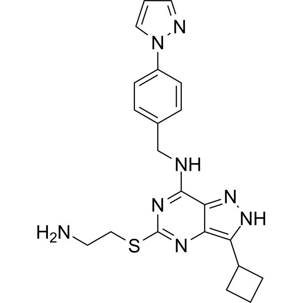
CDK-IN-9 (compound 24) is a potent CDK inhibitor, also as a molecular glue inducing an interaction between CDK12 and DDB1, with an IC50 values of 4 nM for CDK2/E. CDK-IN-9 leads to polyubiquitination of cyclin K and its subsequent degradation. CDK-IN-9 induce apoptosis through dephosphorylation of retinoblastoma protein and RNA polymerase II.
Nos produits utilisent uniquement pour la recherche. Nous ne vendons pas aux patients.
- CAS No.: 3031561-92-6
- Formule: C21H24N8S
- Masse moléculaire:420.53
-
Stockage:
Please store the product under the recommended conditions in the Certificate of Analysis.

To place orders, for customer services and technical support, please contact: MedChemExpress USA
Tel: 609-228-6898 E-mail: [email protected] [email protected]


-
 Activité biologique
-
Chemical Information
- Protocole
- Pureté et documentation
- Références
-
 Help & FAQs


Voir tous les produits spécifiques à Isoform DNA/RNA Synthesis
More

Activité biologique
Description
IC50 & Target
[1]|
RNA Polymerase |
CDK2/E 4 nM (IC50) |
Cdk5/p25 39 nM (IC50) |
CDK9/T1 20 nM (IC50) |
CDK12/K 64 nM (IC50) |
CDK13/K 22 nM (IC50) |
Cellular Effect
|
Cell Line

|
Type | Value | Description | References |
|---|---|---|---|---|
| DB | GI50 |
0.02 μM
Compound: 24
|
Antiproliferative activity against human DB cells assessed as cell growth inhibition measured after 72 hrs by resazurin dye based microplate reader analysis
Antiproliferative activity against human DB cells assessed as cell growth inhibition measured after 72 hrs by resazurin dye based microplate reader analysis
|
[PMID: 35749742] |
| NU-DUL-1 | GI50 |
0.015 μM
Compound: 24
|
Antiproliferative activity against human NU-DUL-1 cells assessed as cell growth inhibition measured after 72 hrs by resazurin dye based microplate reader analysis
Antiproliferative activity against human NU-DUL-1 cells assessed as cell growth inhibition measured after 72 hrs by resazurin dye based microplate reader analysis
|
[PMID: 35749742] |
| OCI-LY19 | GI50 |
0.465 μM
Compound: 24
|
Antiproliferative activity against human OCILY19 cells assessed as cell growth inhibition measured after 72 hrs by resazurin dye based microplate reader analysis
Antiproliferative activity against human OCILY19 cells assessed as cell growth inhibition measured after 72 hrs by resazurin dye based microplate reader analysis
|
[PMID: 35749742] |
| SUD4 | GI50 |
0.028 μM
Compound: 24
|
Antiproliferative activity against human SU-DHL-4 cells assessed as cell growth inhibition measured after 72 hrs by resazurin dye based microplate reader analysis
Antiproliferative activity against human SU-DHL-4 cells assessed as cell growth inhibition measured after 72 hrs by resazurin dye based microplate reader analysis
|
[PMID: 35749742] |
| U2932 | GI50 |
0.055 μM
Compound: 24
|
Antiproliferative activity against human U2932 cells assessed as cell growth inhibition measured after 72 hrs by resazurin dye based microplate reader analysis
Antiproliferative activity against human U2932 cells assessed as cell growth inhibition measured after 72 hrs by resazurin dye based microplate reader analysis
|
[PMID: 35749742] |
In Vitro
CDK-IN-9 (compound 24) (0.005, 0.05, 0.5, or 5 μM; 2 h) potently decreases the level of cyclin K in MINO cells at 5 nM, and makes cyclin K disappear completely at 50 nM[1].
CDK-IN-9 makes siRNA silencing of DDB1 effectively stabilizes cyclin K at the protein level in treated MINO cells[1].
CDK-IN-9 (2.5-40 nM; 24 h) activates caspases 3/7/9 and decreases anti-apoptotic proteins Mcl-1 and XIAP in MINO cells[1].
MedChemExpress (MCE) has not independently confirmed the accuracy of these methods. They are for reference only.
In Vivo
MedChemExpress (MCE) has not independently confirmed the accuracy of these methods. They are for reference only.


Chemical Information
-
CAS No. 3031561-92-6
-
Masse moléculaire 420.53
-
Formule C21H24N8S
-
SMILES
NCCSC1=NC2=C(NN=C2C(NCC3=CC=C(C=C3)N4C=CC=N4)=N1)C5CCC5
-
Livraison
Room temperature in continental US; may vary elsewhere.
-
Stockage
Please store the product under the recommended conditions in the Certificate of Analysis.


Pureté et documentation
Références

Calculators
Concentration (start) × Volume (start) = Concentration (final) × Volume (final)


CDK-IN-9 Related Classifications
HY-150641 Related Classifications
 Powered by Bioz