



Chondroitin sulfate (from fish)
Chondroitin sulfate (from fish), one of five classes of glycosaminoglycans, has been widely used in the treatment of osteoarthritis. Chondroitin sulfate (from fish) reduces inflammation mediators and the apoptotic process and is able to reduce protein production of inflammatory cytokines, iNOS and MMPs.
For research use only. We do not sell to patients.
- CAS No.: 9007-28-7
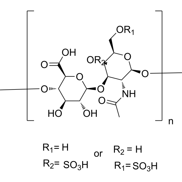
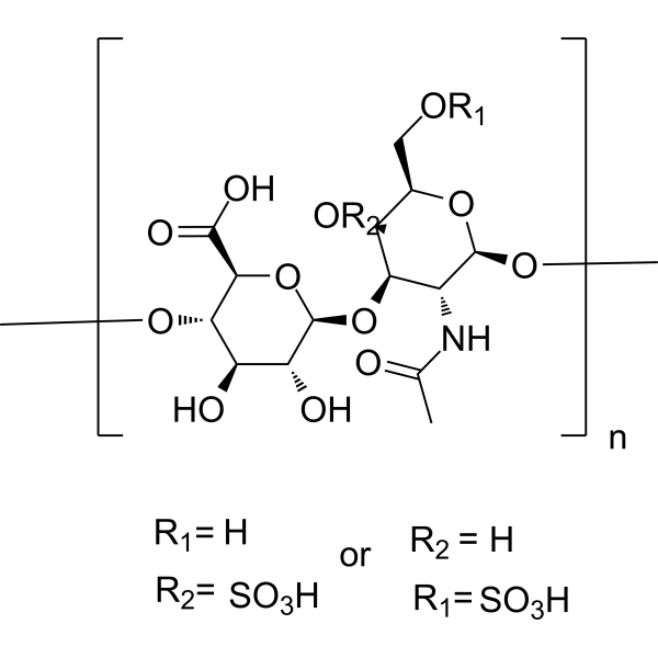
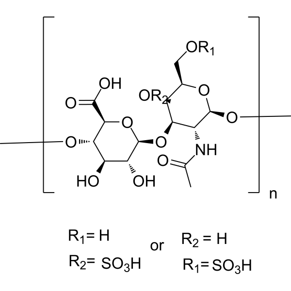
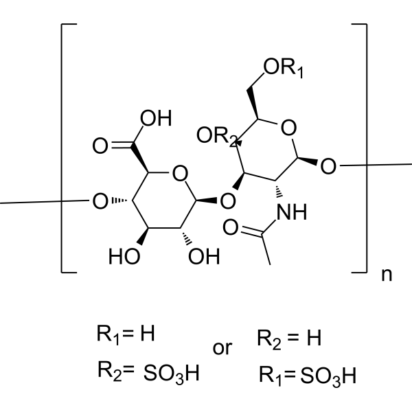
- Formula: (C14H21NO14S)n
-
Storage:
Please store the product under the recommended conditions in the Certificate of Analysis.


To place orders, for customer services and technical support, please contact: MedChemExpress USA
Tel: 609-228-6898 E-mail: [email protected] [email protected]


-
 Biological Activity
-
Chemical Information
- Protocols
- Purity & Documentation
- References
-
 Help & FAQs


-
HY-B2162
Chondroitin sulfateGlycosaminoglycan -
HY-B2162D
Chondroitin sulfate (from chicken)Sulfated Linear Polysaccharide -
HY-B2162C
Chondroitin sulfate (from pig)Sulfated Linear Polysaccharide -
HY-B2162E
Chondroitin sulfate (from shark cartilage)Biochemical Reagent -
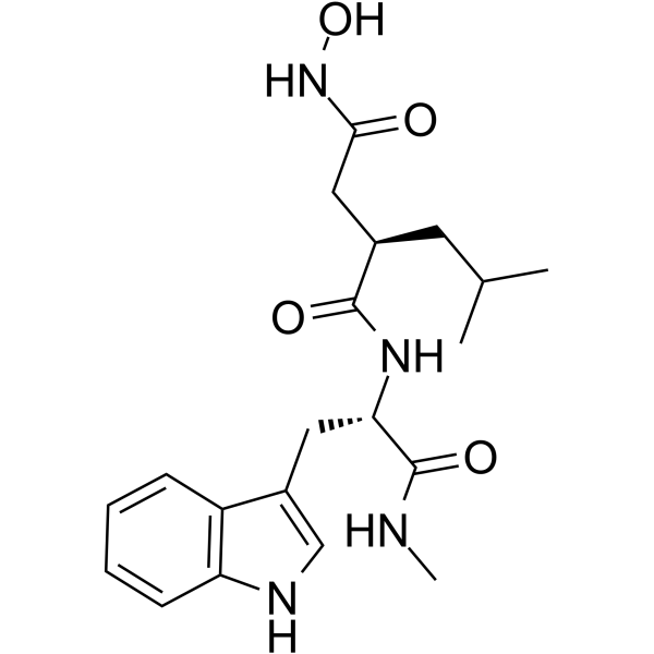
HY-15768
IlomastatMMP Inhibitor -
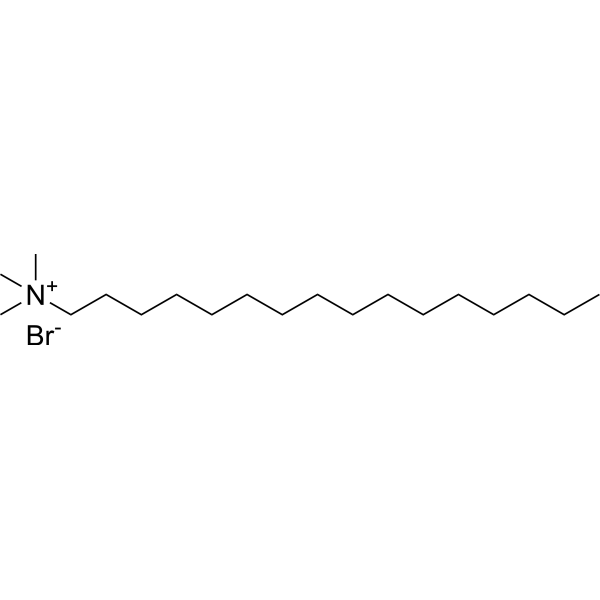
HY-B1260
Cetrimonium bromideCationic Surfactant -
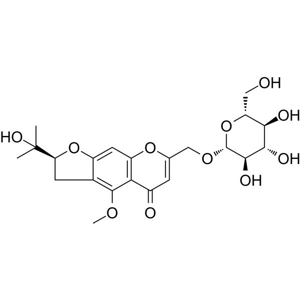
HY-N0635
Prim-O-glucosylcimifuginCOX Inhibitor -
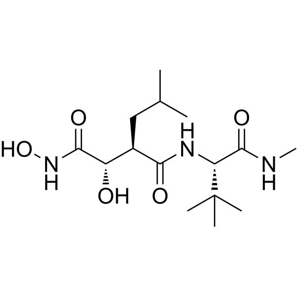
HY-12169
MarimastatMMP Inhibitor -
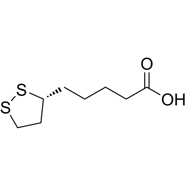
HY-18733
Lipoic acidAntioxidant -
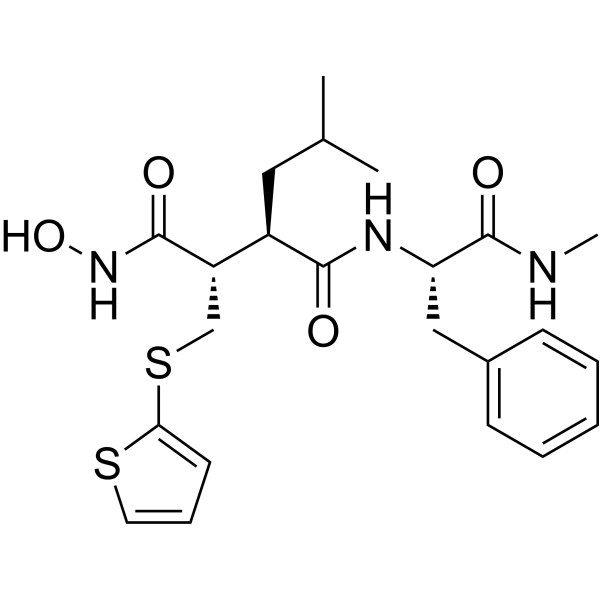
HY-13564
BatimastatMMP Inhibitor -
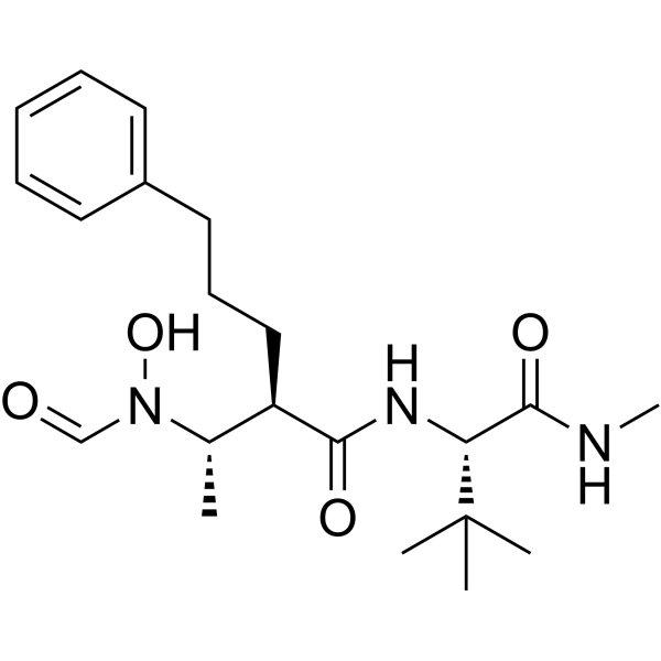
HY-19956
GI254023XADAM10/MMP9 Inhibitor -
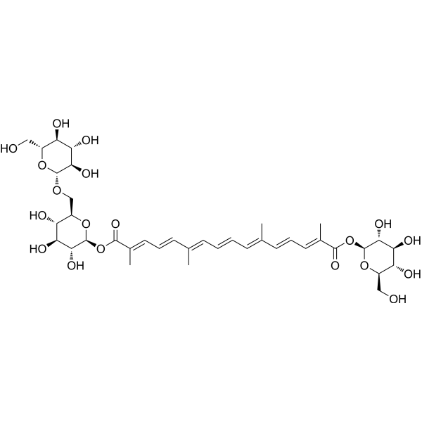
HY-N0698
Crocin IIα-synuclein Inhibitor
All Endogenous Metabolite Isoforms
More

Biological Activity
Description


Chemical Information
-
CAS No. 9007-28-7
-
Formula (C14H21NO14S)n
-
SMILES
O=S(O)=O.O=S(O)=O.OC([C@@H]1[C@@H](OC)[C@H](O)[C@@H](O)[C@H](O[C@@H]2[C@@H](NC(C)=O)[C@H](OC)O[C@H](CO[R])[C@@H]2O[R])O1)=O.[R].[R].[=].[=].[R2=].[R1=].[n].[or]
-
Synonyms
Chondroitin polysulfate (from fish)
-
Shipping
Room temperature in continental US; may vary elsewhere.
-
Storage
Please store the product under the recommended conditions in the Certificate of Analysis.

Protocols
-
Apoptosis
Apoptosis, also called programmed cell death, is generally characterized by distinct morphological characteristics.
-
LPS-Induced Endotoxemia/Systemic Inflammation
Lipopolysaccharide (LPS)-induced endotoxemia is a widely used in vivo model of acute systemic inflammation in which LPS, a Gram-negative bacterial endotoxin, activates innate immune signaling primarily through TLR4, leading to rapid and transient induction of pro-inflammatory cytokines such as TNF-α, IL-6, and IL-1β in circulation and tissues. This cytokine surge is commonly used as a measurable readout of systemic inflammatory activation and immune dysregulation, and is typically assessed within hours after intraperitoneal LPS administration in mouse models of endotoxemia. The model captures key features of systemic inflammatory response syndrome, including cytokine release, immune cell activation, and downstream tissue responses, and has been used to evaluate anti-inflammatory interventions such as cytokine modulation, lipid mediators, and immune cell-targeting therapies.
-
Protocol for Fluorescence In Situ Hybridization (FISH)
Fluorescence in situ hybridization detects specific DNA or RNA sequences inside fixed cells or tissue sections by hybridizing fluorescently labeled nucleic-acid probes to complementary target sequences, allowing the target’s copy number, chromosomal position, spatial distribution, or transcript abundance to be visualized microscopically. DNA-FISH detects genomic loci, chromosomal gains/losses, amplifications, deletions, and rearrangements, while RNA-FISH detects RNA molecules or transcript localization; in cancer cells, mouse tumors, neurons, organoids, macrophages, or drug-screening samples, the readout is fluorescent puncta, fusion/split signals, or localized RNA signal interpreted relative to validated controls.
-
Apoptosis Solutions
Apoptosis is a regulated, generally non-lytic cell-death pathway that removes unwanted, damaged, infected, or abnormal cells through coordinated morphological changes, caspase activation, DNA fragmentation, and membrane remodeling. The intrinsic apoptosis pathway is controlled mainly by mitochondrial outer membrane permeabilization, BCL-2 family proteins, cytochrome c release, apoptosome formation, caspase-9 activation, and downstream executioner caspase-3/7 activation. The extrinsic apoptosis pathway is initiated by death receptors such as Fas, TNFR, and TRAIL receptors, which recruit adaptor proteins and activate caspase-8 before engaging executioner caspases or mitochondrial amplification through BID cleavage. Apoptosis is linked to many phenotypes, including cancer cell killing, tissue homeostasis, immune regulation, neurodegeneration, infection response, and treatment-induced cytotoxicity; unresolved questions include how apoptosis interacts with necroptosis, pyroptosis, ferroptos
-
Research Protocol for Inflammation-related Diseases
The NLRP3 inflammasome is a cytosolic innate immune signaling platform that integrates priming signals and danger-signal activation to promote caspase-1 activation, maturation of IL-1β and IL-18, and gasdermin D-mediated pyroptotic cell death. The core experimental logic is to determine whether inflammatory disease phenotypes are driven by increased NLRP3 expression, ASC-containing inflammasome assembly, caspase-1 cleavage, GSDMD cleavage, and extracellular release of IL-1β/IL-18 rather than by nonspecific cell injury alone. The pathway is strongly linked to inflammation-related disease phenotypes because monosodium urate crystals activate NALP3/NLRP3 inflammasome signaling in gout-like crystal inflammation, cholesterol crystals activate NLRP3 inflammasomes in atherogenesis models, and DSS-induced intestinal inflammation has been reported to involve NLRP3 inflammasome activity. However, experimental colitis studies also show context-dependent protective effects of NLRP3 inflammasome co
-
Protocol For Protein Expression And Purification
Recombinant protein expression in Escherichia coli followed by purification of a His-tagged soluble protein by immobilized metal affinity chromatography (IMAC), with optional MBP fusion and TEV tag removal when the construct includes these elements. The biological readout is production of the encoded target protein, detected as an inducible band at the expected molecular mass by SDS-PAGE and quantified by total protein assay or chromatographic absorbance; the purification readout is enrichment of the target protein in elution fractions after selective binding of polyhistidine residues to immobilized Ni2+/metal-chelate resin and elution by imidazole-containing buffer. Expression is driven by an inducible bacterial expression system, commonly T7/lac-based, in which IPTG or lactose/auto-induction activates transcription and translation of the cloned gene; lower induction temperature, lower inducer concentration, induction timing, and solubility-enhancing fusion tags can influence the frac


Purity & Documentation
References
[1]. Mikami T, et al. Biosynthesis and function of chondroitin sulfate. Biochim Biophys Acta. 2013 Oct;1830(10):4719-33. [Content Brief]
[2]. Monfort J, et al. Biochemical basis of the effect of chondroitin sulphate on osteoarthritis articular tissues. Ann Rheum Dis. 2008 Jun;67(6):735-40. [Content Brief]
[3]. Martel-Pelletier J, et al.Discrepancies in composition and biological effects of different formulations of chondroitin sulfate. Molecules. 2015 Mar 6;20(3):4277-89. [Content Brief]
[4]. Campo GM, et al. Glycosaminoglycans modulate inflammation and apoptosis in LPS-treated chondrocytes. J Cell Biochem. 2009 Jan 1;106(1):83-92. [Content Brief]
[5]. Henrotin Y, et al. Chondroitin sulfate in the treatment of osteoarthritis: from in vitro studies to clinicalrecommendations. Ther Adv Musculoskelet Dis. 2010 Dec;2(6):335-48. [Content Brief]

Calculators
Concentration (start) × Volume (start) = Concentration (final) × Volume (final)


Chondroitin sulfate (from fish) Related Classifications
HY-B2162H Related Classifications
 Powered by Bioz