



LL-K8-22

Based on 1 Customer Validation
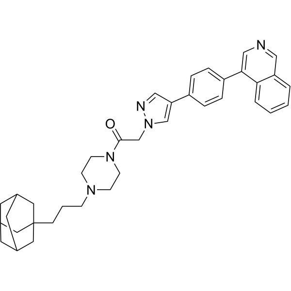
LL-K8-22 is a dual degrader of CDK8 and cyclin C HyT, with DC50 values of 2.52 μM and 2.64 μM, respectively. LL-K8-22 inhibits phosphorylation of STAT1 Ser 727, phosphorylation of the C-terminal domain Ser 2/5 of RNA polymerase II, and phosphorylation of Rb. LL-K8-22 represses E2F- and MYC-driven transcriptional programs and exhibits antiproliferative effects. LL-K8-22 synchronously induces proteasome-dependent selective degradation of CDK8 and cyclin C without degrading CDK19, other CDK family members, or other cyclins. LL-K8-22 can be used in the research of triple-negative breast cancer and colon cancer.
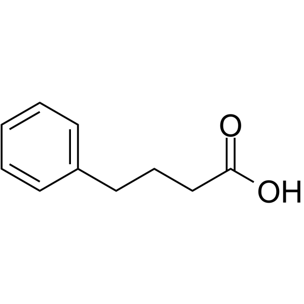
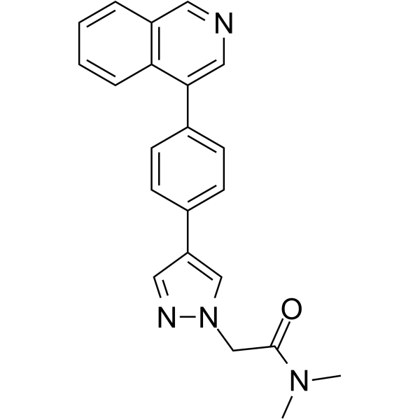
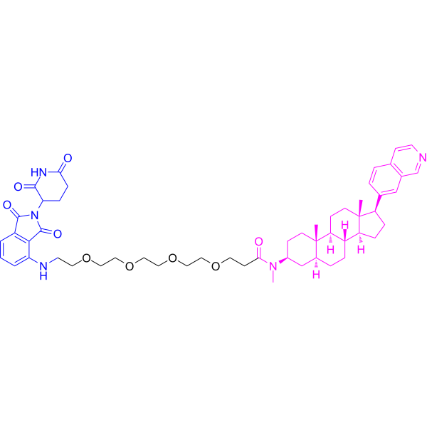
(Pink: CDK8 ligand (HY-168683); Blue: hyt ligand (HY-N2427); Black: linker).
Nur für Forschungszwecke. Wir verkaufen nicht an Patienten.
- Reinheit : 99.06%
- CAS. Nr.: 3034487-84-5
- Formel: C37H43N5O
- Molecular Weight:573.77
-
Speicherung:Powder -20°C, 3 years ; In solvent -80°C, 6 months , -20°C, 1 month

To place orders, for customer services and technical support, please contact: MedChemExpress USA
Tel: 609-228-6898 E-mail: [email protected] [email protected]


-
 Biologische Aktivität
-
Chemical Information
-
Lösungsmittel & Löslichkeit
- Protokoll
- Reinheit & Dokumentation
- Verweise
-
 Help & FAQs




Biologische Aktivität
Beschreibung
IC50 & Target
[1]|
CDK8 2.52 μM (DC50) |
cyclin C 2.64 μM (DC50) |
STAT1 |
Rb |
E2F |
MYC |
Cellular Effect
|
Cell Line

|
Type | Value | Description | References |
|---|---|---|---|---|
| HCT-116 | IC50 |
5.87 μM
Compound: LL-K8-22
|
Antiproliferative activity against human HCT-116 cells assessed as inhibition of cell proliferation incubated for 4 days by Celltiter glo luminescent assay
Antiproliferative activity against human HCT-116 cells assessed as inhibition of cell proliferation incubated for 4 days by Celltiter glo luminescent assay
|
[PMID: 36930701] |
| MDA-MB-231 | IC50 |
6.22 μM
Compound: LL-K8-22
|
Antiproliferative activity against human MDA-MB-231 cells assessed as inhibition of cell proliferation incubated for 4 days by Celltiter glo luminescent assay
Antiproliferative activity against human MDA-MB-231 cells assessed as inhibition of cell proliferation incubated for 4 days by Celltiter glo luminescent assay
|
[PMID: 36930701] |
| MDA-MB-468 | IC50 |
5.73 μM
Compound: LL-K8-22
|
Antiproliferative activity against human MDA-MB-468 cells assessed as inhibition of cell proliferation incubated for 4 days by Celltiter glo luminescent assay
Antiproliferative activity against human MDA-MB-468 cells assessed as inhibition of cell proliferation incubated for 4 days by Celltiter glo luminescent assay
|
[PMID: 36930701] |
In Vitro
LL-K8-22 (0.625-10 μM; 0-72 h) induces effective, dose-dependent, simultaneous, and durable degradation of CDK8 and cyclin C in HCT-116 and MDA-MB-231 cells, with no hook effect[1].
LL-K8-22 (1.1-10 μM) does not alter CDK8 or cyclin C mRNA levels but downregulates CDK1 mRNA levels in MDA-MB-468 cells[1].
LL-K8-22 (5 μM; 12 h) selectively reduces CDK8 and cyclin C protein levels (without affecting CDK19) and downregulates CDK8 downstream targets in MDA-MB-468 cells, with affected proteins enriched in cancer-relevant pathways[1].
LL-K8-22 binds to the CDK8-cyclin C complex such that its hydrophobic tag occupies a positively charged cavity on CDK8, potentially destabilizing the complex to enable degradation[1].
LL-K8-22 (5-20 μM; up to 5 days) exerts greater anti-proliferative effects on MDA-MB-468 cells than BI-1347 over a 5-day period at concentrations of 5, 10, and 20 μM[1].
LL-K8-22 (1.25-20 μM; 3 weeks) potently inhibits colony formation of MDA-MB-468 cells at concentrations from 1.25 to 20 μM, with greater activity than BI-1347[1].
LL-K8-22 (4 μM; 24 h) represses E2F- and MYC-driven carcinogenic transcriptional programs and downregulates cancer-associated genes more significantly than BI-1347 in MDA-MB-468 cells, without altering CDK8 or cyclin C mRNA levels[1].
MedChemExpress (MCE) has not independently confirmed the accuracy of these methods. They are for reference only.
-
Cell Line:MDA-MB-468 cells
-
Concentration:0.38-12 μM (dose-dependent degradation analysis); 0.625-10 μM (24 h incubation); 1.1, 3.3, 10 μM (48 h incubation; 24 h incubation with MG132 co-treatment); 3 μM (24 h incubation with 17-AAG co-treatment); 10 μM (0-20 h incubation with cycloheximide co-treatment); 5 μM (0-72 h incubation); 2-8 μM (24 h incubation); 4 μM (24 h incubation followed by wash-off and 0-24 h drug-free incubation)
-
Incubation Time:48 h; 24 h; 0-20 h (cycloheximide co-treatment); 0-72 h; 0-24 h (drug-free after wash-off)
-
Result:Induced dose-dependent degradation of CDK8 and cyclin C with DC50 values of 2.52 μM for CDK8 and 2.64 μM for cyclin C.
Did not reduce protein levels of CDK2, 4, 5, 6, 7, 9, 12, or 19 at 0.625-10 μM, and only slightly reduced CDK1 at 10 μM; did not degrade cyclin A2, B1, E1, H, K, or T1, only affecting cyclin C.
Accelerated degradation of CDK8 and cyclin C when co-treated with cycloheximide, while MG132 reversed this degradation; 17-AAG potentiated the degradation.
Induced simultaneous degradation of CDK8 and cyclin C over time, with significant degradation observed after 20-24 h of 5 μM treatment, nearly complete degradation after 36-48 h, and durable degradation still present at 72 h.
Reduced phosphorylation of STAT1 at Ser727 (without affecting Tyr701), phosphorylation of Rpb1 CTD at Ser2 and Ser5, and phosphorylation of Rb at Ser780 in a dose-dependent manner, with more pronounced effects than BI-1347.
Maintained ~70% of maximum inhibition of STAT1 Ser727 phosphorylation for up to 24 h after wash-off of 4 μM treatment, while BI-1347-mediated inhibition reversed by 12 h post-wash-off.
-
Cell Line:MDA-MB-231 cells
-
Concentration:1.1 μM; 3.3 μM; 10 μM
-
Incubation Time:24 h
-
Result:Did not alter CDK8 or cyclin C mRNA levels but downregulates CDK1 mRNA levels in MDA-MB-468 cells.
-
Cell Line:HCT-116 and MDA-MB-231 cells
-
Concentration:0.625-10 μM (48 h incubation); 5 μM (0-72 h incubation)
-
Incubation Time:48 h; 0-72 h
-
Result:Effectively degraded CDK8 and cyclin C in a dose-dependent manner, with no observed hook effect.
Induced simultaneous, durable degradation of CDK8 and cyclin C over time, similar to MDA-MB-468 cells.
-
Cell Line:MDA-MB-468, HCT-116, and MDA-MB-231 cells
-
Concentration:Gradient concentrations
-
Incubation Time:4 days
-
Result:Exhibited potent antiproliferative activity with IC50 values of 5.73 μM, 5.87 μM, and 6.22 μM for MDA-MB-468, HCT-116, and MDA-MB-231 cells, respectively.
Was 5.03-, 5.61-, and 4.61-fold more potent than BI-1347.
-
Cell Line:MDA-MB-468 cells
-
Concentration:5 μM, 10 μM, 20 μM
-
Incubation Time:up to 5 days
-
Result:Significantly reduced MDA-MB-468 cell growth and viability over 5 days at all tested concentrations, with more pronounced effects than BI-1347.
-
Cell Line:MDA-MB-468 cells
-
Concentration:1.25 μM, 2.5 μM, 5 μM, 10 μM, 20 μM
-
Incubation Time:3 weeks
-
Result:Significantly reduced the total number and size of MDA-MB-468 cell colonies at all tested concentrations, with strong inhibition even at 1.25 μM, and more potent effects than BI-1347.


Chemical Information
-
CAS. Nr. 3034487-84-5
-
Appearance Solid
-
Molecular Weight 573.77
-
Formel C37H43N5O
-
Color White to off-white
-
SMILES
O=C(CN1C=C(C2=CC=C(C3=CN=CC4=CC=CC=C43)C=C2)C=N1)N5CCN(CCCC67CC8CC(CC(C8)C7)C6)CC5
-
Versand
Room temperature in continental US; may vary elsewhere.
-
Speicherung
Powder -20°C 3 years In solvent -80°C 6 months -20°C 1 month


Lösungsmittel & Löslichkeit
In Vitro:
DMSO : 125 mg/mL (217.86 mM; Need ultrasonic; Hygroscopic DMSO has a significant impact on the solubility of product, please use newly opened DMSO)
Please refer to the solubility information to select the appropriate solvent. Once prepared, please aliquot and store the solution to prevent product inactivation from repeated freeze-thaw cycles.
Storage method and period of stock solution: -80°C, 6 months; -20°C, 1 month. When stored at -80°C, please use it within 6 months. When stored at -20°C, please use it within 1 month.
Please refer to the solubility information to select the appropriate solvent. Once prepared, please aliquot and store the solution to prevent product inactivation from repeated freeze-thaw cycles.
Storage method and period of stock solution: -80°C, 6 months; -20°C, 1 month. When stored at -80°C, please use it within 6 months. When stored at -20°C, please use it within 1 month.
Konzentration (Stammlösung) × Volumen (Stammlösung) = Konzentration (Ziellösung) × Volumen (Ziellösung)


Reinheit & Dokumentation
-
Data Sheet (282 KB)
-
SDS (252 KB)
- English - EN (252 KB)
- Français - FR (252 KB)
- Deutsch - DE (252 KB)
- Norwegian - NO (252 KB)
- Español - ES (252 KB)
- Swedish - SV (252 KB)
- Italian - IT (252 KB)
- Korean - KR (252 KB)
- Portuguese - PT (252 KB)
-
Handling Instructions (2659 KB)
Verweise


Complete Stock Solution Preparation Table
Please refer to the solubility information to select the appropriate solvent. Once prepared, please aliquot and store the solution to prevent product inactivation from repeated freeze-thaw cycles.
Storage method and period of stock solution: -80°C, 6 months; -20°C, 1 month. When stored at -80°C, please use it within 6 months. When stored at -20°C, please use it within 1 month.
| Optional Solvent | Concentration Solvent Mass | 1 mg | 5 mg | 10 mg | 25 mg |
|---|---|---|---|---|---|
| DMSO | 1 mM | 1.7429 mL | 8.7143 mL | 17.4286 mL | 43.5715 mL |
| 5 mM | 0.3486 mL | 1.7429 mL | 3.4857 mL | 8.7143 mL | |
| 10 mM | 0.1743 mL | 0.8714 mL | 1.7429 mL | 4.3571 mL | |
| 15 mM | 0.1162 mL | 0.5810 mL | 1.1619 mL | 2.9048 mL | |
| 20 mM | 0.0871 mL | 0.4357 mL | 0.8714 mL | 2.1786 mL | |
| 25 mM | 0.0697 mL | 0.3486 mL | 0.6971 mL | 1.7429 mL | |
| 30 mM | 0.0581 mL | 0.2905 mL | 0.5810 mL | 1.4524 mL | |
| 40 mM | 0.0436 mL | 0.2179 mL | 0.4357 mL | 1.0893 mL | |
| 50 mM | 0.0349 mL | 0.1743 mL | 0.3486 mL | 0.8714 mL | |
| 60 mM | 0.0290 mL | 0.1452 mL | 0.2905 mL | 0.7262 mL | |
| 80 mM | 0.0218 mL | 0.1089 mL | 0.2179 mL | 0.5446 mL | |
| 100 mM | 0.0174 mL | 0.0871 mL | 0.1743 mL | 0.4357 mL |


LL-K8-22 Related Classifications
HY-149209 Related Classifications
 Powered by Bioz