



MI-192

Based on 1 Customer Validation
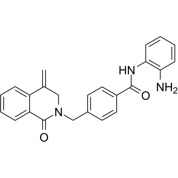
MI-192 is a selective HDAC2 and HDAC3 inhibitor with IC50s of 30 nM and 16 nM, respectively. MI-192 is more selective for HDAC2/3 than other HDAC isomers.MI-192 induces myeloid leukaemic cells apoptosis. Anticaner and neuroprotective activities.
商品は「研究用試薬」です。人や動物の医療用・臨床診断用・食品用の製品ではありません。
研究用途以外に使用した場合、当社は一切の責任を負いかねます。
- 純度 : 99.9%
- CAS 番号: 1415340-63-4
- 分子式: C24H21N3O2
- 分子量:383.44
-
保管条件:Powder -20°C, 3 years ; In solvent -80°C, 6 months , -20°C, 1 month

To place orders, for customer services and technical support, please contact: MedChemExpress USA
Tel: 609-228-6898 E-mail: [email protected] [email protected]


-
 生物活性
生物活性
-
化学情報
-
溶剤 & 溶解度
- プロトコル
- 純度とドキュメンテーション
- 参考文献
-
 Help & FAQs




生物活性
製品説明
体外実験
MI-192 (0.15-1 μM; 72 h) induces differentiation and is cytotoxic through promotion of apoptosis in acute myeloid leukaemic cell lines U937, HL60 and Kasumi-1[1].
MedChemExpress (MCE) has not independently confirmed the accuracy of these methods. They are for reference only.
-
Cell Line:HL60 and Kasumi-1 cells
-
Concentration:150 nM, 300 nM, 500 nM, 1 μM
-
Incubation Time:72 h
-
Result:Induced a substantial degree of apoptosis in both HL60 and Kasumi-1 cells.
体内実験
MedChemExpress (MCE) has not independently confirmed the accuracy of these methods. They are for reference only.
-
Animal Model:Adult male outbred mice CD-1 (20-25 g) with photothrombotic stroke (PTS)[2]
-
Dosage:40 mg/kg
-
Administration:i.p; once a day; for 3 days
-
Result:Reduced the volume of the PTS-induced infarction core in the mouse brain, partly restored the functional symmetry in the forelimb use, decreased the level of PTS-induced apoptosis and acetylation of α-tubulin characteristic for stable microtubules, and increased the expression of GAP-43 in the cerebral cortex of the damaged hemisphere.


化学情報
-
CAS 番号 1415340-63-4
-
性状 Solid
-
分子量 383.44
-
分子式 C24H21N3O2
-
Color Off-white to light yellow
-
SMILES
O=C1C2=CC=CC=C2C(CN1CC3=CC=C(C(NC4=C(C=CC=C4)N)=O)C=C3)=C
-
輸送条件
Room temperature in continental US; may vary elsewhere.
-
保管条件
Powder -20°C 3 years In solvent -80°C 6 months -20°C 1 month


溶剤 & 溶解度
体外:
DMSO : 5 mg/mL (13.04 mM; Need ultrasonic and warming; Hygroscopic DMSO has a significant impact on the solubility of product, please use newly opened DMSO)
Please refer to the solubility information to select the appropriate solvent. Once prepared, please aliquot and store the solution to prevent product inactivation from repeated freeze-thaw cycles.
Storage method and period of stock solution: -80°C, 6 months; -20°C, 1 month. When stored at -80°C, please use it within 6 months. When stored at -20°C, please use it within 1 month.
Please refer to the solubility information to select the appropriate solvent. Once prepared, please aliquot and store the solution to prevent product inactivation from repeated freeze-thaw cycles.
Storage method and period of stock solution: -80°C, 6 months; -20°C, 1 month. When stored at -80°C, please use it within 6 months. When stored at -20°C, please use it within 1 month.
濃度 (開始) × 体積 (開始) = 濃度 (終了) × 体積 (終了)


純度とドキュメンテーション
-
データシート (274 KB)
-
SDS (394 KB)
- English - EN (394 KB)
- Français - FR (394 KB)
- Deutsch - DE (394 KB)
- Norwegian - NO (394 KB)
- Español - ES (394 KB)
- Swedish - SV (394 KB)
- Italian - IT (394 KB)
- Korean - KR (394 KB)
- Portuguese - PT (394 KB)
-
取扱説明書 (2659 KB)
参考文献
[1]. Marjorie Boissinot, et al. Induction of differentiation and apoptosis in leukaemic cell lines by the novel benzamide family histone deacetylase 2 and 3 inhibitor MI-192. Leuk Res. 2012 Oct;36(10):1304-10. [Content Brief]
[2]. S V Demyanenko, et al. The Neuroprotective Effect of the HDAC2/3 Inhibitor MI192 on the Penumbra After Photothrombotic Stroke in the Mouse Brain. Mol Neurobiol. 2020 Jan;57(1):239-248. [Content Brief]


Complete Stock Solution Preparation Table
Please refer to the solubility information to select the appropriate solvent. Once prepared, please aliquot and store the solution to prevent product inactivation from repeated freeze-thaw cycles.
Storage method and period of stock solution: -80°C, 6 months; -20°C, 1 month. When stored at -80°C, please use it within 6 months. When stored at -20°C, please use it within 1 month.
| Optional Solvent | Concentration Solvent Mass | 1 mg | 5 mg | 10 mg | 25 mg |
|---|---|---|---|---|---|
| DMSO | 1 mM | 2.6080 mL | 13.0398 mL | 26.0797 mL | 65.1992 mL |
| 5 mM | 0.5216 mL | 2.6080 mL | 5.2159 mL | 13.0398 mL | |
| 10 mM | 0.2608 mL | 1.3040 mL | 2.6080 mL | 6.5199 mL |


MI-192 関連分類
HY-110264 関連分類
 Powered by Bioz