



NR-7h

Based on 1 Customer Validation
NR-7h is a selective p38α/p38β MAPK PROTAC degrader with multiple activities including anti-leishmanial, anti-malarial, and anti-Mayaro virus properties. NR-7h induces specific degradation of p38α/p38β isoforms and p38-MAPK via the ubiquitin-proteasome system. NR-7h reduces the load of Leishmania donovani, modulates the cytokine profile toward a pro-inflammatory phenotype, and enhances the oxidative burst of macrophages. NR-7h inhibits the growth of Plasmodium falciparum in human red blood cells and merozoite invasion. NR-7h reduces the replication of Mayaro virus and the expression of E1 protein in primary human dermal fibroblasts. NR-7h can be used in studies related to parasitic infections, viral infections, and breast cancer.
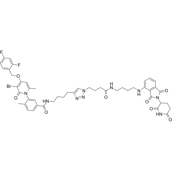
(Pink: p38α and p38β ligand (HY-403098); Blue: Cereblon ligand (HY-103596); Black: linker).
Nos produits utilisent uniquement pour la recherche. Nous ne vendons pas aux patients.
- Pureté : 98.31%
- CAS No.: 2550399-06-7
- Formule: C48H50BrF2N9O8
- Masse moléculaire:998.87
-
Stockage:Powder -20°C, 3 years ; In solvent -80°C, 6 months , -20°C, 1 month

To place orders, for customer services and technical support, please contact: MedChemExpress USA
Tel: 609-228-6898 E-mail: [email protected] [email protected]


-
 Activité biologique
-
Chemical Information
- Protocole
- Pureté et documentation
- Références
-
 Help & FAQs


Voir tous les produits spécifiques à Isoform PROTACs
MoreVoir tous les produits spécifiques à Isoform Parasite
More

Activité biologique
Description
|
p38α |
p38β |
p38 MAPK |
Leishmania |
Plasmodium |
In Vitro
NR-7h (0.01-10 μM; 1-24 h) specifically degrades p38-MAPK in PMA-differentiated human THP-1 macrophages in a concentration- and time-dependent manner via the ubiquitin-proteasome pathway, with a DC50 of 162.9 nM[1].
NR-7h specifically degrades p38-MAPK in human primary erythrocytes via the ubiquitin-proteasome pathway, and exerts no effect on ERK1/2 levels at a concentration of 1 μM[1].
NR-7h (0.05-1 μM; 2-24 h) rapidly and potently induces the degradation of p38α and p38β isoforms in primary human dermal fibroblasts, with only extremely low cytotoxicity[2].
NR-7h (0.1-1 μM; 6 h pre-treatment, followed by 12-60 h infection) reduces Leishmania donovani load in PMA-differentiated human THP-1 macrophages in a dose- and time-dependent manner via degradation of host p38-MAPK; this compound does not directly affect the viability of promastigotes[1].
NR-7h (0.5-1 μM; 6 h pre-treatment, followed by 24 h co-treatment) acts synergistically with Amphotericin B (HY-B0221) to enhance the clearance of intracellular Leishmania donovani in PMA-differentiated human THP-1 macrophages[1].
NR-7h (156 nM-20 μM; 72 h) inhibits the growth of Plasmodium falciparum strain 3D7 in primary human red blood cells, with an IC50 of 1.047 μM; it also reduces the invasion rate of malaria parasites in a dose-dependent manner[1].
NR-7h (1 μM; 6 h pre-treatment, followed by 10 min-24 h infection) mediates the degradation of p38-MAPK in PMA (HY-18739)-differentiated human THP-1 macrophages, thereby reshaping the host immune response to Leishmania donovani infection: it upregulates pro-inflammatory cytokines and oxidative stress mediators (ROS, NO, NOS2), while downregulating the anti-inflammatory factor IL-10[1].
NR-7h (0.05-1 μM; 3 h pre-incubation, 24 h post-infection) reduces Mayaro virus replication levels in primary human dermal fibroblasts in a dose-dependent manner, and significantly decreases viral titers and the number of antigen-positive cells[2].
NR-7h (0.05-1 μM; 3 h pre-incubation, 16-24 h post-infection) triggers the degradation of p38α/p38β, which downregulates the expression of Mayaro virus E1 structural protein in primary human dermal fibroblasts[2].
NR-7h (24 h) induces the degradation of p38α and p38β proteins in human breast cancer T47D and MB-MDA-231 cells; the DC50 values of p38α in the two cell lines are 24 nM and 27.2 nM, respectively, while the corresponding DC50 values of p38β are 48.47 nM and 48.90 nM[3].
NR-7h (1 µM; 16 h) selectively downregulates p38α in human breast cancer cell line MB-MDA-231 without affecting other cellular proteins[3].
NR-7h not only inhibits UV-induced activation of the p38α pathway in human breast cancer MB-MDA-231 cells for at least two days after administration, but also blocks p38α pathway activation in LPS/IFN-γ-stimulated primary BMDMs[3].
MedChemExpress (MCE) has not independently confirmed the accuracy of these methods. They are for reference only. Further protocols information, click here.
-
Cell Line:PMA-differentiated THP-1 human macrophages
-
Concentration:0.01, 0.05, 0.1, 0.25, 0.5, 1, 5, 10 μM (24 h)
0.25, 1 μM (1-6 h)
1 μM (6 h, with/without 20 μM MG132 (HY-13259)) -
Incubation Time:24 h
1 h, 3 h, 6 h
6 h (with/without 20 μM MG132) -
Result:Induced ~2-fold degradation of p38-MAPK across 0.25-10 μM at 24 h, with no effect on ERK1/2 levels across all tested concentrations.
Achieved maximal degradation with 1 μM at 6 h, in a concentration- and time-dependent manner.
Confirmed reduced p38-MAPK and phospho-p38-MAPK levels via imaging flow cytometry at 1 μM.
Exhibited a half-maximal degradation concentration (DC50) of 162.9 nM for p38-MAPK.
Had its p38-MAPK degradation blocked by proteasome inhibition with MG132, confirming a ubiquitin-proteasome-dependent mechanism.
-
Cell Line:PMA-differentiated THP-1 human macrophages
-
Concentration:0.1, 0.2, 0.5, 0.75, 1, 2, 2.5, 5, 10 μM
-
Incubation Time:72 h
-
Result:Showed no significant reduction in THP-1 macrophage viability at all tested concentrations.


Chemical Information
-
CAS No. 2550399-06-7
-
Appearance Solid
-
Masse moléculaire 998.87
-
Formule C48H50BrF2N9O8
-
Color Light yellow to yellow
-
SMILES
O=C(NCCCCC1=CN(CCCC(NCCCCNC2=CC=CC(C(N3C(CC4)C(NC4=O)=O)=O)=C2C3=O)=O)N=N1)C5=CC=C(C)C(N6C(C)=CC(OCC7=CC=C(F)C=C7F)=C(Br)C6=O)=C5
-
Livraison
Room temperature in continental US; may vary elsewhere.
-
Stockage
Powder -20°C 3 years In solvent -80°C 6 months -20°C 1 month

Protocole
-
RNA extraction experimental
By lysing cells, releasing RNA, and removing impurities such as proteins and DNA, high-purity RNA products are finally obtained. The commonly used traditional method is the guanidine isothiocyanate/phenol/chloroform method (Trizol), which is suitable for a variety of animal materials including animal tissues, microorganisms, cultured cells, etc., and most plant materials.
-
Cell invasion
Cell invasion is the ability of cells to migrate from one area to another via the extracellular matrix. Cell invasion is the response of normal and cancer cells to chemical and mechanical stimuli. Before migrating to a new region, the extracellular matrix is degraded by proteases within the cell. Cell invasion often occurs during wound repair, vascularization and inflammation, abnormal tissue invasion, and tumor cell metastasis.
-
Research Protocol for Inflammation-related Diseases
The NLRP3 inflammasome is a cytosolic innate immune signaling platform that integrates priming signals and danger-signal activation to promote caspase-1 activation, maturation of IL-1β and IL-18, and gasdermin D-mediated pyroptotic cell death. The core experimental logic is to determine whether inflammatory disease phenotypes are driven by increased NLRP3 expression, ASC-containing inflammasome assembly, caspase-1 cleavage, GSDMD cleavage, and extracellular release of IL-1β/IL-18 rather than by nonspecific cell injury alone. The pathway is strongly linked to inflammation-related disease phenotypes because monosodium urate crystals activate NALP3/NLRP3 inflammasome signaling in gout-like crystal inflammation, cholesterol crystals activate NLRP3 inflammasomes in atherogenesis models, and DSS-induced intestinal inflammation has been reported to involve NLRP3 inflammasome activity. However, experimental colitis studies also show context-dependent protective effects of NLRP3 inflammasome co
-
Breast Cancer Modeling
Breast cancer is a heterogeneous cancer, and it has been distinguished into four subtypes: luminal A, luminal B, HER2-positive and basal-like. Molecular mutations, epigenetic alterations, hormone exposure and immune microenvironment are related to the progression of breast cancer.


Pureté et documentation
-
Fiche technique (288 KB)
-
SDS (252 KB)
- English - EN (252 KB)
- Français - FR (252 KB)
- Deutsch - DE (252 KB)
- Norwegian - NO (252 KB)
- Español - ES (252 KB)
- Swedish - SV (252 KB)
- Italian - IT (252 KB)
- Korean - KR (252 KB)
- Portuguese - PT (252 KB)
-
Instruction de manipulation (2659 KB)
Références
[1]. Singhal J, et al. Chemical degradation of Human p38-MAPK reveals it as a potential host target to combat parasitic infections by Leishmania donovani and Plasmodium falciparum. Communications biology. 2026 Jun 11. [Content Brief]
[2]. Sugasti-Salazar M, et al. Inhibition of p38 Mitogen-Activated Protein Kinase Impairs Mayaro Virus Replication in Human Dermal Fibroblasts and HeLa Cells. Viruses. 2021 Jun 17;13(6):1156. [Content Brief]
[3]. Donoghue C, et al. Optimal linker length for small molecule PROTACs that selectively target p38α and p38β for degradation. European journal of medicinal chemistry. 2020 Sep 01;201:112451. [Content Brief]

Calculators
Concentration (start) × Volume (start) = Concentration (final) × Volume (final)


NR-7h Related Classifications
HY-153932 Related Classifications
 Powered by Bioz