



Nuvisertib hydrochloride
Based on 4 publication(s) in Google Scholar
Nuvisertib hydrochloride (TP-3654 hydrochloride) is an orally active second-generation pan-PIM kinase inhibitor with Ki values of 5 nM, 239 nM, and 42 nM against PIM-1, PIM-2, and PIM-3, respectively. Nuvisertib hydrochloride inhibits JAK-independent inflammatory and survival signaling pathways, reduces the production of proinflammatory cytokines, restores apoptotic sensitivity, inhibits mTORC1, MYC, and TGF-β signaling pathways, and decreases the expression of fibrosis markers. Nuvisertib hydrochloride selectively impairs the transport function of ABCG2, resensitizes multidrug-resistant cancer cells to cytotoxic drugs, and overcomes JAK2 inhibitor resistance. Nuvisertib hydrochloride can be used in research related to myelofibrosis, renal cell carcinoma, multidrug-resistant cancer, advanced solid tumors, urothelial carcinoma, and prostate adenocarcinoma.
For research use only. We do not sell to patients.
- CAS No.: 1418143-09-5
- Formula: C22H26ClF3N4O
- Molecular Weight:454.92
-
Storage:
Please store the product under the recommended conditions in the Certificate of Analysis.


To place orders, for customer services and technical support, please contact: MedChemExpress USA
Tel: 609-228-6898 E-mail: [email protected] [email protected]


-
 Biological Activity
-
Chemical Information
- Protocols
- Purity & Documentation
- References
-
 Help & FAQs


Publications Citing Use of MedChemExpress (MCE) Nuvisertib hydrochloride
More Customer Validation & Images

-
WB


Biological Activity
Description
|
PIM1 5 nM (Ki) |
PIM2 239 nM (Ki) |
PIM3 42 nM (Ki) |
JAK2 |
mTORC1 |
MYC |
TGF-β |
ABCG2 |
Cellular Effect
|
Cell Line

|
Type | Value | Description | References |
|---|---|---|---|---|
| HEK293 | IC50 |
2.55 μM
|
Cytotoxicity against human R482-HEK293 cells assessed as reduction in cell viability incubated for 72 hrs by MTT assay.
Cytotoxicity against human R482-HEK293 cells assessed as reduction in cell viability incubated for 72 hrs by MTT assay.
|
34502348 |
| OVCAR-8 | IC50 |
34.33 μM
|
Cytotoxicity against human OVCAR-8 cells assessed as reduction in cell viability incubated for 72 hrs by MTT assay.
Cytotoxicity against human OVCAR-8 cells assessed as reduction in cell viability incubated for 72 hrs by MTT assay.
|
34502348 |
| KB 3-1 | IC50 |
19.07 nM
|
Cytotoxicity against human KB-3-1 cells assessed as reduction in cell viability incubated for 72 hrs by MTT assay.
Cytotoxicity against human KB-3-1 cells assessed as reduction in cell viability incubated for 72 hrs by MTT assay.
|
34502348 |
| KB-V1 | IC50 |
24.50 nM
|
Cytotoxicity against human KB-V-1 cells assessed as reduction in cell viability incubated for 72 hrs by MTT assay.
Cytotoxicity against human KB-V-1 cells assessed as reduction in cell viability incubated for 72 hrs by MTT assay.
|
34502348 |
| NCI/ADR-RES | IC50 |
28 nM
|
Cytotoxicity against human NCI-ADR-RES cells assessed as reduction in cell viability incubated for 72 hrs by MTT assay.
Cytotoxicity against human NCI-ADR-RES cells assessed as reduction in cell viability incubated for 72 hrs by MTT assay.
|
34502348 |
| S1 | IC50 |
4.10 μM
|
Cytotoxicity against human S1 cells assessed as reduction in cell viability incubated for 72 hrs by MTT assay.
Cytotoxicity against human S1 cells assessed as reduction in cell viability incubated for 72 hrs by MTT assay.
|
34502348 |
| S1-M1-80 | IC50 |
3.39 nM
|
Cytotoxicity against human S1-M1-80 cells assessed as reduction in cell viability incubated for 72 hrs by MTT assay.
Cytotoxicity against human S1-M1-80 cells assessed as reduction in cell viability incubated for 72 hrs by MTT assay.
|
34502348 |
| T-24 | EC50 |
1.1 μM
|
Inhibition of colony formation in T24 bladder cancer cells quantified via absorbance at 560 nm after 6 or 10 days of incubation.
Inhibition of colony formation in T24 bladder cancer cells quantified via absorbance at 560 nm after 6 or 10 days of incubation.
|
24953177 |
| UMUC3 | EC50 |
2.2 μM
|
Inhibition of colony formation in UM-UC-3 bladder cancer cells quantified via absorbance at 560 nm after 6 or 10 days of incubation.
Inhibition of colony formation in UM-UC-3 bladder cancer cells quantified via absorbance at 560 nm after 6 or 10 days of incubation.
|
24953177 |
In Vitro
Nuvisertib hydrochloride (TP-3654 hydrochloride) synergizes with Dordaviprone (HY-15615A) to reduce renal cell carcinoma (RCC) cell viability more potently than either agent alone[4].
Nuvisertib (2.55-34.33 μM; 72 h) hydrochloride is equally cytotoxic to ABCB1-overexpressing KB-V-1, NCI-ADR-RES, MDR19-HEK293 cells; ABCG2-overexpressing S1-M1-80, H460-MX20, R482-HEK293 cells; and their respective drug-sensitive parental cells, with IC50 values ranging from 2.55 to 34.33 μM[6].
Nuvisertib (100-500 nM; 72 h) hydrochloride selectively resensitizes ABCG2-overexpressing S1-M1-80, H460-MX20, and R482-HEK293 cells to ABCG2 substrate drugs Topotecan (HY-13768), SN-38 (HY-13704), and Mitoxantrone (HY-13502) in a concentration-dependent manner, with fold-reversal values ranging from 1.2 to 31.9[6].
Nuvisertib (0-1 μM) hydrochloride selectively inhibits ABCG2-mediated drug efflux in R482-HEK293, S1-M1-80, and H460-MX20 cells with IC50 values of 140 nM, 240 nM, and 200 nM, respectively, and does not affect ABCB1-mediated drug efflux[6].
Nuvisertib (0.1-0.5 μM; 72 h) hydrochloride does not affect ABCG2 protein expression in ABCG2-overexpressing S1-M1-80 or H460-MX20 cancer cells when incubated at concentrations from 0.1 to 0.5 μM for 72 h[6].
Nuvisertib hydrochloride binds to the substrate-binding pocket of human ABCG2 (PDB: 6VXH) with a binding energy of -58.23 kcal/mol, forming hydrophobic interactions and one hydrogen bond with specific amino acid residues[6].
Nuvisertib (0.03-3 μM; 12 h) hydrochloride dose-dependently reduces phospho-BADS112 levels in UM-UC-3 bladder cancer cells without altering phospho-4EBP1Th37/46 levels[7].
Nuvisertib (0.01-100 μM; 6 or 10 days) hydrochloride inhibits colony formation in T24 and UM-UC-3 bladder cancer cells, with average EC50 values of 1.1 μM and 2.2 μM, respectively[7].
Nuvisertib hydrochloride significantly inhibits colony growth of human MPN/MF CD34+ hematopoietic progenitor cells[8].
Nuvisertib (0.5 μM; 48 h) hydrochloride enhances Topotecan-induced apoptosis in ABCG2-overexpressing S1-M1-80 colon cancer cells, increasing total apoptosis from approximately 4% to 23%, without inducing apoptosis on its own[6].
Nuvisertib (0.25-1.0 μM) hydrochloride potently reduces proliferation and induces apoptosis in JAK2V617F- or MPLW515L-expressing hematopoietic cells (including Ruxolitinib (HY-50856)-resistant cells), while having only modest effects on wild-type JAK2-expressing Ba/F3-EpoR cells[8].
Nuvisertib (10 μM starting concentration with three-fold serial dilutions; up to 120 minutes) hydrochloride potently inhibits purified PIM-1, PIM-2, and PIM-3 kinases with Ki values of 5 nM, 239 nM, and 42 nM, respectively, and exhibits at least 10-fold selectivity for PIM-1 over other tested kinases[7].
Nuvisertib (30 μM starting concentration with three-fold serial dilutions) hydrochloride does not inhibit hERG potassium channels in CHO cells stably expressing hERG, with an IC50 greater than 30 μM[7].
Nuvisertib (0.1 nM‑100 μM) hydrochloride potently inhibits PIM-1-mediated BAD phosphorylation at serine 112 in transfected HEK-293 cells, with an average EC50 of 67 nM[7].
MedChemExpress (MCE) has not independently confirmed the accuracy of these methods. They are for reference only. Further protocols information, click here.
-
Cell Line:KB-3-1, KB-V-1, OVCAR-8, NCI-ADR-RES, S1, S1-M1-80, H460, H460-MX20, pcDNA3.1-HEK293, MDR19-HEK293, R482-HEK293
-
Concentration:2.55-34.33 μM
-
Incubation Time:72 h
-
Result:Exhibited similar cytotoxicity in drug-sensitive parental cells and their ABCB1- or ABCG2-overexpressing multidrug-resistant variants, with IC50 values ranging from 2.55 μM to 34.33 μM.
-
Cell Line:S1-M1-80, H460-MX20, R482-HEK293, S1, H460, pcDNA3.1-HEK293
-
Concentration:100-500 nM
-
Incubation Time:72 h
-
Result:Resensitized ABCG2-overexpressing cells to ABCG2 substrate drugs in a concentration-dependent manner.
Achieved fold-reversal values of 2.1-3.9 for Topotecan, 2.6-5.9 for SN-38, and 3.3-27.0 for Mitoxantrone in S1-M1-80 cells.
Achieved fold-reversal values of 4.6-19.9 for Topotecan, 8.8-31.9 for SN-38, and 1.2-6.5 for Mitoxantrone in H460-MX20 cells.
Achieved fold-reversal values of 2.9-8.8 for Topotecan, 3.3-7.0 for SN-38, and 2.5-6.0 for Mitoxantrone in R482-HEK293 cells.
-
Cell Line:S1, S1-M1-80
-
Concentration:0.5 μM (alone or in combination with 5 μM Topotecan)
-
Incubation Time:48 h
-
Result:Did not induce significant apoptosis in either S1 or S1-M1-80 cells when used alone.
Increased total apoptosis in S1-M1-80 cells from approximately 4% to 23% when combined with Topotecan.
-
Cell Line:S1-M1-80, H460-MX20
-
Concentration:0.1-0.5 μM
-
Incubation Time:72 h
-
Result:Did not significantly alter the protein expression of ABCG2 in either S1-M1-80 or H460-MX20 cells at any tested concentration.
-
Cell Line:UM-UC-3 bladder cancer cells
-
Concentration:0.03 μM; 0.3 μM; 1 μM; 3 μM
-
Incubation Time:12 h
-
Result:Dose-dependently reduced levels of phospho-BAD (S112) without causing appreciable changes in phospho-4EBP1 (Th37/46) levels.
In Vivo
Nuvisertib (150 mg/kg; p.o.; once daily; 12 weeks) hydrochloride preferentially inhibits Jak2V617F mutant hematopoietic progenitors, reducing leukocytosis and thrombocytosis in chimeric C57BL/6 mice with myelofibrosis[5].
Nuvisertib (150 mg/kg; p.o.; once daily; 3 weeks) hydrochloride reduces leukocytosis, splenomegaly, and bone marrow fibrosis, and improves survival in MPLW515L-induced myelofibrosis in BALB/c mice[5].
Nuvisertib (25 mg/kg; i.p.; 5 days on, 2 days off; 3 weeks) hydrochloride inhibits PIM-1-driven prostate adenocarcinoma xenograft growth in female Nu/Nu mice[7].
Nuvisertib (25 mg/kg; i.p.; 5 days on, 2 days off; 3 weeks) hydrochloride inhibits PIM-2-driven fibrosarcoma xenograft growth in female Nu/Nu mice[7].
Nuvisertib (200 mg/kg; p.o.; 5 days on, 2 days off; 3 weeks) hydrochloride inhibits bladder carcinoma xenograft growth in female Nu/Nu mice[7].
Nuvisertib (200 mg/kg; p.o.; 5 days on, 2 days off; 3 weeks) hydrochloride inhibits prostate adenocarcinoma xenograft growth in male Nu/Nu mice[7].
MedChemExpress (MCE) has not independently confirmed the accuracy of these methods. They are for reference only.
-
Animal Model:C57BL/6 (lethally irradiated; transplanted with BM cells from pI-pC induced Mx1Cre; Jak2VF/VF mice; treatment started 6 weeks post-transplant)[5]
-
Dosage:150 mg/kg
-
Administration:p.o.; once daily; 6 weeks
-
Result:Significantly reduced white blood cell (WBC) and neutrophil counts.
Significantly reduced myeloid (Gr-1+/Mac-1+) cells in bone marrow (BM) and spleens.
Significantly reduced LSK, LK, and GMP populations in BM and spleens.
Significantly reduced CFU-GM colonies in BM.
Significantly reduced spleen size/weight.
Significantly attenuated BM fibrosis.
-
Animal Model:C57BL/6 (lethally irradiated; transplanted with 1:1 mixed BM cells from Mx1Cre; Jak2VF/VF GFP+ mice and WT C57BL/6 mice; treatment started 6 weeks post-transplant)[5]
-
Dosage:150 mg/kg
-
Administration:p.o.; once daily; 12 weeks
-
Result:Significantly reduced WBC, neutrophil, and platelet counts.
Significantly reduced percentage of Jak2V617F mutant GFP+ LSK, LK, myeloid (Gr-1+), and megakaryocytic (CD41+) cells in BM.
-
Animal Model:BALB/c (lethally irradiated; transplanted with WT BALB/c BM cells transduced with MPLW515L-expressing retrovirus; treatment started 3 weeks post-transplant)[5]
-
Dosage:150 mg/kg
-
Administration:p.o.; once daily; 3 weeks
-
Result:Significantly reduced WBC and neutrophil counts.
Significantly reduced myeloid precursors (Gr-1+/Mac-1+) in BM and spleens.
Significantly increased survival.
Significantly reduced splenomegaly.
Significantly reduced BM fibrosis.
-
Animal Model:Nu/Nu (female; prostate adenocarcinoma xenograft model)[7]
-
Dosage:25 mg/kg
-
Administration:i.p.; 5 days on, 2 days off; 3 weeks
-
Result:Significantly reduced tumor growth compared to vehicle-treated mice.
Showed no significant changes in body weight.
-
Animal Model:Nu/Nu (female; fibrosarcoma xenograft model)[7]
-
Dosage:25 mg/kg
-
Administration:i.p.; 5 days on, 2 days off; 3 weeks
-
Result:Significantly reduced tumor growth compared to vehicle-treated mice.
Showed no significant changes in body weight.
-
Animal Model:Nu/Nu (female; bladder carcinoma xenograft model)[7]
-
Dosage:200 mg/kg
-
Administration:p.o.; 5 days on, 2 days off; 3 weeks
-
Result:Significantly reduced tumor volume and final tumor weight compared to vehicle-treated mice.
Showed no significant changes in body weight or gross adverse toxicity.
-
Animal Model:Nu/Nu (male; prostate adenocarcinoma xenograft model)[7]
-
Dosage:200 mg/kg
-
Administration:p.o.; 5 days on, 2 days off; 3 weeks
-
Result:Significantly reduced tumor volume and final tumor weight compared to vehicle-treated mice.
Showed no significant changes in body weight or gross adverse toxicity.


Chemical Information
-
CAS No. 1418143-09-5
-
Molecular Weight 454.92
-
Formula C22H26ClF3N4O
-
SMILES
FC(F)(F)C1=CC=CC(C2=CN=C(C=C3)N2N=C3N[C@H]4CC[C@@H](CC4)C(C)(O)C)=C1.Cl
-
Synonyms
TP-3654 hydrochloride
-
Shipping
Room temperature in continental US; may vary elsewhere.
-
Storage
Please store the product under the recommended conditions in the Certificate of Analysis.


Publications (4)
-
Journal Impact Factor
-
Most Recent
-
Arterioscler Thromb Vasc Biol
PIM1 (Moloney Murine Leukemia Provirus Integration Site) Inhibition Decreases the Nonhomologous End-Joining DNA Damage Repair Signaling Pathway in Pulmonary Hypertension. [Abstract]2020 Mar;40(3):783-801. PMID: 31969012 -
Cancer Res Commun
Pim kinase inhibitors increase gilteritinib cytotoxicity in FLT3-ITD acute myeloid leukemia through GSK-3β activation and c-Myc and Mcl-1 proteasomal degradation. [Abstract]2024 Feb 16;4(2):431-445. PMID: 38284896 -
Biochem Biophys Res Commun
β-catenin stimulates Tcf7l1 degradation through recruitment of casein kinase 2 in mouse embryonic stem cells. [Abstract]2020 Apr 2;524(2):280-287. PMID: 31987502
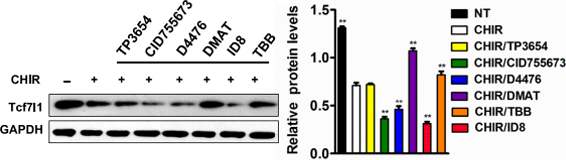
Nuvisertib hydrochloride purchased from MedChemExpress. Usage Cited in: Biochem Biophys Res Commun. 2020 Apr 2;524(2):280-287. [Abstract]
Western blot analysis of Tcf7l1 protein levels in mESCs pre-treated with the indicative different small molecules for 1 h and then treated with CHIR for 24 h.
-
bioRxiv
Radiation synergizes with BET inhibition to stimulate durable, systemic anti-tumor immunity in murine cancer models. [Abstract]2026 Feb 18:2026.02.16.706212. PMID: 41757027

Protocols
-
Fibrosis/Collagen Morphometry
Fibrosis and collagen morphometry is based on the quantitative visualization of fibrillar collagen deposition in tissue sections using histochemical stains such as Sirius Red (Picrosirius Red) or Masson's trichrome, followed by image-based or polarization-enhanced analysis to estimate collagen proportional area as a surrogate of extracellular matrix accumulation during fibrotic remodeling. Sirius Red combined with polarized light microscopy enhances detection of collagen fibers due to birefringence properties, enabling more specific visualization of collagen type I and III fibrils compared to conventional bright-field histology, while whole-section or region-restricted digital morphometry reduces field-selection bias in fibrosis assessment. Alternative quantitative approaches include second harmonic generation (SHG) and two-photon excited fluorescence microscopy, which enable label-free detection of fibrillar collagen and have been validated against histological staining and biochemica
-
RNA extraction experimental
By lysing cells, releasing RNA, and removing impurities such as proteins and DNA, high-purity RNA products are finally obtained. The commonly used traditional method is the guanidine isothiocyanate/phenol/chloroform method (Trizol), which is suitable for a variety of animal materials including animal tissues, microorganisms, cultured cells, etc., and most plant materials.
-
Collagen: Sirius Red Staining
Sirius Red or picrosirius red staining is a histochemical method for visualizing collagen-rich extracellular matrix in tissue sections, and collagen fibers are detected as red-stained structures under bright-field microscopy with enhanced birefringence under polarized light. Picrosirius red is useful for assessing total collagen organization, distribution, and fibrosis burden, but polarized color should not be interpreted as a definitive collagen type I versus type III readout because color is affected by fiber orientation, thickness, and packing.
-
Apoptosis
Apoptosis, also called programmed cell death, is generally characterized by distinct morphological characteristics.
-
Connective Tissue: Masson's Trichrome/Collagen Trichrome Staining
Masson’s Trichrome (collagen/trichrome staining) is a histological technique that differentially stains tissue compartments using sequential acidic dyes to distinguish collagen from muscle and cytoplasmic components based on dye affinity and tissue permeability differences, enabling visualization of fibrosis and connective tissue architecture in histological sections. The classical formulation typically uses Weigert's iron hematoxylin for nuclear staining, Biebrich scarlet-acid fuchsin for cytoplasm and muscle, and aniline blue (or light green variants) for collagen, producing a characteristic blue/green collagen signal contrasted against red cytoplasm and dark nuclei. The staining principle relies on selective displacement of smaller dye molecules by larger anionic dyes in collagen-rich regions under controlled acidified conditions, which enhances collagen-specific dye retention. This property makes the method widely used for fibrosis assessment in organs such as heart, liver, lung, a
-
Apoptosis Solutions
Apoptosis is a regulated, generally non-lytic cell-death pathway that removes unwanted, damaged, infected, or abnormal cells through coordinated morphological changes, caspase activation, DNA fragmentation, and membrane remodeling. The intrinsic apoptosis pathway is controlled mainly by mitochondrial outer membrane permeabilization, BCL-2 family proteins, cytochrome c release, apoptosome formation, caspase-9 activation, and downstream executioner caspase-3/7 activation. The extrinsic apoptosis pathway is initiated by death receptors such as Fas, TNFR, and TRAIL receptors, which recruit adaptor proteins and activate caspase-8 before engaging executioner caspases or mitochondrial amplification through BID cleavage. Apoptosis is linked to many phenotypes, including cancer cell killing, tissue homeostasis, immune regulation, neurodegeneration, infection response, and treatment-induced cytotoxicity; unresolved questions include how apoptosis interacts with necroptosis, pyroptosis, ferroptos
-
Cell Viability Determination by MTT Colorimetric Assay
The following protocol uses the MTT colorimetric assay as a classic literature-established method for assessing cell viability/metabolic activity in cultured mammalian cells. MTT[3-(4,5-dimethylthiazol-2-yl)-2,5-diphenyltetrazolium bromide] is reduced by metabolically active cells to a colored formazan product; the amount of formazan is quantified spectrophotometrically and provides an indirect measure of metabolically active viable cells. Importantly, MTT reduction reflects cellular oxidoreductase/metabolic activity rather than an absolute direct count of living cells, so changes in cellular metabolism can alter the signal independently of cell number.


Purity & Documentation
References

Calculators
Concentration (start) × Volume (start) = Concentration (final) × Volume (final)


Nuvisertib hydrochloride Related Classifications
HY-101126A Related Classifications
 Powered by Bioz