



Phenoxodiol
Based on 3 publication(s) in Google Scholar
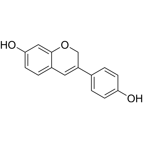
Phenoxodiol (Idronoxil), a synthetic analog of Genestein, activates the mitochondrial caspase system, inhibits XIAP (an apoptosis inhibitor), and sensitizes the cancer cells to Fas-mediated apoptosis. Phenoxodiol also inhibits DNA topoisomerase II by stabilizing the cleavable complex. Phenoxodiol induces cell cycle arrest in the G1/S phase of the cell cycle and upregulates p21WAF1 via a p53 independent manner.
For research use only. We do not sell to patients.
- Purity : 99.89%
- CAS No.: 81267-65-4
- Formula: C15H12O3
- Molecular Weight:240.25
-
Storage:Powder -20°C, 3 years , 4°C, 2 years ; In solvent -80°C, 6 months , -20°C, 1 month

To place orders, for customer services and technical support, please contact: MedChemExpress USA
Tel: 609-228-6898 E-mail: [email protected] [email protected]


-
 Biological Activity
-
Chemical Information
-
Solvent & Solubility
- Protocols
- Purity & Documentation
- References
-
 Help & FAQs


-

HY-18739
Phorbol 12-myristate 13-acetatePKC Activator -
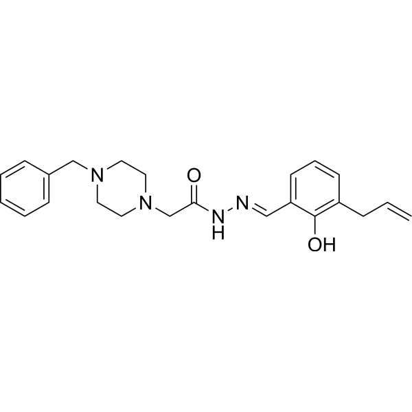
HY-13523
PAC-1Caspase Activator -
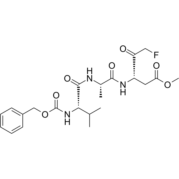
HY-16658
Z-VAD(OMe)-FMKPan-caspase Inhibitor -
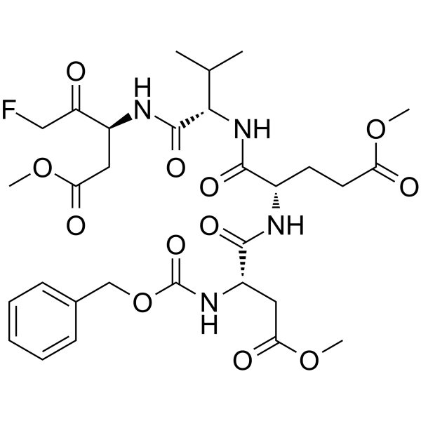
HY-12466
Z-DEVD-FMKCaspase-3 Inhibitor -
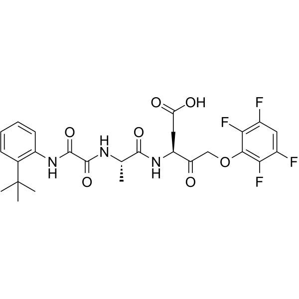
HY-10396
Emricasanpan-Caspase Inhibitor -
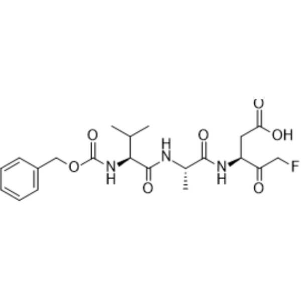
HY-16658B
Z-VAD-FMKPan-Caspase Inhibitor -
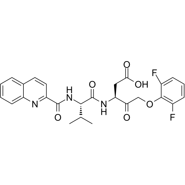
HY-12305
Q-VD-OPhPan-caspase Inhibitor -
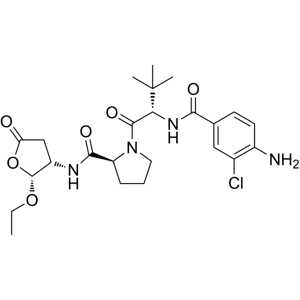
HY-13205
BelnacasanCaspase-1 Inhibitor -
HY-P1001
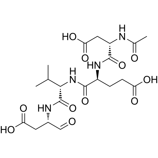
Ac-DEVD-CHOCaspase-3 Inhibitor -

HY-13721R
Phenoxodiol (Standard)Anticancer Agent -
HY-P0109A
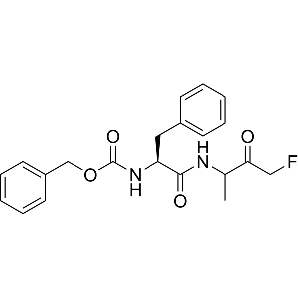
Z-FA-FMKCathepsin B/L Inhibitor -
HY-101297
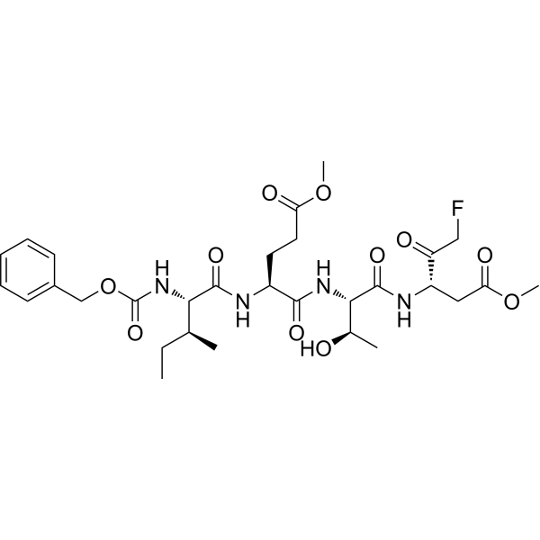
Z-IETD-FMKCaspase-8 Inhibitor
Publications Citing Use of MedChemExpress (MCE) Phenoxodiol
MoreAll Caspase Isoforms
MoreAll Topoisomerase Isoforms
More

Biological Activity
Description
IC50 & Target
Cellular Effect
|
Cell Line

|
Type | Value | Description | References |
|---|---|---|---|---|
| HMEC-1 | GI50 |
4.1 μM
Compound: 1, Isoflavene
|
Antiproliferative activity against human HMEC1 cells by measuring metabolic activity of cells after 72 hrs by spectrophotometric analysis
Antiproliferative activity against human HMEC1 cells by measuring metabolic activity of cells after 72 hrs by spectrophotometric analysis
|
[PMID: 23462711] |
| MDA-MB-231 | GI50 |
31.3 μM
Compound: 1
|
Growth inhibition of human MDA-MB-231 cells after 72 hrs by Alamar blue assay
Growth inhibition of human MDA-MB-231 cells after 72 hrs by Alamar blue assay
|
[PMID: 28408225] |
| MDA-MB-231 | GI50 |
62.9 μM
Compound: 1, Isoflavene
|
Antiproliferative activity against human MDA-MB-231 cells by measuring metabolic activity of cells after 72 hrs by spectrophotometric analysis
Antiproliferative activity against human MDA-MB-231 cells by measuring metabolic activity of cells after 72 hrs by spectrophotometric analysis
|
[PMID: 23462711] |
| MDA-MB-231 | IC50 |
33 μM
Compound: 1
|
Antiproliferative activity against human MDA-MB-231 cells after 72 hrs by Alamar blue/spectrophotometric analysis
Antiproliferative activity against human MDA-MB-231 cells after 72 hrs by Alamar blue/spectrophotometric analysis
|
[PMID: 25189689] |
| MRC5 | GI50 |
109 μM
Compound: 1, Isoflavene
|
Antiproliferative activity against human MRC5 cells by measuring metabolic activity of cells after 72 hrs by spectrophotometric analysis
Antiproliferative activity against human MRC5 cells by measuring metabolic activity of cells after 72 hrs by spectrophotometric analysis
|
[PMID: 23462711] |
| MRC5 | IC50 |
108.5 μM
Compound: 1
|
Cytotoxicity against human MRC5 cells after 72 hrs by Alamar blue assay
Cytotoxicity against human MRC5 cells after 72 hrs by Alamar blue assay
|
[PMID: 28408225] |
| U-87MG ATCC | GI50 |
>100 μM
Compound: 1
|
Growth inhibition of human U87 cells after 72 hrs by Alamar blue assay
Growth inhibition of human U87 cells after 72 hrs by Alamar blue assay
|
[PMID: 28408225] |
In Vitro
Phenoxodiol (Idronoxil) (0-10 μg/mL; 24 h) decreases cell viability of primary ovarian cancer cells[1].
Phenoxodiol (0-10 μg/mL; 24 h) induces apoptosis and restores sensitivity to Fas-mediated apoptosis in ovarian cancer cells[1].
Phenoxodiol (0-10 μg/mL; 24 h) induces caspase-8 activation and FLIP downregulation through the Akt-pathway. Phenoxodiol-induced apoptosis involves activation of the mitochondrial pathway and is caspase dependent. Phenoxodiol treatment results in downregulation and cleavage of XIAP[1].
Phenoxodiol (10 and 30 μM; 24 and 48 h) induces cell cycle arrest in the G1/S phase of the cell cycle in prostate cancer cells[2].
MedChemExpress (MCE) has not independently confirmed the accuracy of these methods. They are for reference only.
-
Cell Line:R182s, R127, Hey, CP70, A2780, R187, R188, R207 and OSE cells
-
Concentration:0, 0.01, 0.1, 1 and 10 μg/mL
-
Incubation Time:24 h
-
Result:A significant decrease in cell viabilityin all the ovarian cancer cell cultures was observed at a concentration of 10 μg/mL (41.6 μM) and did not affect ovarian surface epithelial (OSE) cells’ viability. In CP70 cells, the IC50 was 1.35 μM.
-
Cell Line:CP70 and OSE cells
-
Concentration:10 μg/mL
-
Incubation Time:24 h
-
Result:Induced apoptosis and resulted in a twofold increase in caspase-3 activity. No change in caspase-3 activity was found in normal OSE cells.
-
Cell Line:Ovarian cancer cells
-
Concentration:10 μg/mL
-
Incubation Time:24 h
-
Result:Induced caspase-8 activation, characterized bycleavage of procaspase-8 into its p43/41 and p28 forms and in downregulation of the p43 form of FLIPC in all the primarycultures as well as in the CP70 and Hey cell lines. Decreased the levels of Akt expression. Resulted in XIAP downregulation and cleavage to its 30 kDa inactive form.
-
Cell Line:LNCaP, DU145 and PC3 cells
-
Concentration:10 and 30 μM
-
Incubation Time:24 and 48 h
-
Result:Induced significantly decreased G2 phase cell populations versus DMSO vehicle control, over 24 hours for both 10 μM and 30 μM treatments. The S phase cell population was found to increase versus DMSO vehicle control.
-
Cell Line:LNCaP, DU145 and PC3 cells
-
Concentration:10 and 30 μM
-
Incubation Time:24 and 48 h
-
Result:PC3 cells were found to significantly increase the expression of c-Myc at 30 μM after 48 h. Decreased the expression of Cyclin-D1 after 24 hours of treatment with 30 μM in DU145 and PC3 cells. Decreased the expression of Ki-67 after 24 hours of treatment with 10 and 30 μM in LNCaP and PC3 cells. Increased the expression of p21 in LNCaP and PC3 cells.
- EdU Incorporation Assay (Click Chemistry-Based DNA Synthesis Measurement)
- Detection of Apoptosis
- TUNEL staining for apoptotic DNA fragmentation
- Flow cytometric DNA-content cell-cycle staining
- Annexin V plus membrane-impermeant dye apoptosis staining
- BrdU Incorporation Assay
- Apoptosis Solutions
- Protocol for Cell Cycle
Clinical Trial
| NCT Number | Sponsor | Condition | Start Date |
Phase
|
|---|---|---|---|---|
| NCT01329991 | 2011-05 | PHASE1 |


Chemical Information
-
CAS No. 81267-65-4
-
Appearance Solid
-
Molecular Weight 240.25
-
Formula C15H12O3
-
Color Off-white to brown
-
SMILES
OC1=CC=C2C=C(C3=CC=C(O)C=C3)COC2=C1
-
Synonyms
Idronoxil; Dehydroequol; Haginin E
-
Shipping
Room temperature in continental US; may vary elsewhere.
-
Storage
Powder -20°C 3 years 4°C 2 years In solvent -80°C 6 months -20°C 1 month


Publications (3)
-
Journal Impact Factor
-
Most Recent
-
Int J Mol Sci
Ecto-NOX Disulfide-Thiol Exchanger 2 (ENOX2/tNOX) Is a Potential Prognostic Marker in Primary Malignant Melanoma and May Serve as a Therapeutic Target. [Abstract]2024 Nov 4;25(21):11853. PMID: 39519404 -
Cell Biochem Biophys
Dihydroceramide Desaturase Functions as an Inducer and Rectifier of Apoptosis: Effect of Retinol Derivatives, Antioxidants and Phenolic Compounds. [Abstract]2021 Sep;79(3):461-475. PMID: 33991313 -


Solvent & Solubility
In Vitro:
DMSO : ≥ 100 mg/mL (416.23 mM; Hygroscopic DMSO has a significant impact on the solubility of product, please use newly opened DMSO)
* "≥" means soluble, but saturation unknown.
Please refer to the solubility information to select the appropriate solvent. Once prepared, please aliquot and store the solution to prevent product inactivation from repeated freeze-thaw cycles.
Storage method and period of stock solution: -80°C, 6 months; -20°C, 1 month. When stored at -80°C, please use it within 6 months. When stored at -20°C, please use it within 1 month.
Please refer to the solubility information to select the appropriate solvent. Once prepared, please aliquot and store the solution to prevent product inactivation from repeated freeze-thaw cycles.
Storage method and period of stock solution: -80°C, 6 months; -20°C, 1 month. When stored at -80°C, please use it within 6 months. When stored at -20°C, please use it within 1 month.
Concentration (start) × Volume (start) = Concentration (final) × Volume (final)
In Vivo:
Select the appropriate dissolution method based on your experimental animal and administration route.
- For the following dissolution methods, please ensure to first prepare a clear stock solution using an In Vitro approach and then sequentially add co-solvents:
- To ensure reliable experimental results, the clarified stock solution can be appropriately stored based on storage conditions. As for the working solution for In Vivo experiments, it is recommended to prepare freshly and use it on the same day.
- The percentages shown for the solvents indicate their volumetric ratio in the final prepared solution. If precipitation or phase separation occurs during preparation, heat and/or sonication can be used to aid dissolution.
Add each solvent one by one: 10% DMSO 40% PEG300 5% Tween-80 45% Saline
Solubility: ≥ 2.5 mg/mL (10.41 mM); Clear solution
This protocol yields a clear solution of ≥ 2.5 mg/mL (saturation unknown).
Taking 1 mL working solution as an example, add 100 μL DMSO stock solution (25.0 mg/mL) to 400 μL PEG300, and mix evenly; then add 50 μL Tween-80 and mix evenly; then add 450 μL Saline to adjust the volume to 1 mL.
Preparation of Saline: Dissolve 0.9 g sodium chloride in ddH₂O and dilute to 100 mL to obtain a clear Saline solution.
Add each solvent one by one: 10% DMSO 90% (20% SBE-β-CD in Saline)
Solubility: ≥ 2.5 mg/mL (10.41 mM); Clear solution
This protocol yields a clear solution of ≥ 2.5 mg/mL (saturation unknown).
Taking 1 mL working solution as an example, add 100 μL DMSO stock solution (25.0 mg/mL) to 900 μL 20% SBE-β-CD in Saline, and mix evenly.
Preparation of 20% SBE-β-CD in Saline (4°C, storage for one week): 2 g SBE-β-CD powder is dissolved in 10 mL Saline, completely dissolve until clear.
In Vivo Dissolution Calculator
Please enter the basic information of animal experiments:
-
-
-
-
Recommended: Prepare an additional quantity of animals to account for potential losses during experiments.
Please enter your animal formula composition:
-
%DMSO +
Recommended: Keep the proportion of DMSO in working solution below 2% if your animal is weak.
-
%+
-
+%Tween-80 + +
-
%Saline +
The co-solvents required include: DMSO, . All of co-solvents are available by MedChemExpress (MCE). , Tween 80. All of co-solvents are available by MedChemExpress (MCE).
Working solution concentration: 0.22 mg/mL
Method for preparing stock solution: mg drug dissolved in μL DMSO. Stock solution concentration: mg/mL.
1. Take μL DMSO stock solution;
2. Add μL .
μL , mix evenly;
3. Then add μL Tween 80, mix evenly;
4. Then add μL
Please ensure that the stock solution in the first step is dissolved to a clear state, and add co-solvents in sequence. You can use ultrasonic heating (ultrasonic cleaner, recommended frequency 20-40 kHz), vortexing, etc. to assist dissolution.


Purity & Documentation
-
Data Sheet (277 KB)
-
SDS (396 KB)
- English - EN (396 KB)
- Français - FR (396 KB)
- Deutsch - DE (396 KB)
- Norwegian - NO (396 KB)
- Español - ES (396 KB)
- Swedish - SV (396 KB)
- Italian - IT (396 KB)
- Korean - KR (396 KB)
- Portuguese - PT (396 KB)
-
Handling Instructions (2659 KB)
References
[1]. Kamsteeg M, et al. Phenoxodiol--an isoflavone analog--induces apoptosis in chemoresistant ovarian cancer cells. Oncogene. 2003 May 1;22(17):2611-20. [Content Brief]
[2]. Mahoney S, et al. The effects of phenoxodiol on the cell cycle of prostate cancer cell lines. Cancer Cell Int. 2014 Nov 8;14(1):110. [Content Brief]


Complete Stock Solution Preparation Table
Please refer to the solubility information to select the appropriate solvent. Once prepared, please aliquot and store the solution to prevent product inactivation from repeated freeze-thaw cycles.
Storage method and period of stock solution: -80°C, 6 months; -20°C, 1 month. When stored at -80°C, please use it within 6 months. When stored at -20°C, please use it within 1 month.
| Optional Solvent | Concentration Solvent Mass | 1 mg | 5 mg | 10 mg | 25 mg |
|---|---|---|---|---|---|
| DMSO | 1 mM | 4.1623 mL | 20.8117 mL | 41.6233 mL | 104.0583 mL |
| 5 mM | 0.8325 mL | 4.1623 mL | 8.3247 mL | 20.8117 mL | |
| 10 mM | 0.4162 mL | 2.0812 mL | 4.1623 mL | 10.4058 mL | |
| 15 mM | 0.2775 mL | 1.3874 mL | 2.7749 mL | 6.9372 mL | |
| 20 mM | 0.2081 mL | 1.0406 mL | 2.0812 mL | 5.2029 mL | |
| 25 mM | 0.1665 mL | 0.8325 mL | 1.6649 mL | 4.1623 mL | |
| 30 mM | 0.1387 mL | 0.6937 mL | 1.3874 mL | 3.4686 mL | |
| 40 mM | 0.1041 mL | 0.5203 mL | 1.0406 mL | 2.6015 mL | |
| 50 mM | 0.0832 mL | 0.4162 mL | 0.8325 mL | 2.0812 mL | |
| 60 mM | 0.0694 mL | 0.3469 mL | 0.6937 mL | 1.7343 mL | |
| 80 mM | 0.0520 mL | 0.2601 mL | 0.5203 mL | 1.3007 mL | |
| 100 mM | 0.0416 mL | 0.2081 mL | 0.4162 mL | 1.0406 mL |


Phenoxodiol Related Classifications
HY-13721 Related Classifications
 Powered by Bioz