



Pretilachlor

Based on 1 Customer Validation
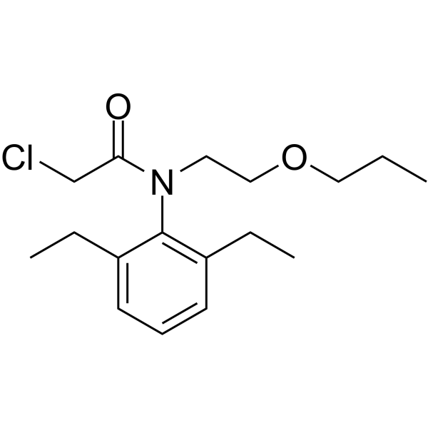
Pretilachlor is a chloroacetamide herbicide with biological activities including endocrine disruption, oxidative stress induction, apoptosis induction, and immunotoxicity. Pretilachlor exerts its effects by interfering with hormone metabolism, inducing oxidative stress, activating apoptotic pathways, and inhibiting immune functions. Pretilachlor upregulates the transcription of P53, Mdm2, and Bbc3, and increases the activities of Caspase3 and Caspase9; it upregulates the transcription of genes in the HPG/HPT axis and the activity of aromatase; it induces oxidative stress, elevates ROS levels, and upregulates CAT, SOD, and GPX. Pretilachlor downregulates the transcription of CXCL-C1C, IL-1β, and IL-8. Pretilachlor disrupts the normal physiological processes and embryonic development of fish, exhibiting significant toxicity. Pretilachlor can be used in studies related to weeding, environmental pollution, and behavioral toxicity in fish.
For research use only. We do not sell to patients.
- Purity : 99.25%
- CAS No.: 51218-49-6
- Formula: C17H26ClNO2
- Molecular Weight:311.85
-
Storage:
Please store the product under the recommended conditions in the Certificate of Analysis.

To place orders, for customer services and technical support, please contact: MedChemExpress USA
Tel: 609-228-6898 E-mail: [email protected] [email protected]


-
 Biological Activity
-
Chemical Information
-
Solvent & Solubility
- Protocols
- Purity & Documentation
- References
-
 Help & FAQs


-

HY-B2035R
Pretilachlor (Standard)Herbicide -
HY-B1880
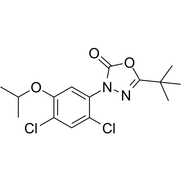
OxadiazonHerbicide -
HY-B0863
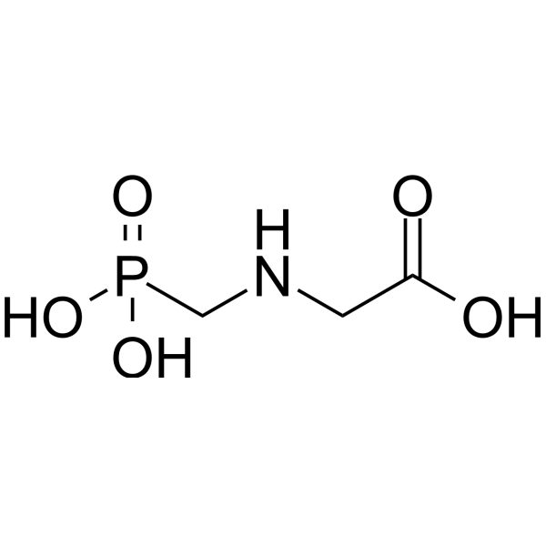
GlyphosateEPSPS Inhibitor -

HY-B1880R
Oxadiazon (Standard)Herbicide -
HY-B0863R
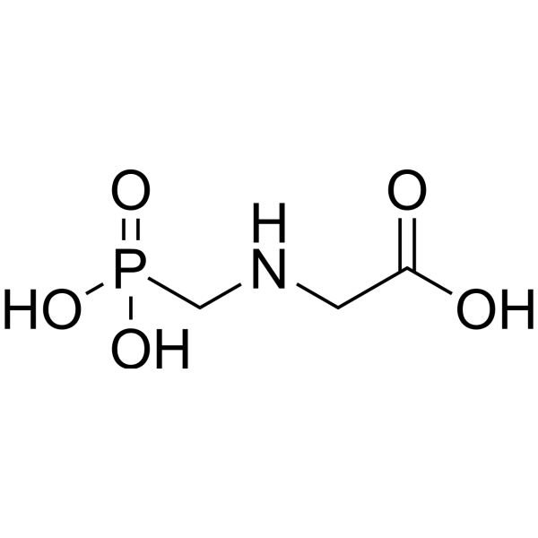
Glyphosate (Standard)Herbicide -
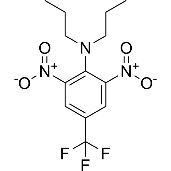
HY-B2050
TrifluralinPre-Emergence Herbicide -
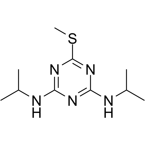
HY-121324
PrometrynHerbicide -
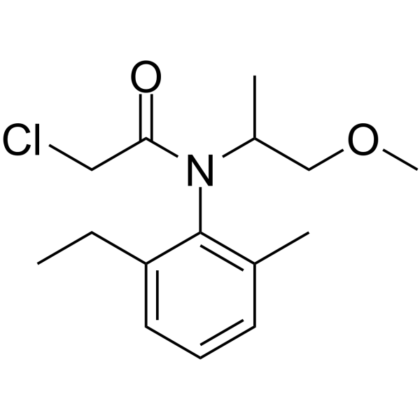
HY-B1871
MetolachlorHerbicide -
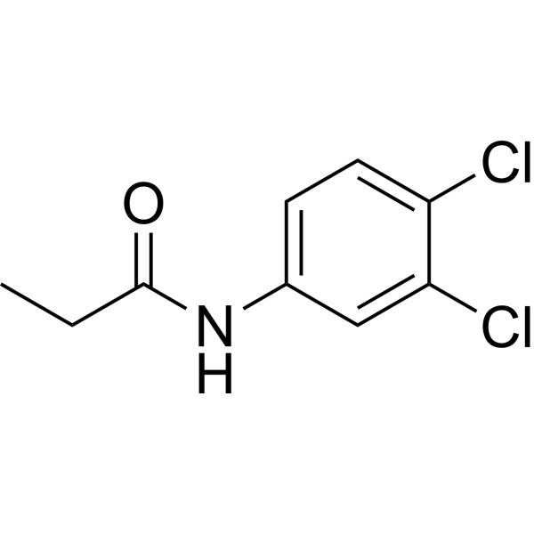
HY-B2030
PropanilHerbicide -

HY-B1871R
Metolachlor (Standard)Chloroacetanilide Herbicide -

HY-B2030R
Propanil (Standard)Herbicide -

HY-B2050R
Trifluralin (Standard)Pre-Emergence Herbicide
All Caspase Isoforms
More

Biological Activity
Description
IC50 & Target
[1]|
Caspase 3 |
Caspase 9 |
Aromatase |
IL-8 |
IL-1β |
In Vivo
MedChemExpress (MCE) has not independently confirmed the accuracy of these methods. They are for reference only.
-
Animal Model:AB strain (embryos from adult females with average weight 0.93 g, males with average weight 0.65 g)[1]
-
Dosage:50 μg/L, 100 μg/L, 200 μg/L
-
Administration:aqueous exposure; continuous; 96 hours
-
Result:Upregulated VTG1 mRNA by 3.45-fold (50 μg/L), 2.74-fold (100 μg/L) and 2.43-fold (200 μg/L).
Upregulated CYP19a mRNA by 4.29-fold (50 μg/L); upregulated CYP19b mRNA by 3.29-fold (50 μg/L) and raised its level at 200 μg/L.
Elevated ERα mRNA at 200 μg/L; suppressed ERβ1 mRNA at 100 μg/L and 200 μg/L.
Raised TRα mRNA at 50 μg/L and 200 μg/L; upregulated Dio2 mRNA by 1.5-fold (50 μg/L).
Boosted VTG protein contents and aromatase activity at 50 μg/L and 100 μg/L.
Elevated T3 content at 100 μg/L; increased T4 content at 50 μg/L and 100 μg/L.
Suppressed CXCL-C1C mRNA across all tested concentrations.
Reduced IL-1β mRNA to 0.172-fold (50 μg/L), 0.350-fold (100 μg/L) and 0.400-fold (200 μg/L) in a dose-dependent manner.
Lowered IL-8 mRNA at 100 μg/L.
Upregulated CAT and Cu/Zn-SOD mRNA at 50 μg/L; upregulated GPX mRNA at 200 μg/L.
Increased CAT protein contents at 50 μg/L and 100 μg/L; enhanced CAT activity at 100 μg/L.
Raised SOD protein content at 50 μg/L; elevated GPX protein contents at 50 μg/L and 200 μg/L.
Elevated ROS levels at all tested concentrations.
Upregulated P53 mRNA at 50 μg/L and 200 μg/L; upregulated Mdm2 mRNA at 50 μg/L; upregulated Bbc3 mRNA by 2.08-fold (200 μg/L).
Enhanced Caspase3 activity at 100 μg/L and 200 μg/L; increased Caspase9 activity at 100 μg/L.


Chemical Information
-
CAS No. 51218-49-6
-
Appearance Liquid (Density: 1.074±0.06 g/cm3)
-
Molecular Weight 311.85
-
Formula C17H26ClNO2
-
Color Colorless to light yellow
-
SMILES
O=C(N(C1=C(CC)C=CC=C1CC)CCOCCC)CCl
-
Shipping
Room temperature in continental US; may vary elsewhere.
-
Storage
Please store the product under the recommended conditions in the Certificate of Analysis.


Solvent & Solubility
In Vitro:
DMSO : 200 mg/mL (641.33 mM; Need ultrasonic; Hygroscopic DMSO has a significant impact on the solubility of product, please use newly opened DMSO)
Concentration (start) × Volume (start) = Concentration (final) × Volume (final)

Protocols
-
Apoptosis
Apoptosis, also called programmed cell death, is generally characterized by distinct morphological characteristics.
-
TUNEL staining for apoptotic DNA fragmentation
TUNEL staining detects DNA strand breaks by using terminal deoxynucleotidyl transferase to add labeled nucleotides to exposed 3′-OH DNA termini, generating either microscopic staining in fixed cells or tissue sections, or fluorescence/cytometric signal in cell suspensions. TUNEL positivity reflects DNA fragmentation but should not be interpreted alone as definitive apoptosis, because TUNEL can also label necrotic, autolytic, mechanically damaged, or DNA-repair-associated DNA breaks.
-
Annexin V plus membrane-impermeant dye apoptosis staining
Annexin V-based apoptosis assays rely on the detection of phosphatidylserine (PS) externalization from the inner leaflet of the plasma membrane to the outer leaflet, an early biochemical hallmark of apoptosis. Fluorescently labeled Annexin V binds PS in a calcium-dependent manner, enabling identification of early apoptotic cells by flow cytometry or fluorescence microscopy. When combined with a membrane-impermeant DNA-binding dye (e. g. , propidium iodide), this approach allows discrimination between viable (Annexin V−/dye−), early apoptotic (Annexin V+/dye−), and late apoptotic or necrotic (Annexin V+/dye+) cell populations by assessing membrane integrity and PS exposure.
-
ROS/oxidative-stress fluorescent staining
ROS/oxidative-stress fluorescent staining uses cell-permeant fluorogenic probes that become fluorescent after oxidation inside cells or tissues; commonly used examples include DCFH-DA/DCFDA for broad cellular oxidant detection, DHE for superoxide-related signal detection, MitoSOX for mitochondrial superoxide-related signal detection, and CellROX probes for oxidative-stress-associated fluorescence readouts. The assay detects probe oxidation rather than a single ROS species unless the probe and analysis method have been chemically validated for that species. DCFH-DA enters cells, is deacetylated by intracellular esterases to DCFH, and produces fluorescent DCF after oxidation, so the readout is used as an operational measure of total cellular oxidative stress rather than a species-specific ROS measurement. DHE and MitoSOX can report superoxide-related oxidation, but red fluorescence alone can include non-specific ethidium-like oxidation products; HPLC or optimized spectral approaches are
-
Protocol for Fluorescence In Situ Hybridization (FISH)
Fluorescence in situ hybridization detects specific DNA or RNA sequences inside fixed cells or tissue sections by hybridizing fluorescently labeled nucleic-acid probes to complementary target sequences, allowing the target’s copy number, chromosomal position, spatial distribution, or transcript abundance to be visualized microscopically. DNA-FISH detects genomic loci, chromosomal gains/losses, amplifications, deletions, and rearrangements, while RNA-FISH detects RNA molecules or transcript localization; in cancer cells, mouse tumors, neurons, organoids, macrophages, or drug-screening samples, the readout is fluorescent puncta, fusion/split signals, or localized RNA signal interpreted relative to validated controls.
-
Research Protocol for Endocrine Diseases
Endocrine diseases often arise from disrupted hormone production, hormone signaling, or target-tissue responsiveness; for diabetes-focused endocrine disease models, insulin signaling regulates glucose uptake, hepatic glucose output, lipid metabolism, and β-cell compensation. Type 2 diabetes develops through interacting defects in insulin resistance, β-cell dysfunction, adipose inflammation, hepatic glucose overproduction, altered incretin signaling, and ectopic lipid metabolism. A major unresolved question is whether endocrine dysfunction is driven primarily by target-tissue insulin resistance, intrinsic β-cell failure, immune/inflammatory stress, or combined multi-organ failure that differs by disease stage.
-
Apoptosis Solutions
Apoptosis is a regulated, generally non-lytic cell-death pathway that removes unwanted, damaged, infected, or abnormal cells through coordinated morphological changes, caspase activation, DNA fragmentation, and membrane remodeling. The intrinsic apoptosis pathway is controlled mainly by mitochondrial outer membrane permeabilization, BCL-2 family proteins, cytochrome c release, apoptosome formation, caspase-9 activation, and downstream executioner caspase-3/7 activation. The extrinsic apoptosis pathway is initiated by death receptors such as Fas, TNFR, and TRAIL receptors, which recruit adaptor proteins and activate caspase-8 before engaging executioner caspases or mitochondrial amplification through BID cleavage. Apoptosis is linked to many phenotypes, including cancer cell killing, tissue homeostasis, immune regulation, neurodegeneration, infection response, and treatment-induced cytotoxicity; unresolved questions include how apoptosis interacts with necroptosis, pyroptosis, ferroptos
-
Pyroptosis Solutions
Pyroptosis is a lytic inflammatory cell-death pathway executed by gasdermin pores, most classically through inflammasome-mediated activation of caspase-1, cleavage of gasdermin D, membrane pore formation, LDH release, and secretion of IL-1β and IL-18. The canonical pathway is commonly modeled by priming cells with an inflammatory signal such as LPS to induce pro-IL-1β and inflammasome components, followed by an activation signal such as ATP or nigericin to activate NLRP3, ASC speck formation, caspase-1 cleavage, GSDMD cleavage, cytokine release, and pyroptotic membrane rupture. The non-canonical pathway is triggered when cytosolic LPS activates mouse caspase-11 or human caspase-4/5, leading to GSDMD cleavage and pyroptosis, and this can secondarily activate NLRP3-dependent IL-1β release. Pyroptosis is linked to inflammatory injury, infection, cancer, liver disease, ocular disease, placental inflammation, and other disease phenotypes, but unresolved questions include which gasdermin fam


Purity & Documentation
-
Data Sheet (275 KB)
-
SDS (421 KB)
- English - EN (421 KB)
- Français - FR (421 KB)
- Deutsch - DE (421 KB)
- Norwegian - NO (421 KB)
- Español - ES (421 KB)
- Swedish - SV (421 KB)
- Italian - IT (421 KB)
- Korean - KR (421 KB)
- Portuguese - PT (421 KB)
-
Handling Instructions (2659 KB)
References


Complete Stock Solution Preparation Table
| Optional Solvent | Concentration Solvent Mass | 1 mg | 5 mg | 10 mg | 25 mg |
|---|---|---|---|---|---|
| DMSO | 1 mM | 3.2067 mL | 16.0333 mL | 32.0667 mL | 80.1667 mL |
| 5 mM | 0.6413 mL | 3.2067 mL | 6.4133 mL | 16.0333 mL | |
| 10 mM | 0.3207 mL | 1.6033 mL | 3.2067 mL | 8.0167 mL | |
| 15 mM | 0.2138 mL | 1.0689 mL | 2.1378 mL | 5.3444 mL | |
| 20 mM | 0.1603 mL | 0.8017 mL | 1.6033 mL | 4.0083 mL | |
| 25 mM | 0.1283 mL | 0.6413 mL | 1.2827 mL | 3.2067 mL | |
| 30 mM | 0.1069 mL | 0.5344 mL | 1.0689 mL | 2.6722 mL | |
| 40 mM | 0.0802 mL | 0.4008 mL | 0.8017 mL | 2.0042 mL | |
| 50 mM | 0.0641 mL | 0.3207 mL | 0.6413 mL | 1.6033 mL | |
| 60 mM | 0.0534 mL | 0.2672 mL | 0.5344 mL | 1.3361 mL | |
| 80 mM | 0.0401 mL | 0.2004 mL | 0.4008 mL | 1.0021 mL | |
| 100 mM | 0.0321 mL | 0.1603 mL | 0.3207 mL | 0.8017 mL |


Pretilachlor Related Classifications
HY-B2035 Related Classifications
 Powered by Bioz