



Ro 106-9920
Based on 2 publication(s) in Google Scholar
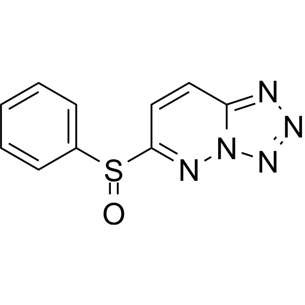
Ro 106-9920 is an orally active NF-κB inhibitor. Ro 106-9920 prevents ubiquitination of IκBα, blocks nuclear translocation of NF-κB, and inhibits NF-κB activation. Ro 106-9920 suppresses TNF-α-induced tissue factor expression and procoagulant activity, enhances TNF-α-mediated cytotoxicity against cancer cells, and eliminates the formation of neutrophil extracellular traps. Ro 106-9920 inhibits NLRP3 inflammasome activation, reduces inflammatory cytokine secretion, decreases myeloperoxidase activity, attenuates renal cell apoptosis, and improves renal function. Ro 106-9920 blocks ANG II (Angiotensin II human) (HY-13948)-induced nuclear localization of p65, internalization of AT1A receptor, colocalization of β-arrestin-2, and expression of COX-2, and alters the formation of β-arrestin endosomes. Ro 106-9920 can be used in research related to non-small cell lung cancer, acute kidney injury, diabetes mellitus, and osteoporosis.
For research use only. We do not sell to patients.
- CAS No.: 62645-28-7
- Formula: C10H7N5OS
- Molecular Weight:245.26
-
Storage:Powder -20°C, 3 years , 4°C, 2 years ; In solvent -80°C, 6 months , -20°C, 1 month

To place orders, for customer services and technical support, please contact: MedChemExpress USA
Tel: 609-228-6898 E-mail: [email protected] [email protected]


-
 Biological Activity
-
Chemical Information
-
Solvent & Solubility
- Protocols
- Purity & Documentation
- References
-
 Help & FAQs


-
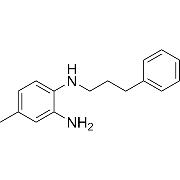
HY-13982
JSH-23NF-κB Inhibitor -
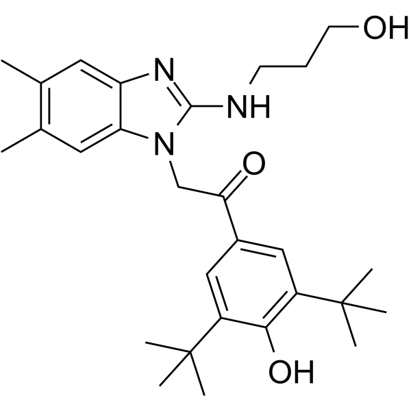
HY-15530
CID-2858522NF-κB Inhibitor -
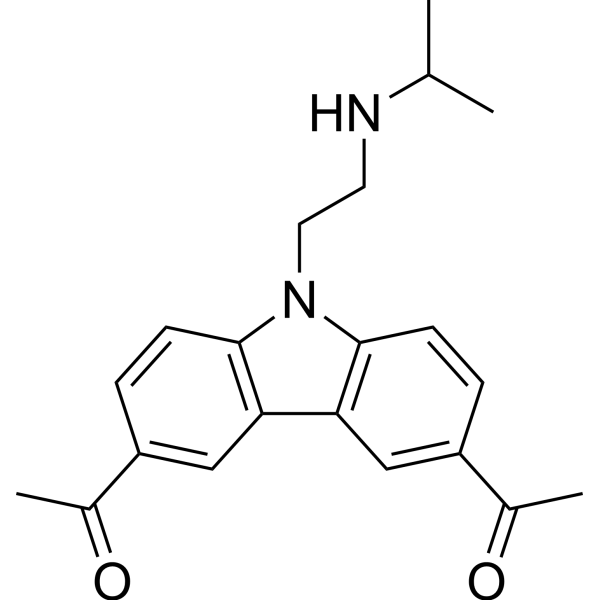
HY-18935
CBL0137FACT Inhibitor -
HY-18738
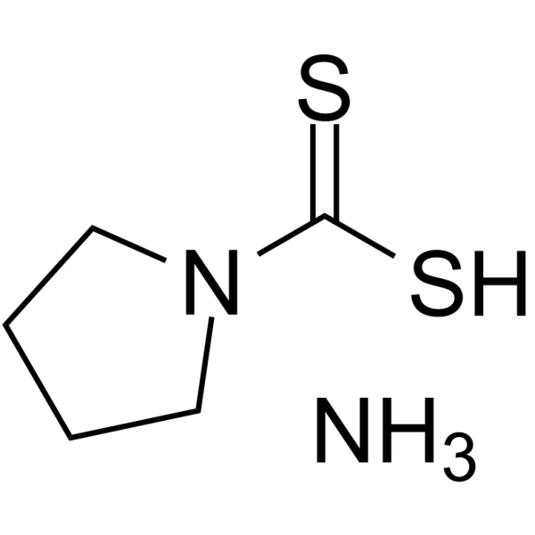
Pyrrolidinedithiocarbamate ammoniumSelective NF-κB Inhibitor -
HY-10496
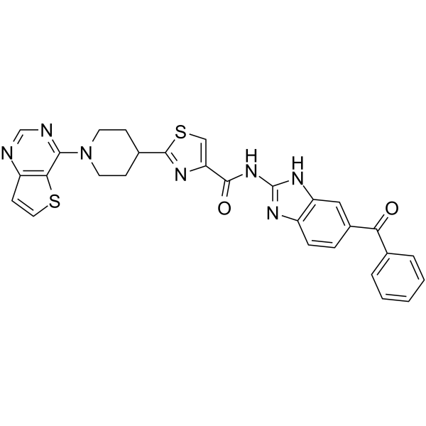
SC75741NF-κB Inhibitor -
HY-N2065
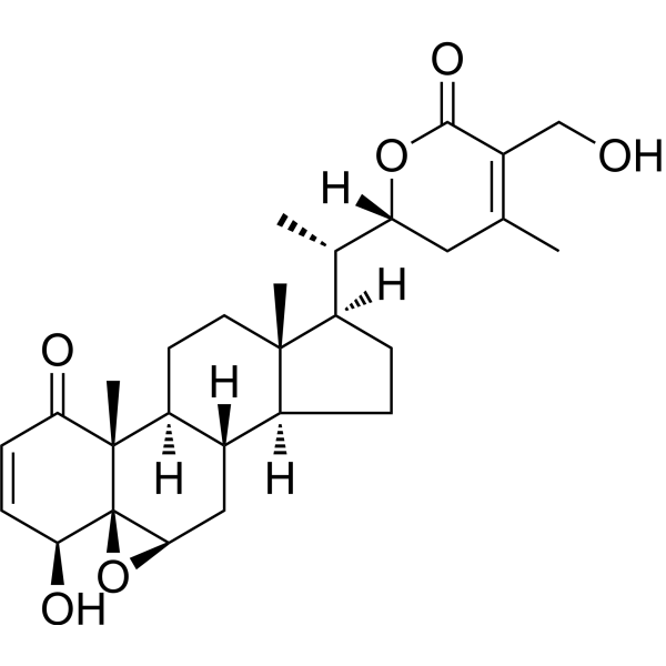
Withaferin ANF-κB Inhibitor -
HY-N1378
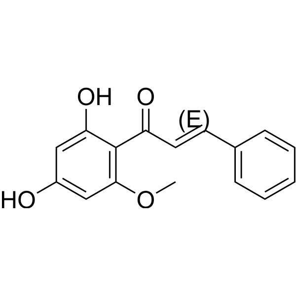
(E)-CardamoninhTRPA1 Antagonist -
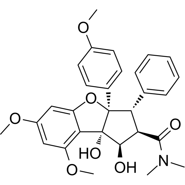
HY-19356
RocaglamideNF-κB Inhibitor -
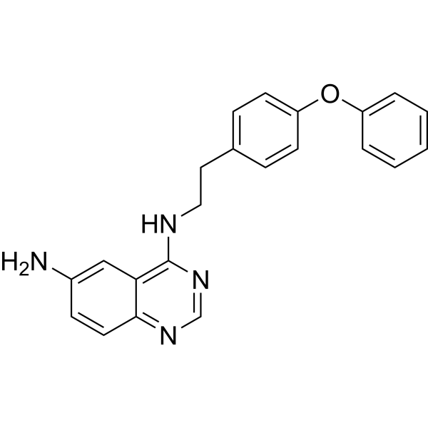
HY-13812
QNZTNF Receptor Inhibitor -
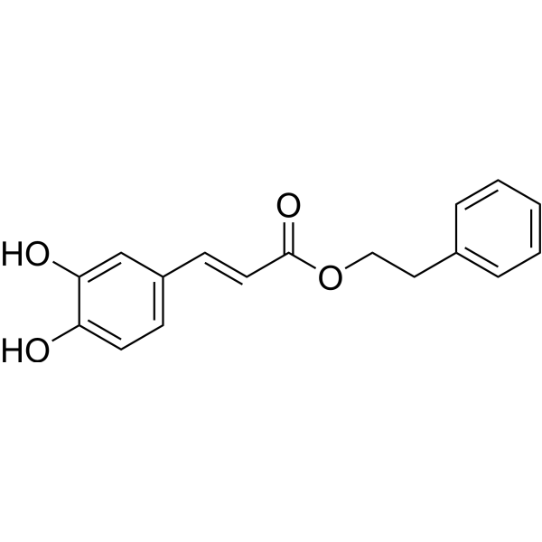
HY-N0274
Caffeic acid phenethyl esterApoptosis Inducer -
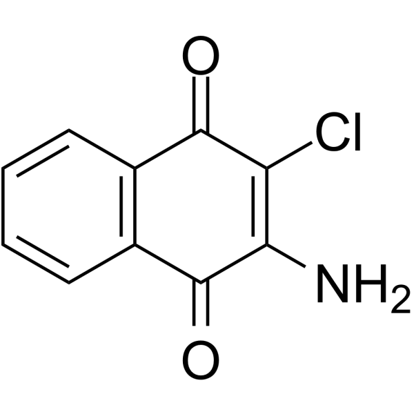
HY-121632
QuinoclamineNF-κB Inhibitor -
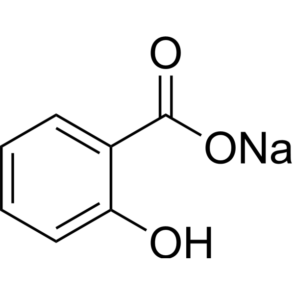
HY-B0167A
Sodium SalicylateCOX Inhibitor
Publications Citing Use of MedChemExpress (MCE) Ro 106-9920
MoreAll Angiotensin Receptor Isoforms
MoreAll Arrestin Isoforms
More

Biological Activity
Description
|
NF-κB |
IκBα |
NLRP3 |
p65 |
AT1A |
Arrestin-3/β-Arrestin 2 |
COX-2 |
In Vitro
Ro 106-9920 (10 μM; 1 h pre-incubation prior to 12 h TNF-α exposure) prevents TNF-α-induced tissue factor protein expression in human non-small cell lung cancer H1299 and A549 cells[1].
Ro 106-9920 (10 μM; 1 h pre-incubation prior to 48 h TNF-α exposure) enhances TNF-α-mediated cytotoxicity against human non-small cell lung cancer H1299 and A549 cells[1].
Ro 106-9920 (10 μM; 1 h pre-incubation prior to 24 h TNF-α exposure) reverses TNF-α-induced procoagulant activity, restored to control levels, in human non-small cell lung cancer H1299 and A549 cells[1].
Ro 106-9920 (1 μM; 3 days) reduces CuLDL-stimulated cell-associated RANKL expression in MG63 human osteoblast-like cells[4].
Ro 106-9920 (10 μM; 30 min) potently reduces ANG II-induced AT1A receptor internalization in rat aortic vascular smooth muscle cells (RASMC), lowering internalization from ~70% to ~20%[5].
Ro 106-9920 (10 μM; 30 min) blocks ANG II-induced internalization of AT1A/GFP receptors in HEK-293 cells, preventing movement from the plasma membrane to the nuclear membrane area[5].
Ro 106-9920 (10 μM; 30 min) inhibits ANG II- and SII-ANG II-induced β-arrestin-2 recruitment to the human AT1 receptor in CHO-K1 cells, reducing recruitment to basal levels[5].
Ro 106-9920 (10 μM; 60 min) inhibits ANG II-induced colocalization of AT1A/GFP receptors with β-arrestin-2/RFP in HEK-293 cells, preventing the approximately fourfold increase in colocalization seen with ANG II alone[5].
Ro 106-9920 (10 μM; 60 min) alters ANG II-induced β-arrestin-2 endosome formation in HEK-293 cells, producing significantly larger endosomes compared to ANG II alone[5].
Ro 106-9920 (6.25-25 μM; 1 min pre-incubation) dose-dependently inhibits thrombin-induced IκBα degradation without altering IκBα phosphorylation in human leukocyte-free washed platelets[6].
Ro 106-9920 (12.5 μM; 1 min pre-incubation) reduces thrombin-induced fibrinogen binding to αIIbβ3 integrin in human washed platelets[6].
Ro 106-9920 (12.5 μM; 1 min pre-incubation) inhibits thrombin-induced platelet spreading on immobilized fibrinogen in human washed platelets[6].
Ro 106-9920 (12.5 μM) does not alter thrombin-induced intracellular Ca2+ mobilization in human washed platelets[6].
Ro 106-9920 (12.5 μM) inhibits thrombin-induced ATP release but does not affect Arachidonic acid (HY-109590)-induced ATP release in human washed platelets[6].
Ro 106-9920 (12.5 μM) reduces thrombin-induced P-selectin expression in human washed platelets[6].
Ro 106-9920 (25 μM) inhibits thrombin-induced cPLA2 activity in human washed platelets[6].
Ro 106-9920 (12.5-25 μM; 1 min pre-incubation) dose-dependently inhibits thrombin-induced ERK2 phosphorylation in human washed platelets[6].
Ro 106-9920 (2.5 μM; 12 h) inhibits NET formation and NLRP3 inflammasome activation in mouse neutrophil-RAW264.7 macrophage cocultures, and mitigates NET-induced impairments in viability, migration, and angiogenic function of NIH/3T3 fibroblasts, HaCaT keratinocytes, and HUVEC endothelial cells[3].
Ro 106-9920 (1 μM; 1 h) reduces CuLDL-stimulated NFkappaB DNA binding activity in MG63 human osteoblast-like cells[4].
Ro 106-9920 (10 μM; 30 min) inhibits ANG II (Angiotensin II human) (HY-13948)- and TNFα-induced p65 NF-κB nuclear localization in rat aortic vascular smooth muscle cells (RASMC)[5].
Ro 106-9920 (1-10 μM; 30 min) inhibits ANG II-induced COX-2 protein expression in rat aortic vascular smooth muscle cells (RASMC) in a concentration-dependent manner, reducing the sixfold increase in COX-2 expression to baseline at 10 μM[5].
MedChemExpress (MCE) has not independently confirmed the accuracy of these methods. They are for reference only. Further protocols information, click here.
-
Cell Line:human non-small cell lung cancer H1299 and A549 cells
-
Concentration:10 μM
-
Incubation Time:1 h (pre-incubation); 12 h (TNF-α exposure)
-
Result:Prevented TNF-α-induced TF protein expression in both H1299 and A549 cells.
-
Cell Line:human non-small cell lung cancer H1299 and A549 cells
-
Concentration:10 μM
-
Incubation Time:1 h (pre-incubation); 48 h (TNF-α exposure)
-
Result:Enhanced TNF-α-mediated cytotoxicity, leading to a marked reduction in cell viability in both H1299 and A549 cells compared to Ro 106-9920 alone or TNF-α alone.
-
Cell Line:MG63 human osteoblast-like cells
-
Concentration:1 μM
-
Incubation Time:1 h
-
Result:Reduced the CuLDL-induced increase in NFkappaB DNA binding activity.
-
Cell Line:rat aortic vascular smooth muscle cells (RASMC)
-
Concentration:10 μM
-
Incubation Time:30 min (pretreatment)
-
Result:Blocked ANG II- and TNFα-induced nuclear accumulation of p65 NF-κB.
-
Cell Line:rat aortic vascular smooth muscle cells (RASMC)
-
Concentration:1-10 μM
-
Incubation Time:30 min (pretreatment)
-
Result:Inhibited ANG II-induced COX-2 protein expression in a concentration-dependent manner, reducing the approximately sixfold increase in COX-2 expression to baseline values with 10 μM pretreatment.
Showed no effect on COX-2 expression when used alone.
-
Cell Line:rat aortic vascular smooth muscle cells (RASMC)
-
Concentration:10 μM
-
Incubation Time:30 min (pretreatment)
-
Result:Did not inhibit ANG II-induced phosphorylation and activation of p42/44 ERK, unlike losartan which blocked this effect.
-
Cell Line:human leukocyte-free washed platelets
-
Concentration:6.25 μM; 12.5 μM; 25 μM
-
Incubation Time:1 min pre-incubation
-
Result:Inhibited thrombin-induced IκBα degradation in a dose-dependent manner without altering the phosphorylation level of IκBα in leukocyte-free human washed platelets.
In Vivo
Ro 106-9920 (5 mg/kg; s.c.) enhances diabetic wound healing by suppressing neutrophil extracellular trap formation and NLRP3 inflammasome activation, while promoting angiogenesis and re-epithelialization without altering blood glucose levels or causing organ toxicity[3].
MedChemExpress (MCE) has not independently confirmed the accuracy of these methods. They are for reference only.
-
Animal Model:C57BL/6J (male, 6-8 weeks old, 18-22 g, bilateral renal ischemia/reperfusion injury model)[2]
-
Dosage:5 mg/kg/day; 10 mg/kg/day; 20 mg/kg/day
-
Administration:i.g.; daily; 7 days
-
Result:Reduced ischemia/reperfusion-induced elevations in serum biomarkers in a dose-dependent manner, with serum creatinine AUC values of 410.625, 280.875, and 205.088 for 5, 10, and 20 mg/kg, respectively.
Reduced serum cystatin C AUC values to 3036.875, 2326.375, and 1527.625 for 5, 10, and 20 mg/kg, respectively.
Reduced serum BUN AUC values to 138.675, 125.663, and 96.094 for 5, 10, and 20 mg/kg, respectively.
Lowered serum NGAL levels to 1614.776 pg/mL, 1139.937 pg/mL, and 776.175 pg/mL at 168 h post-reperfusion for 5, 10, and 20 mg/kg, respectively.
Lowered serum FGF-23 levels to 2836.791 pg/mL, 2277.763 pg/mL, and 841.909 pg/mL for 5, 10, and 20 mg/kg, respectively.
Reduced renal KIM-1 protein expression in a dose-dependent manner.
Lowered renal histopathological scores to 7.000, 3.333, and 1.000 for 5, 10, and 20 mg/kg, respectively.
Reduced renal tubular epithelial cell apoptosis rates to 70.750%, 50.767%, and 1.310% for 5, 10, and 20 mg/kg, respectively.
Lowered serum IL-1β levels to 99.540 pg/mL, 62.247 pg/mL, and 36.848 pg/mL for 5, 10, and 20 mg/kg, respectively.
Lowered serum IL-6 levels to 217.051 pg/mL, 147.485 pg/mL, and 76.842 pg/mL for 5, 10, and 20 mg/kg, respectively.
Lowered serum TNF-α levels to 117.583 pg/mL, 92.040 pg/mL, and 58.023 pg/mL for 5, 10, and 20 mg/kg, respectively.
Reduced renal mRNA expression of IL-1β to 3.980, 3.330, and 2.007 relative units; IL-6 to 100.217, 75.847, and 36.930 relative units; and TNF-α to 4.413, 3.330, and 1.973 relative units for 5, 10, and 20 mg/kg, respectively.
Did not significantly reduce NF-κB activation at 5 mg/kg; reduced renal NF-κB IHC score to 4.667, cytosolic p-NF-κB relative expression to 0.944, and nuclear NF-κB relative expression to 0.944 at 10 mg/kg; reduced renal NF-κB IHC score to 2.000, cytosolic p-NF-κB relative expression to 0.722, and nuclear NF-κB relative expression to 0.722 at 20 mg/kg.
Reduced renal MPO activity to 1.537, 1.045, and 0.738 relative units for 5, 10, and 20 mg/kg, respectively.
Reduced renal MPO protein expression to 1.154, 1.029, and 0.815 relative units; Cit-H3 protein expression to 0.979, 0.762, and 0.522 relative units for 5, 10, and 20 mg/kg, respectively.
-
Animal Model:C57BL/6J mice (male, 8-10 weeks old, 17-18 g, diabetic induced by high-fat diet + Streptozotocin (HY-13753), with dorsal excisional wounds)[3]
-
Dosage:5 mg/kg
-
Administration:s.c.
-
Result:Promoted significantly faster wound closure over 14 days.
Reduced scar width and improved tissue architecture in wound tissues.
Increased CD31-positive vascular loops (angiogenesis) in wound tissues.
Reduced expression of NET markers (citrullinated histone H3, peptidyl arginine deiminase 4) in wound tissues.
Suppressed NLRP3 inflammasome activation (lowered NLRP3 and mature IL-1β protein levels, with accumulated pro-IL-1β) in wound tissues.
Showed no effect on blood glucose levels or body weight.
Caused no hepatotoxicity or nephrotoxicity.


Chemical Information
-
CAS No. 62645-28-7
-
Appearance Solid
-
Molecular Weight 245.26
-
Formula C10H7N5OS
-
Color White to off-white
-
SMILES
O=S(C1=CC=CC=C1)C2=NN3N=NN=C3C=C2
-
Shipping
Room temperature in continental US; may vary elsewhere.
-
Storage
Powder -20°C 3 years 4°C 2 years In solvent -80°C 6 months -20°C 1 month


Publications (2)
-
Journal Impact Factor
-
Most Recent
-
Front Biosci (Landmark Ed)
Neutrophil Extracellular Trap Formation Suppressed by Ro 106-9920 Enhances Diabetic Wound Healing by Blocking NLRP3 Inflammasome Activation. [Abstract]2025 May 19;30(5):37393. PMID: 40464514 -
Ren Fail
Suppression of nF-κB by ro 106-9920 alleviates ischemia/reperfusion-induced renal dysfunction and inflammation via modulation of neutrophil extracellular trap formation in acute kidney injury mice. [Abstract]2025 Dec;47(1):2545983. PMID: 40796802


Solvent & Solubility
In Vitro:
DMSO : 20 mg/mL (81.55 mM; Need ultrasonic; Hygroscopic DMSO has a significant impact on the solubility of product, please use newly opened DMSO)
Please refer to the solubility information to select the appropriate solvent. Once prepared, please aliquot and store the solution to prevent product inactivation from repeated freeze-thaw cycles.
Storage method and period of stock solution: -80°C, 6 months; -20°C, 1 month. When stored at -80°C, please use it within 6 months. When stored at -20°C, please use it within 1 month.
Please refer to the solubility information to select the appropriate solvent. Once prepared, please aliquot and store the solution to prevent product inactivation from repeated freeze-thaw cycles.
Storage method and period of stock solution: -80°C, 6 months; -20°C, 1 month. When stored at -80°C, please use it within 6 months. When stored at -20°C, please use it within 1 month.
Concentration (start) × Volume (start) = Concentration (final) × Volume (final)

Protocols
-
RNA extraction experimental
By lysing cells, releasing RNA, and removing impurities such as proteins and DNA, high-purity RNA products are finally obtained. The commonly used traditional method is the guanidine isothiocyanate/phenol/chloroform method (Trizol), which is suitable for a variety of animal materials including animal tissues, microorganisms, cultured cells, etc., and most plant materials.
-
Nephrotoxicity Study
This protocol assesses nephrotoxicity by combining functional kidney injury readouts, urinary/tissue injury biomarkers, and renal histopathology. Serum creatinine and BUN reflect impaired kidney function, while KIM-1, NGAL, clusterin, osteopontin, IL-18, cystatin C, nephrin, Oat5, urinary protein, glucose, and alkaline phosphatase have been used to detect tubular injury in cisplatin-, gentamicin-, and acetaminophen-induced nephrotoxicity models.
-
Mammalian live/dead viability and cytotoxicity staining
Live/dead viability and cytotoxicity staining assays are based on the simultaneous detection of intracellular esterase activity in metabolically active (viable) cells and membrane integrity loss in non-viable cells. In commonly used dual-staining approaches, membrane-permeant fluorogenic substrates are converted by intracellular esterases into fluorescent products in live cells, while impermeant DNA-binding dyes selectively enter cells with compromised plasma membranes and label nucleic acids in dead or dying cells, enabling discrimination between viable and non-viable populations by fluorescence microscopy or flow cytometry.
-
Annexin V plus membrane-impermeant dye apoptosis staining
Annexin V-based apoptosis assays rely on the detection of phosphatidylserine (PS) externalization from the inner leaflet of the plasma membrane to the outer leaflet, an early biochemical hallmark of apoptosis. Fluorescently labeled Annexin V binds PS in a calcium-dependent manner, enabling identification of early apoptotic cells by flow cytometry or fluorescence microscopy. When combined with a membrane-impermeant DNA-binding dye (e. g. , propidium iodide), this approach allows discrimination between viable (Annexin V−/dye−), early apoptotic (Annexin V+/dye−), and late apoptotic or necrotic (Annexin V+/dye+) cell populations by assessing membrane integrity and PS exposure.
-
Research Protocol for Inflammation-related Diseases
The NLRP3 inflammasome is a cytosolic innate immune signaling platform that integrates priming signals and danger-signal activation to promote caspase-1 activation, maturation of IL-1β and IL-18, and gasdermin D-mediated pyroptotic cell death. The core experimental logic is to determine whether inflammatory disease phenotypes are driven by increased NLRP3 expression, ASC-containing inflammasome assembly, caspase-1 cleavage, GSDMD cleavage, and extracellular release of IL-1β/IL-18 rather than by nonspecific cell injury alone. The pathway is strongly linked to inflammation-related disease phenotypes because monosodium urate crystals activate NALP3/NLRP3 inflammasome signaling in gout-like crystal inflammation, cholesterol crystals activate NLRP3 inflammasomes in atherogenesis models, and DSS-induced intestinal inflammation has been reported to involve NLRP3 inflammasome activity. However, experimental colitis studies also show context-dependent protective effects of NLRP3 inflammasome co
-
Research Protocol for Metabolic Diseases
AMP-activated protein kinase, AMPK, is a conserved cellular energy sensor that responds to reduced cellular energy status and coordinates metabolism by increasing ATP-generating catabolic pathways while suppressing ATP-consuming anabolic processes. In metabolic disease research, the AMPK pathway is experimentally relevant because it regulates hepatic lipid synthesis, fatty acid oxidation, glucose production, skeletal-muscle glucose disposal, mTORC1-linked biosynthesis, autophagy, mitochondrial homeostasis, and whole-body energy balance. The central pathway logic is that energy stress, metformin, exercise-like stimulation, or direct AMPK activators increase AMPKα Thr172 phosphorylation and downstream substrate phosphorylation, including ACC and RAPTOR. Phosphorylation of ACC suppresses lipogenesis and supports fatty acid oxidation, whereas phosphorylation of RAPTOR suppresses mTORC1 signaling and links cellular energy status to growth and protein synthesis control. The pathway is linked
-
Cell Viability Determination by MTT Colorimetric Assay
The following protocol uses the MTT colorimetric assay as a classic literature-established method for assessing cell viability/metabolic activity in cultured mammalian cells. MTT[3-(4,5-dimethylthiazol-2-yl)-2,5-diphenyltetrazolium bromide] is reduced by metabolically active cells to a colored formazan product; the amount of formazan is quantified spectrophotometrically and provides an indirect measure of metabolically active viable cells. Importantly, MTT reduction reflects cellular oxidoreductase/metabolic activity rather than an absolute direct count of living cells, so changes in cellular metabolism can alter the signal independently of cell number.
-
Cytoplasmic-Nuclear Fractionated Protein Extraction
Cytoplasmic-nuclear fractionated protein extraction separates soluble cytoplasmic proteins from nuclear-enriched proteins by mild plasma-membrane permeabilization, differential centrifugation, washing of nuclei, and extraction of nuclear proteins for downstream immunoblotting or related molecular analysis. The readout is the relative abundance of a protein in cytoplasmic and nuclear fractions, commonly assessed by western blotting together with compartment markers such as tubulin or pyruvate kinase for cytoplasm and lamin, nucleoporin, hnRNP, H2AX, or Lamin B for nuclear fractions.
-
Apoptosis
Apoptosis, also called programmed cell death, is generally characterized by distinct morphological characteristics.
-
TUNEL staining for apoptotic DNA fragmentation
TUNEL staining detects DNA strand breaks by using terminal deoxynucleotidyl transferase to add labeled nucleotides to exposed 3′-OH DNA termini, generating either microscopic staining in fixed cells or tissue sections, or fluorescence/cytometric signal in cell suspensions. TUNEL positivity reflects DNA fragmentation but should not be interpreted alone as definitive apoptosis, because TUNEL can also label necrotic, autolytic, mechanically damaged, or DNA-repair-associated DNA breaks.
-
Cell Cytotoxicity Assay
Cytotoxicity assays are usually based on the assessment of cell membrane damage, which can also be indirectly detected by measuring cell viability. Detection methods include MTT assay, CKK-8 assay, LDH assay and ATP assay, etc.
-
Apoptosis Solutions
Apoptosis is a regulated, generally non-lytic cell-death pathway that removes unwanted, damaged, infected, or abnormal cells through coordinated morphological changes, caspase activation, DNA fragmentation, and membrane remodeling. The intrinsic apoptosis pathway is controlled mainly by mitochondrial outer membrane permeabilization, BCL-2 family proteins, cytochrome c release, apoptosome formation, caspase-9 activation, and downstream executioner caspase-3/7 activation. The extrinsic apoptosis pathway is initiated by death receptors such as Fas, TNFR, and TRAIL receptors, which recruit adaptor proteins and activate caspase-8 before engaging executioner caspases or mitochondrial amplification through BID cleavage. Apoptosis is linked to many phenotypes, including cancer cell killing, tissue homeostasis, immune regulation, neurodegeneration, infection response, and treatment-induced cytotoxicity; unresolved questions include how apoptosis interacts with necroptosis, pyroptosis, ferroptos
-
Pyroptosis Solutions
Pyroptosis is a lytic inflammatory cell-death pathway executed by gasdermin pores, most classically through inflammasome-mediated activation of caspase-1, cleavage of gasdermin D, membrane pore formation, LDH release, and secretion of IL-1β and IL-18. The canonical pathway is commonly modeled by priming cells with an inflammatory signal such as LPS to induce pro-IL-1β and inflammasome components, followed by an activation signal such as ATP or nigericin to activate NLRP3, ASC speck formation, caspase-1 cleavage, GSDMD cleavage, cytokine release, and pyroptotic membrane rupture. The non-canonical pathway is triggered when cytosolic LPS activates mouse caspase-11 or human caspase-4/5, leading to GSDMD cleavage and pyroptosis, and this can secondarily activate NLRP3-dependent IL-1β release. Pyroptosis is linked to inflammatory injury, infection, cancer, liver disease, ocular disease, placental inflammation, and other disease phenotypes, but unresolved questions include which gasdermin fam


Purity & Documentation
-
Data Sheet (302 KB)
-
SDS (251 KB)
- English - EN (251 KB)
- Français - FR (251 KB)
- Deutsch - DE (251 KB)
- Norwegian - NO (251 KB)
- Español - ES (251 KB)
- Swedish - SV (251 KB)
- Italian - IT (251 KB)
- Korean - KR (251 KB)
- Portuguese - PT (251 KB)
-
Handling Instructions (2659 KB)
References
[2]. Wang M, et al. Suppression of nF-κB by ro 106-9920 alleviates ischemia/reperfusion-induced renal dysfunction and inflammation via modulation of neutrophil extracellular trap formation in acute kidney injury mice. Renal failure. 2025 Dec;47(1):2545983. [Content Brief]
[3]. Li H, et al. Neutrophil Extracellular Trap Formation Suppressed by Ro 106-9920 Enhances Diabetic Wound Healing by Blocking NLRP3 Inflammasome Activation. Frontiers in bioscience (Landmark edition). 2025 May 19;30(5):37393. [Content Brief]
[6]. Malaver E, et al. NF-kappaB inhibitors impair platelet activation responses. J Thromb Haemost. 2009 Aug;7(8):1333-43. [Content Brief]


Complete Stock Solution Preparation Table
Please refer to the solubility information to select the appropriate solvent. Once prepared, please aliquot and store the solution to prevent product inactivation from repeated freeze-thaw cycles.
Storage method and period of stock solution: -80°C, 6 months; -20°C, 1 month. When stored at -80°C, please use it within 6 months. When stored at -20°C, please use it within 1 month.
| Optional Solvent | Concentration Solvent Mass | 1 mg | 5 mg | 10 mg | 25 mg |
|---|---|---|---|---|---|
| DMSO | 1 mM | 4.0773 mL | 20.3865 mL | 40.7731 mL | 101.9326 mL |
| 5 mM | 0.8155 mL | 4.0773 mL | 8.1546 mL | 20.3865 mL | |
| 10 mM | 0.4077 mL | 2.0387 mL | 4.0773 mL | 10.1933 mL | |
| 15 mM | 0.2718 mL | 1.3591 mL | 2.7182 mL | 6.7955 mL | |
| 20 mM | 0.2039 mL | 1.0193 mL | 2.0387 mL | 5.0966 mL | |
| 25 mM | 0.1631 mL | 0.8155 mL | 1.6309 mL | 4.0773 mL | |
| 30 mM | 0.1359 mL | 0.6796 mL | 1.3591 mL | 3.3978 mL | |
| 40 mM | 0.1019 mL | 0.5097 mL | 1.0193 mL | 2.5483 mL | |
| 50 mM | 0.0815 mL | 0.4077 mL | 0.8155 mL | 2.0387 mL | |
| 60 mM | 0.0680 mL | 0.3398 mL | 0.6796 mL | 1.6989 mL | |
| 80 mM | 0.0510 mL | 0.2548 mL | 0.5097 mL | 1.2742 mL |


Ro 106-9920 Related Classifications
HY-107665 Related Classifications
 Powered by Bioz