



Rolipram GMP is Rolipram (HY-16900) produced by using GMP guidelines. GMP small molecules works appropriately as an auxiliary reagent for cell therapy manufacture. Rolipram is a PDE4 inhibitor, with blood-brain barrier permeability, that reverses β-amyloid-induced learning and memory impairment in rats. Rolipram elevates intracellular cAMP and clevels and regulates the cAMP/CREB signaling pathway, thereby alleviating neuroinflammation and apoptotic responses. Rolipram promotes neuronal differentiation of human bone marrow mesenchymal stem cells and inhibits Methamphetamine- and morphine-induced hyperlocomotion in mice. Rolipram also reduces the viability of glioblastoma stem-like cells and enhances Bevacizumab (HY-P9906)-induced cell death. Rolipram inhibits the expression of proinflammatory cytokines and enhances central noradrenergic transmission. Rolipram is mainly used in studies related to various central nervous system diseases including Alzheimer's disease, major depressive disorder, glioblastoma multiforme, and multiple sclerosis.
For research use only. We do not sell to patients.
- CAS No.: 61413-54-5
- Formula: C16H21NO3
- Molecular Weight:275.34
-
Storage:
Please store the product under the recommended conditions in the Certificate of Analysis.


To place orders, for customer services and technical support, please contact: MedChemExpress USA
Tel: 609-228-6898 E-mail: [email protected] [email protected]


-
 Biological Activity
-
Chemical Information
- Protocols
- Purity & Documentation
- References
-
 Help & FAQs


-
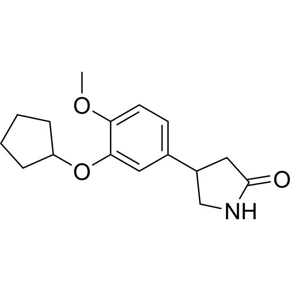
HY-16900
RolipramPDE4 Inhibitor -

HY-16900R
Rolipram (Standard)Reference Standard -
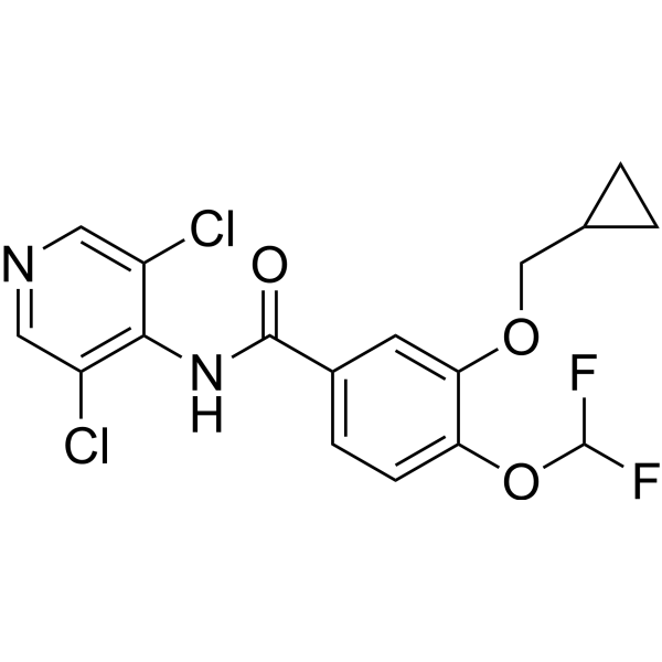
HY-15455
RoflumilastPDE4 Inhibitor -
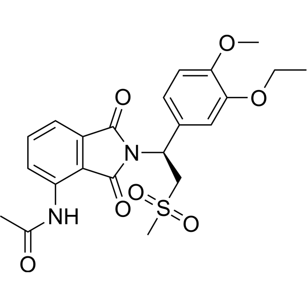
HY-12085
ApremilastPDE-4 Inhibitor -
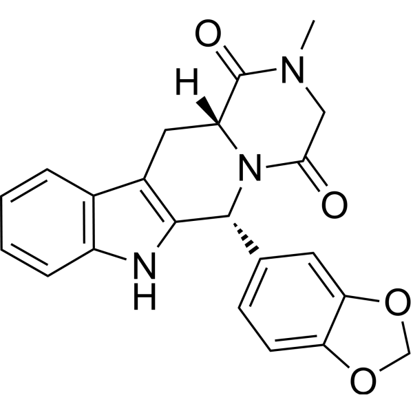
HY-90009A
TadalafilPDE5 Inhibitor -
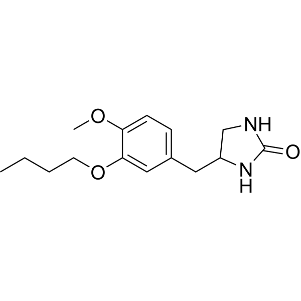
HY-100927
Ro 20-1724PDE4/PDE IV Inhibitor -
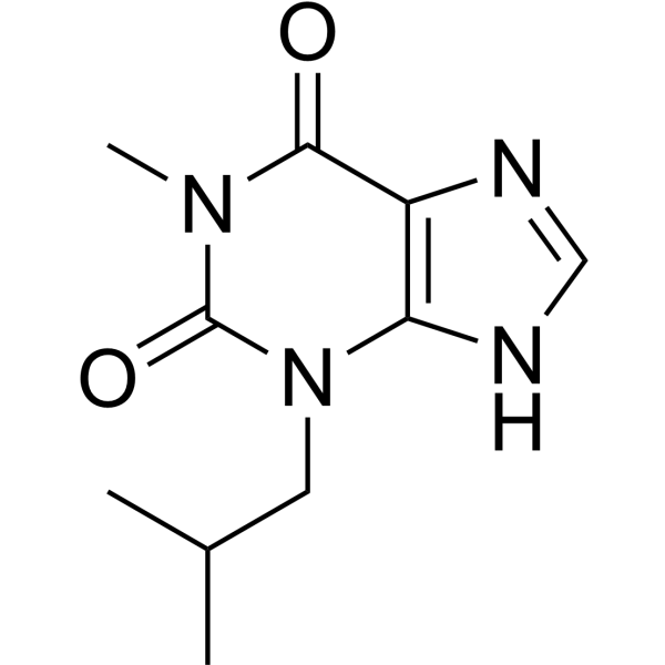
HY-12318
IBMXPDE Inhibitor -
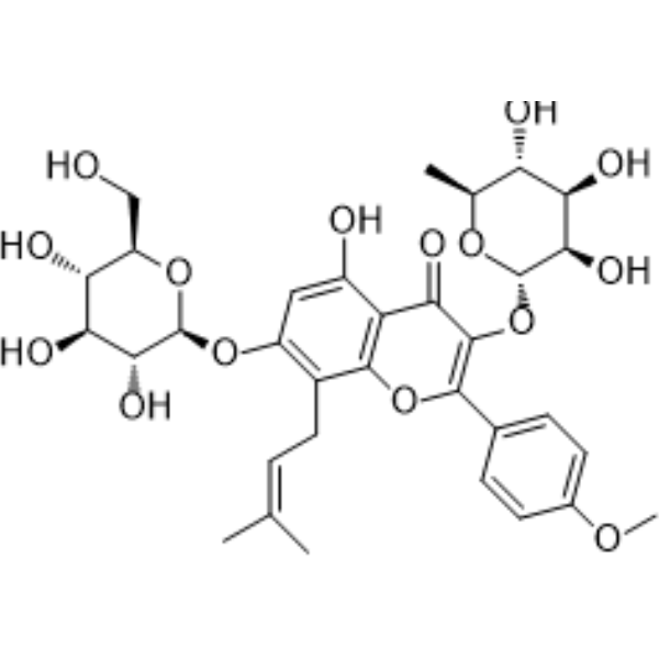
HY-N0014
IcariinPPAR Activator -
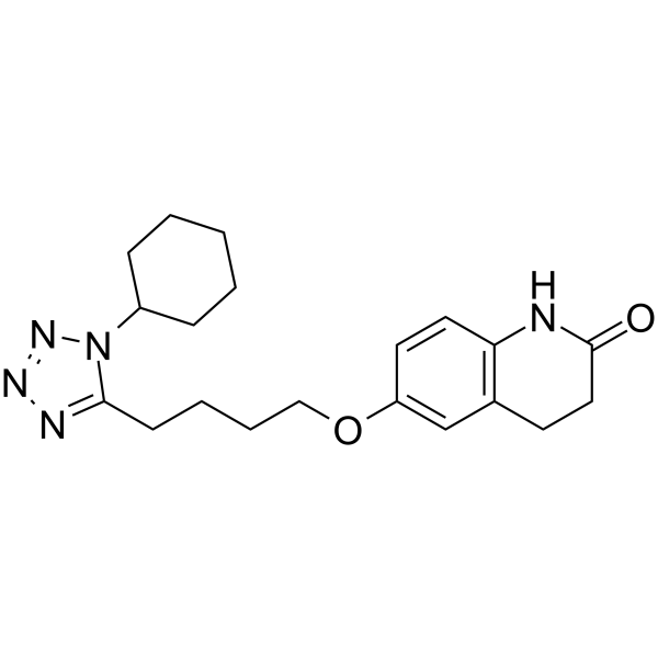
HY-17464
CilostazolPDE3 Inhibitor -
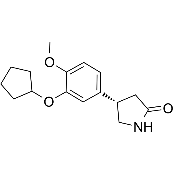
HY-B0392
(S)-(+)-RolipramPDE4 Inhibitor -
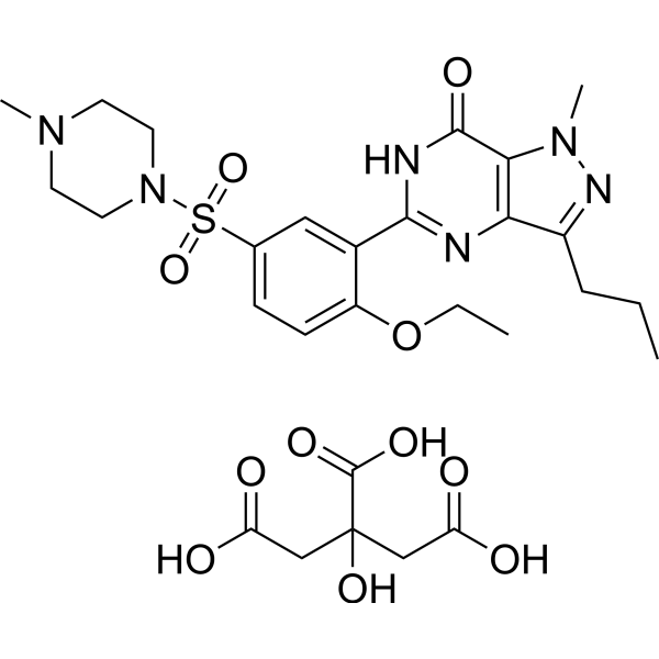
HY-15025A
Sildenafil citratePDE5 Inhibitor -
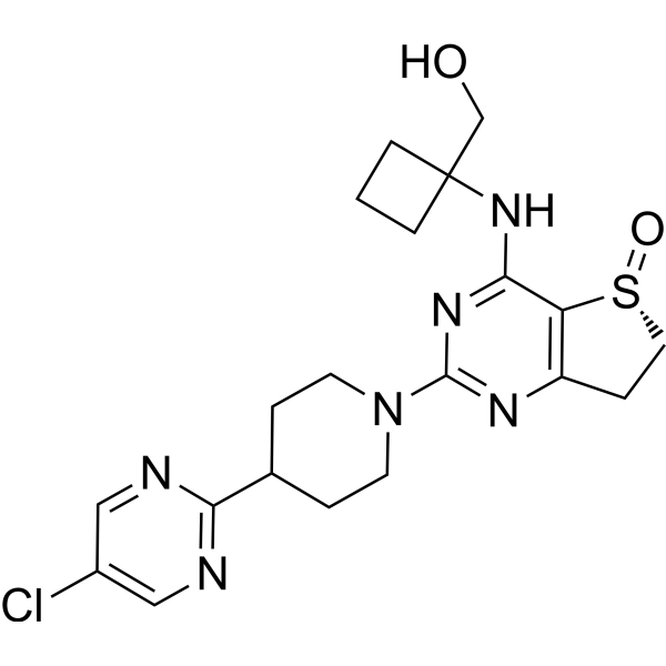
HY-153192
NerandomilastPDE4B Inhibitor


Biological Activity
Description
IC50 & Target
IC50: 3 nM (PDE4A), 130 nM (PDE4B), 240 nM (PDE4D)[1]
Cellular Effect
|
Cell Line

|
Type | Value | Description | References |
|---|---|---|---|---|
| A549 | IC50 |
100 μM
Compound: Rol
|
Antiproliferative activity against human A549 cells after 48 hrs by MTT assay
Antiproliferative activity against human A549 cells after 48 hrs by MTT assay
|
[PMID: 34311158] |
| COS-7 | IC50 |
105 nM
Compound: rolipram
|
Inhibition of PDE4B2 expressed in COS7 cells assessed as cAMP hydrolysis
Inhibition of PDE4B2 expressed in COS7 cells assessed as cAMP hydrolysis
|
[PMID: 18222088] |
| HEK293 | EC50 |
131.5 nM
Compound: 1
|
Inhibition of PDE4 expressed in HEK293 cells coexpressing G-protein coupled receptor and cyclic nucleotide gated ion channel by fluorescence assay
Inhibition of PDE4 expressed in HEK293 cells coexpressing G-protein coupled receptor and cyclic nucleotide gated ion channel by fluorescence assay
|
[PMID: 19464886] |
| HEK293 | EC50 |
0.22 μM
Compound: Rolipram
|
Inhibition of PDE4B expressed in HEK293 cells assessed as fold reduction in forskolin-stimulated cAMP production pretreated for 30 mins measured 4 hrs after forskolin challenge by luciferase reporter gene assay
Inhibition of PDE4B expressed in HEK293 cells assessed as fold reduction in forskolin-stimulated cAMP production pretreated for 30 mins measured 4 hrs after forskolin challenge by luciferase reporter gene assay
|
[PMID: 22386978] |
| HEK293 | EC50 |
2.62 μM
Compound: Rolipram
|
Inhibition of PDE4A4 (unknown origin) transfected in HEK293 cells assessed as forskolin-induced cAMP accumulation pretreated for 30 mins before forskolin challenge by luciferase reporter gene assay
Inhibition of PDE4A4 (unknown origin) transfected in HEK293 cells assessed as forskolin-induced cAMP accumulation pretreated for 30 mins before forskolin challenge by luciferase reporter gene assay
|
10.1039/C2MD00273F |
| HEK293 | EC50 |
0.16 μM
Compound: Rolipram
|
Inhibition of PDE4B1 (unknown origin) transfected in HEK293 cells assessed as forskolin-induced cAMP accumulation pretreated for 30 mins before forskolin challenge by luciferase reporter gene assay
Inhibition of PDE4B1 (unknown origin) transfected in HEK293 cells assessed as forskolin-induced cAMP accumulation pretreated for 30 mins before forskolin challenge by luciferase reporter gene assay
|
10.1039/C2MD00273F |
| HEK293 | EC50 |
0.67 μM
Compound: Rolipram
|
Inhibition of PDE4D3 (unknown origin) transfected in HEK293 cells assessed as forskolin-induced cAMP accumulation pretreated for 30 mins before forskolin challenge by luciferase reporter gene assay
Inhibition of PDE4D3 (unknown origin) transfected in HEK293 cells assessed as forskolin-induced cAMP accumulation pretreated for 30 mins before forskolin challenge by luciferase reporter gene assay
|
10.1039/C2MD00273F |
| MCF7 | IC50 |
100 μM
Compound: Rol
|
Antiproliferative activity against human MCF7 cells after 48 hrs by MTT assay
Antiproliferative activity against human MCF7 cells after 48 hrs by MTT assay
|
[PMID: 34311158] |
| NCI-H460 | IC50 |
100 μM
Compound: Rol
|
Antiproliferative activity against human NCI-H460 cells after 48 hrs by MTT assay
Antiproliferative activity against human NCI-H460 cells after 48 hrs by MTT assay
|
[PMID: 34311158] |
| PBMC | IC50 |
0.5 μM
Compound: 1
|
Inhibition of TNF-alpha release from LPS-stimulated human mononuclear cells
Inhibition of TNF-alpha release from LPS-stimulated human mononuclear cells
|
[PMID: 9784096] |
| PBMC | IC50 |
0.15 μM
Compound: Rolipram
|
Inhibition of LPS stimulated Tumor Necrosis Factor-alpha (TNF-alpha) release by human PBMC
Inhibition of LPS stimulated Tumor Necrosis Factor-alpha (TNF-alpha) release by human PBMC
|
[PMID: 9873600] |
| PBMC | IC50 |
0.16 μM
Compound: Rolipram
|
Antiinflammatory activity in human PBMC assessed as inhibition of LPS-induced TNFalpha release in after 24 hrs by FACS Array analysis
Antiinflammatory activity in human PBMC assessed as inhibition of LPS-induced TNFalpha release in after 24 hrs by FACS Array analysis
|
[PMID: 19303290] |
| PBMC | IC50 |
0.23 μM
Compound: Rolipram
|
Antiinflammatory activity in human PBMC assessed as inhibition of phytohemagglutininin-induced IL2 release after 24 hrs by FACS Array analysis
Antiinflammatory activity in human PBMC assessed as inhibition of phytohemagglutininin-induced IL2 release after 24 hrs by FACS Array analysis
|
[PMID: 19303290] |
| PBMC | IC50 |
0.5 μM
Compound: Rolipram
|
Antiinflammatory activity in human PBMC assessed as inhibition of phytohemagglutininin-induced IL5 release after 48 hrs by FACS Array analysis
Antiinflammatory activity in human PBMC assessed as inhibition of phytohemagglutininin-induced IL5 release after 48 hrs by FACS Array analysis
|
[PMID: 19303290] |
| PBMC | IC50 |
0.88 μM
Compound: Rolipram
|
Antiinflammatory activity in human PBMC assessed as inhibition of phytohemagglutininin-induced IL10 release after 48 hrs by FACS Array analysis
Antiinflammatory activity in human PBMC assessed as inhibition of phytohemagglutininin-induced IL10 release after 48 hrs by FACS Array analysis
|
[PMID: 19303290] |
| PBMC | IC50 |
60 nM
Compound: Rolipram
|
Inhibition of LPS-induced TNFalpha production in human PBMC
Inhibition of LPS-induced TNFalpha production in human PBMC
|
[PMID: 19049349] |
| PBMC | EC50 |
0.13 μM
Compound: rolipram
|
Antiinflammatory activity in human PBMC assessed as inhibition of LPS-induced TNFalpha production after 24 hrs by ELISA
Antiinflammatory activity in human PBMC assessed as inhibition of LPS-induced TNFalpha production after 24 hrs by ELISA
|
[PMID: 22891752] |
| PBMC | IC50 |
0.27 μM
Compound: Rolipram
|
Inhibition of LPS-induced TNF-alpha production in rat PBMC
Inhibition of LPS-induced TNF-alpha production in rat PBMC
|
[PMID: 24852119] |
| PBMC | IC50 |
12.5 μM
Compound: Rolipram
|
Anti-inflammatory activity in human PBMC assessed as suppression of LPS-induced TNF-alpha release preincubated for 30 mins followed by LPS stimulation for 18 hrs by TR-FRET assay
Anti-inflammatory activity in human PBMC assessed as suppression of LPS-induced TNF-alpha release preincubated for 30 mins followed by LPS stimulation for 18 hrs by TR-FRET assay
|
[PMID: 27289320] |
| PBMC | IC50 |
18.6 μM
Compound: Rolipram
|
Anti-inflammatory activity in human PBMC assessed as inhibition of LPS-induced TNFalpha secretion preincubated for 30 mins followed by LPS stimulation for 18 hrs by TR-FRET assay
Anti-inflammatory activity in human PBMC assessed as inhibition of LPS-induced TNFalpha secretion preincubated for 30 mins followed by LPS stimulation for 18 hrs by TR-FRET assay
|
[PMID: 28196708] |
| PBMC | IC50 |
10.85 μM
Compound: 1
|
Inhibition of LPS-induced TNFalpha production in human PBMC pre-incubated for 30 mins before LPS stimulation for 18 hrs by TR-FRET assay
Inhibition of LPS-induced TNFalpha production in human PBMC pre-incubated for 30 mins before LPS stimulation for 18 hrs by TR-FRET assay
|
[PMID: 28888661] |
| PBMC | IC50 |
0.13 μM
Compound: Rolipram
|
Anti-inflammatory activity in human PBMC assessed as inhibition of LPS-induced TNFalpha production after 24 hrs by ELISA
Anti-inflammatory activity in human PBMC assessed as inhibition of LPS-induced TNFalpha production after 24 hrs by ELISA
|
[PMID: 29432948] |
| PBMC | IC50 |
30 μM
Compound: IV
|
Cytotoxicity against human PBMC by MTS assay
Cytotoxicity against human PBMC by MTS assay
|
[PMID: 29173945] |
| RAW264.7 | IC50 |
0.19 μM
Compound: Rolipram
|
Inhibition of LPS-induced TNFalpha production in mouse RAW264.7 cells pretreated for 30 mins before LPS challenge by ELISA assay
Inhibition of LPS-induced TNFalpha production in mouse RAW264.7 cells pretreated for 30 mins before LPS challenge by ELISA assay
|
10.1039/C2MD00273F |
| RAW264.7 | CC50 |
60 μM
Compound: Rolipram
|
Cytotoxicity against mouse RAW264.7 cells measured after 24 hrs by CCK-8 method
Cytotoxicity against mouse RAW264.7 cells measured after 24 hrs by CCK-8 method
|
[PMID: 40423264] |
| RAW264.7 | IC50 |
39.93 μM
Compound: Rolipram
|
Antiinflammatory activity against mouse RAW264.7 cells assessed as inhibition of LPS-induced TNF-alpha release preincubated for 2 hrs followed by LPS stimulation and measured after 4 hrs by RT-PCR analysis
Antiinflammatory activity against mouse RAW264.7 cells assessed as inhibition of LPS-induced TNF-alpha release preincubated for 2 hrs followed by LPS stimulation and measured after 4 hrs by RT-PCR analysis
|
[PMID: 40423264] |
| RAW264.7 | IC50 |
60 μM
Compound: Rolipram
|
Antiinflammatory activity against mouse RAW264.7 cells assessed as inhibition of LPS-induced NO release preincubated for 2 hrs followed by LPS stimulation and measured after 4 hrs by RT-PCR analysis
Antiinflammatory activity against mouse RAW264.7 cells assessed as inhibition of LPS-induced NO release preincubated for 2 hrs followed by LPS stimulation and measured after 4 hrs by RT-PCR analysis
|
[PMID: 40423264] |
| Sf21 | IC50 |
100 μM
Compound: 7
|
Inhibition of His-tagged catalytic domain Trypanosoma brucei brucei PDEB1 expressed in baculovirus infected insect Sf21 cells
Inhibition of His-tagged catalytic domain Trypanosoma brucei brucei PDEB1 expressed in baculovirus infected insect Sf21 cells
|
[PMID: 22023548] |
| Sf21 | IC50 |
95 nM
Compound: 1
|
Inhibition of human full length PDE4A4 expressed in baculovirus infected sf21 cells
Inhibition of human full length PDE4A4 expressed in baculovirus infected sf21 cells
|
[PMID: 23806553] |
| Sf9 | IC50 |
4 nM
Compound: 1
|
Inhibition of recombinant human PDE4A expressed in Sf9 cells
Inhibition of recombinant human PDE4A expressed in Sf9 cells
|
[PMID: 11052785] |
| Sf9 | IC50 |
2.3 μM
Compound: Rolipram
|
Inhibition of human recombinant PDE4 catalytic domain expressed in baculovirus-infected insect Sf9 cells by modified two-step method
Inhibition of human recombinant PDE4 catalytic domain expressed in baculovirus-infected insect Sf9 cells by modified two-step method
|
[PMID: 19303290] |
| Sf9 | IC50 |
0.092 μM
Compound: rolipram
|
Inhibition of human recombinant PDE4D expressed in Sf9 cells
Inhibition of human recombinant PDE4D expressed in Sf9 cells
|
[PMID: 19827751] |
| Sf9 | IC50 |
0.941 μM
Compound: Rolipram
|
Inhibition of PDE4B1 (unknown origin) expressed in sf9 cells using cAMP as substrate preincubated for 15 mins before substrate addition measured after 1 hr by luminescence assay
Inhibition of PDE4B1 (unknown origin) expressed in sf9 cells using cAMP as substrate preincubated for 15 mins before substrate addition measured after 1 hr by luminescence assay
|
[PMID: 23415088] |
| Sf9 | IC50 |
1 μM
Compound: Rolipram
|
Inhibition of recombinant His-tagged PDE4B (unknown origin) expressed in Sf9 cells using cAMP as substrate incubated for 15 mins prior to substrate addition measured after 1 hr by luminescence-based assay
Inhibition of recombinant His-tagged PDE4B (unknown origin) expressed in Sf9 cells using cAMP as substrate incubated for 15 mins prior to substrate addition measured after 1 hr by luminescence-based assay
|
[PMID: 23380174] |
| Sf9 | IC50 |
0.94 μM
Compound: Rolipram
|
Inhibition of His-tagged recombinant human PDE4B1 expressed in baculovirus infected sf9 cells using cAMP as substrate preincubated for 15 mins followed by substrate addition measured after 1 hr by luminescence assay
Inhibition of His-tagged recombinant human PDE4B1 expressed in baculovirus infected sf9 cells using cAMP as substrate preincubated for 15 mins followed by substrate addition measured after 1 hr by luminescence assay
|
10.1039/C2MD00273F |
| Sf9 | IC50 |
0.15 μM
Compound: 4
|
Inhibition of human full length N-terminal GST-tagged PDE4B1 expressed in baculovirus infected Sf9 cells using cAMP as substrate incubated for 1 hr by fluorescence polarization assay
Inhibition of human full length N-terminal GST-tagged PDE4B1 expressed in baculovirus infected Sf9 cells using cAMP as substrate incubated for 1 hr by fluorescence polarization assay
|
[PMID: 30822711] |
| Sf9 | IC50 |
0.3 μM
Compound: 4
|
Inhibition of human N-terminal GST-tagged PDE4D7 (2 to 748 residues) expressed in baculovirus infected Sf9 cells using cAMP as substrate after 1 hr by fluorescence polarization assay
Inhibition of human N-terminal GST-tagged PDE4D7 (2 to 748 residues) expressed in baculovirus infected Sf9 cells using cAMP as substrate after 1 hr by fluorescence polarization assay
|
[PMID: 30822711] |
| Sf9 | IC50 |
0.94 μM
Compound: Rolipram
|
Inhibition of recombinant His-tagged human PDE4B expressed in Sf9 cells using cAMP as substrate preincubated with enzyme for 15 mins followed by substrate addition and incubated for 1 hr by PDELight HTS cAMP phosphodiesterase Kit based luminometry
Inhibition of recombinant His-tagged human PDE4B expressed in Sf9 cells using cAMP as substrate preincubated with enzyme for 15 mins followed by substrate addition and incubated for 1 hr by PDELight HTS cAMP phosphodiesterase Kit based luminometry
|
[PMID: 31035240] |
| Sf9 | IC50 |
288 nM
Compound: CHEMBL63
|
Inhibition of recombinant human PDE4D7 catalytic domain expressed in baculovirus infected sf9 cells using cAMP as substrate
Inhibition of recombinant human PDE4D7 catalytic domain expressed in baculovirus infected sf9 cells using cAMP as substrate
|
[PMID: 26908025] |
| Sf9 | IC50 |
1.03 μM
Compound: Rolipram
|
Inhibition of recombinant human PDE4B expressed in baculovirus infected Sf9 cells using [3H] cAMP as substrate after 30 min by scintillation proximity assay
Inhibition of recombinant human PDE4B expressed in baculovirus infected Sf9 cells using [3H] cAMP as substrate after 30 min by scintillation proximity assay
|
[PMID: 32209292] |
| T-cell | IC50 |
10 μM
Compound: Rolipram
|
Inhibitory concentration against T cell proliferation using concanavalin A stimulated mice spleen cells
Inhibitory concentration against T cell proliferation using concanavalin A stimulated mice spleen cells
|
[PMID: 11229767] |
| U-251 | IC50 |
100 μM
Compound: Rol
|
Antiproliferative activity against human U-251 cells after 48 hrs by MTT assay
Antiproliferative activity against human U-251 cells after 48 hrs by MTT assay
|
[PMID: 34311158] |
| U-937 | IC50 |
0.93 μM
Compound: Rolipram
|
Inhibition of phosphodiesterase type 4 isozyme (PDE4) from the U937 human cell line.
Inhibition of phosphodiesterase type 4 isozyme (PDE4) from the U937 human cell line.
|
[PMID: 11123995] |
| U-937 | IC50 |
0.4 μM
Compound: Rolipram
|
Inhibition of phosphodiesterase 4 from U937 cells
Inhibition of phosphodiesterase 4 from U937 cells
|
[PMID: 9873600] |
| U-937 | IC50 |
3.5 μM
Compound: Rolipram
|
Inhibition of Phosphodiesterase 4 from human U937 cells
Inhibition of Phosphodiesterase 4 from human U937 cells
|
[PMID: 10999488] |
| U-937 | IC50 |
3.5 μM
Compound: Rolipram
|
Inhibition of Phosphodiesterase 4 (PDE-4) from human U937 cells
Inhibition of Phosphodiesterase 4 (PDE-4) from human U937 cells
|
[PMID: 12039574] |
| U-937 | IC50 |
3.5 μM
Compound: Rolipram
|
Inhibition of human phosphodiesterase 4 from U937 cells
Inhibition of human phosphodiesterase 4 from U937 cells
|
[PMID: 11814830] |
| U-937 | EC50 |
1.23 μM
Compound: Rolipram
|
Arbitary maximal response for cAMP elevation from baseline level was evaluated in Human U-937 cells
Arbitary maximal response for cAMP elevation from baseline level was evaluated in Human U-937 cells
|
[PMID: 9632360] |
| U-937 | IC50 |
1.23 μM
Compound: 1
|
Ability to elevate intracellular levels of cAMP in human U937 cells
Ability to elevate intracellular levels of cAMP in human U937 cells
|
10.1016/S0960-894X(97)00097-8 |
| U-937 | EC50 |
1100 nM
Compound: 1, Rolipram
|
cAMP elevation in U937 cells
cAMP elevation in U937 cells
|
[PMID: 16263279] |
| U-937 | IC50 |
0.86 μM
Compound: Rolipram
|
Inhibition of PDE4 in human U937 cells assessed as accumulation of [3H]adenosine by scintillation counting
Inhibition of PDE4 in human U937 cells assessed as accumulation of [3H]adenosine by scintillation counting
|
[PMID: 19303290] |
| U-937 | IC50 |
0.41 μM
Compound: rolipram
|
Inhibition of PDE4 from human U937 cells
Inhibition of PDE4 from human U937 cells
|
[PMID: 19827751] |
| U-937 | IC50 |
880 nM
Compound: (+/-)-Rolipram
|
Inhibition of PDE4 isolated from human U937 cells using [3H]-cAMP as substrate after 30 mins by liquid scintillation counting analysis
Inhibition of PDE4 isolated from human U937 cells using [3H]-cAMP as substrate after 30 mins by liquid scintillation counting analysis
|
10.1039/C0MD00215A |
| U-937 | IC50 |
1.3 μM
Compound: 1
|
Inhibition of PDE4B isolated from human U937 cells using [3H]-cAMP as substrate after 30 mins
Inhibition of PDE4B isolated from human U937 cells using [3H]-cAMP as substrate after 30 mins
|
[PMID: 26320621] |
| U-937 | IC50 |
1.12 μM
Compound: Rolipram
|
Anti-inflammatory activity in human U937 cells assessed as inhibition of LPS-induced TNFalpha production pre-incubated for 1 hr before LPS stimulation for 24 hrs by ELISA method
Anti-inflammatory activity in human U937 cells assessed as inhibition of LPS-induced TNFalpha production pre-incubated for 1 hr before LPS stimulation for 24 hrs by ELISA method
|
[PMID: 28927905] |
| U-937 | IC50 |
1.17 μM
Compound: Rolipram
|
Anti-inflammatory activity in human U937 cells assessed as inhibition of LPS-induced IL6 production pre-incubated for 1 hr before LPS stimulation for 24 hrs by ELISA method
Anti-inflammatory activity in human U937 cells assessed as inhibition of LPS-induced IL6 production pre-incubated for 1 hr before LPS stimulation for 24 hrs by ELISA method
|
[PMID: 28927905] |
In Vitro
Rolipram GMP (0.5-25 μM; 6-24 h) upregulates the expression of neural precursor cell genes in hBM-MSCs in a dose- and time-dependent manner, with the maximal effect observed at 1 μM for 12 h[2].
Rolipram GMP (1-25 μM; 12 h) shows no toxicity to hBM-MSCs at concentrations ≤5 μM, but induces significant cytotoxicity at 10 μM and 25 μM[2].
Rolipram GMP (1 μM; 12 h) significantly increases the neuronal differentiation rate, neurite length, and neurite number of hBM-MSCs following neuronal induction[2].
Rolipram GMP (1 μM; 12 h) enhances neuron-specific gene expression and suppresses non-neuronal lineage gene expression in hBM-MSCs following neuronal induction[2].
Treatment with Rolipram GMP (10 μM; 48 h) alone reduces the viability of CD133+/CD15+ human glioma stem cells (T2), and its cytotoxic effect is stronger when used in combination with bevacizumab than when either drug is used alone[4].
Rolipram GMP downregulates antigen-driven proliferation and the gene expression of IL-5 and IFN-γ in human peripheral blood mononuclear cells, but does not affect the gene expression of IL-4[5].
MedChemExpress (MCE) has not independently confirmed the accuracy of these methods. They are for reference only. Further protocols information, click here.
-
Cell Line:human bone marrow-mesenchymal stem cells (hBM-MSCs)
-
Concentration:0.5 μM; 1 μM; 5 μM; 25 μM
-
Incubation Time:6 h; 12 h; 24 h
-
Result:Increased expression of neuroprogenitor genes Nestin and CD133 in a dose-dependent manner at ≤1 μM for 12 h, with peak expression at 1 μM.
Decreased expression of neuroprogenitor genes Nestin and CD133 at 25 μM for 12 h.
Peaked expression of neuroprogenitor genes Nestin and CD133 at 12 h, then declined at 24 h when treated with 1 μM.
Significantly increased expression of neuroprogenitor genes Nestin, Musashi, CD133, GFAP, BDNF, and Syn1 under the optimal condition of 1 μM for 12 h compared to untreated hBM-MSCs.
-
Cell Line:human bone marrow-mesenchymal stem cells (hBM-MSCs)
-
Concentration:1 μM
-
Incubation Time:12 h (pretreatment, followed by neuronal differentiation)
-
Result:Showed significantly higher expression of neuronal-specific marker genes CD133, NF-M, MAP-2, KCNH5, SCN3A, and CACNA1A compared to dMSCs.
Exhibited significantly decreased expression of adipogenic (Adiponectin, FABP4), chondrogenic (MMP13), and osteogenic (ALP) marker genes compared to undifferentiated hBM-MSCs, similar to dMSCs.
In Vivo
Rolipram GMP (0.5 mg/kg; i.p.; once daily; 14 days) reverses Aβ1-42-induced hippocampal changes in pCREB, NF-κB p65, Bcl-2, and Bax in rats, confirming effects observed with Aβ25-35[1].
Rolipram GMP (1.0-10 mg/kg; i.p.; single dose; 15 minutes pre-methamphetamine) significantly suppresses methamphetamine-induced hyperlocomotion in male ddY mice, with 10 mg/kg producing nearly complete inhibition[3].
Rolipram GMP (10-100 mg/kg/day; p.o.; daily; 5-14 days) induces dose-dependent toxic effects in female rats, including cardiac, vascular, gastrointestinal, and salivary gland changes, with lethal effects at 100 mg/kg/day within 5 days[5].
Rolipram GMP (0.02-400 mg/kg; systemic; single dose) induces a characteristic behavioral syndrome and mild hypothermia in rats, with effects linked to enhanced central adrenergic signal transduction[5].
MedChemExpress (MCE) has not independently confirmed the accuracy of these methods. They are for reference only.
-
Animal Model:Rat[5]
-
Dosage:0.02 mg/kg; 400 mg/kg
-
Administration:systemic; single dose
-
Result:Elicited a head twitch reaction, inhibited spontaneous motility, increased maintenance activity (forepaw shaking, grooming, head twitches), caused vocalization on touch, salivation, increased respiration rate, stimulated the hind limb flexor reflex in the spinal cord, and induced slight hypothermia.
Did not have behavioral effects blocked by cyproheptadine, pizotifen, or phenoxybenzamine.


Chemical Information
-
CAS No. 61413-54-5
-
Molecular Weight 275.34
-
Formula C16H21NO3
-
SMILES
O=C1NCC(C2=CC=C(OC)C(OC3CCCC3)=C2)C1
-
Shipping
Room temperature in continental US; may vary elsewhere.
-
Storage
Please store the product under the recommended conditions in the Certificate of Analysis.

Protocols
-
RNA extraction experimental
By lysing cells, releasing RNA, and removing impurities such as proteins and DNA, high-purity RNA products are finally obtained. The commonly used traditional method is the guanidine isothiocyanate/phenol/chloroform method (Trizol), which is suitable for a variety of animal materials including animal tissues, microorganisms, cultured cells, etc., and most plant materials.
-
Mammalian live/dead viability and cytotoxicity staining
Live/dead viability and cytotoxicity staining assays are based on the simultaneous detection of intracellular esterase activity in metabolically active (viable) cells and membrane integrity loss in non-viable cells. In commonly used dual-staining approaches, membrane-permeant fluorogenic substrates are converted by intracellular esterases into fluorescent products in live cells, while impermeant DNA-binding dyes selectively enter cells with compromised plasma membranes and label nucleic acids in dead or dying cells, enabling discrimination between viable and non-viable populations by fluorescence microscopy or flow cytometry.
-
Protocol for Water Maze
The Morris Water Maze is a rodent spatial learning and memory assay in which a mouse or rat swims in opaque water to find an escape platform; in the hidden-platform version, the animal cannot see the platform and must use distal extra-maze cues to learn its fixed spatial location. The assay primarily measures hippocampus-dependent spatial learning during acquisition trials and spatial reference memory during probe trials after platform removal; readouts include escape latency, swim path length, swim speed, quadrant occupancy, platform-site crossings, and proximity to the former platform location.
-
Cell differentiation
Cell differentiation refers to the process in which cells of the same origin gradually produce cell groups with different morphological structure and functional characteristics.
-
PC12 NGF-induced neuronal-like differentiation
PC12 cells are a rat adrenal pheochromocytoma-derived clonal cell line that responds to nerve growth factor by stopping proliferation and extending neurites, producing a sympathetic neuron-like phenotype used to study neuronal differentiation and neurite outgrowth. NGF acts through TrkA-dependent signaling, and neurite outgrowth is associated with ERK/Akt signaling, microtubule organization, neuronal-marker expression, and increased electrophysiological neuronal features such as sodium-channel density. The main assay readout is morphological differentiation, usually measured as the percentage of neurite-bearing cells, neurite length, neurite number, or total neurite length per cell. Additional readouts include GAP-43, tyrosine hydroxylase, βIII-tubulin, neurofilament, synapsin I, synaptophysin, ERK phosphorylation, Akt phosphorylation, and sodium-channel current density.
-
Research Protocol for Neurological Diseases
PINK1/Parkin-mediated mitophagy pathway is a mitochondrial quality-control signaling axis in which mitochondrial depolarization stabilizes PINK1 on damaged mitochondria, activates Parkin recruitment and E3 ubiquitin ligase activity, promotes ubiquitination of outer mitochondrial membrane proteins, recruits selective autophagy adaptors, and drives lysosomal degradation of damaged mitochondria. In neurological disease research, this pathway is experimentally important because neurons, especially dopaminergic neurons, are highly dependent on mitochondrial integrity, and defective mitochondrial turnover can lead to mitochondrial dysfunction, oxidative stress, impaired neuronal survival, α-synuclein accumulation, and neuroinflammatory damage-associated signals. The genetic disease link is strongest in Parkinson’s disease because mutations in PRKN/parkin cause autosomal recessive juvenile parkinsonism, mutations in PINK1 cause hereditary early-onset Parkinson’s disease, and Drosophila studie
-
Amyloid: Congo Red Amyloid Staining
Congo red amyloid staining is a histochemical method used to detect extracellular amyloid deposits in tissue sections based on the affinity of Congo red dye for β-pleated sheet-rich protein aggregates. When bound to amyloid, Congo red produces characteristic apple-green birefringence under polarized light microscopy, which is widely regarded as a diagnostic feature of amyloid deposition in histopathology. The diagnostic principle relies on the combination of dye binding (congophilia) and optical anisotropy under polarized illumination, which distinguishes amyloid from most non-amyloid eosinophilic extracellular deposits in routine histological evaluation. Amyloid identification by Congo red staining remains a cornerstone in diagnostic pathology despite the availability of adjunct methods such as immunohistochemistry and mass spectrometry, particularly because of its ability to localize deposits directly within tissue architecture. The specificity of Congo red-positive deposits is incre
-
Directly Induced Neuron Culture
Directly induced neuron culture converts somatic cells, most commonly fibroblasts, into induced neurons without passing through a pluripotent or neural progenitor stage; classic evidence shows that mouse fibroblasts can be converted by Ascl1, Brn2/Pou3f2, and Myt1l, human fibroblasts can be converted by defined neuronal transcription factors, and human fibroblasts can also be converted by miR-9/9-124 with neurogenic or subtype-specifying transcription factors. The readout is acquisition of neuronal identity and function, assessed by neuronal morphology, neuronal markers such as Tuj1/βIII-tubulin, MAP2, synapsin, and subtype markers when relevant, together with functional assays such as action-potential firing, synaptic activity, and electrophysiology.
-
Cell Viability Determination by MTT Colorimetric Assay
The following protocol uses the MTT colorimetric assay as a classic literature-established method for assessing cell viability/metabolic activity in cultured mammalian cells. MTT[3-(4,5-dimethylthiazol-2-yl)-2,5-diphenyltetrazolium bromide] is reduced by metabolically active cells to a colored formazan product; the amount of formazan is quantified spectrophotometrically and provides an indirect measure of metabolically active viable cells. Importantly, MTT reduction reflects cellular oxidoreductase/metabolic activity rather than an absolute direct count of living cells, so changes in cellular metabolism can alter the signal independently of cell number.
-
Alzheimer’s Disease Modeling
Alzheimer’s Disease (AD) is a neurodegenerative disorder characterized by a progressive decline in cognitive functions and loss of specific types of neurons and synapses. Alzheimer's symptoms can be simulated in mice by injecting drugs (such as Aβ) or genetically modified.
-
PC12 NGF-Induced Neuronal Differentiation Culture
PC12 cells are a rat adrenal pheochromocytoma clonal line that responds to NGF by stopping proliferation and extending branching neurite-like processes; after longer NGF exposure, cells develop long processes and neuronal-like ultrastructural and functional features. NGF-induced differentiation is read out mainly by neurite outgrowth, reduced proliferation, microtubule assembly, and neuronal differentiation-associated proteins such as MAPs, tau, GAP-43, and synapsin-1.
-
Protocol for Shuttle Box Test (TDPA)
The Shuttle Box Test for TDPA, or temporally dissociated passive avoidance, measures hippocampus-dependent associative learning by testing whether a rodent avoids entering a dark compartment that was previously paired with foot shock after a temporal delay between dark-compartment entry and shock delivery. The main behavioral readout is crossover or step-through latency from the light chamber into the dark chamber; increased latency across training or retention trials reflects learned avoidance memory rather than motor performance alone.
-
iPSC/hPSC-Derived Neuron Differentiation Culture
iPSC/hPSC-derived neuron differentiation culture directs pluripotent cells toward neuroectoderm and then neuronal lineages by suppressing developmental signals that maintain non-neural fates; the classic monolayer dual-SMAD approach blocks BMP and Activin/TGF-β signaling with Noggin or dorsomorphin/LDN193189 plus SB431542, producing PAX6-positive neural progenitors that can be further matured into neurons. The readout is generated by morphology, neural progenitor markers, neuronal markers, subtype markers, and functional assays: PAX6/SOX1/NESTIN indicate neural progenitor induction, βIII-tubulin/TUJ1 and MAP2 indicate neuronal differentiation, cortical programs can be assessed by FOXG1, TBR1, CTIP2, SATB2, and synaptic maturation can be assessed by synaptic proteins, calcium activity, multielectrode arrays, or patch-clamp electrophysiology.
-
Apoptosis
Apoptosis, also called programmed cell death, is generally characterized by distinct morphological characteristics.
-
SH-SY5Y Neuronal Differentiation Culture
SH-SY5Y neuronal differentiation culture uses sequential exposure to retinoic acid and neurotrophic factors to reduce proliferative neuroblastoma-like behavior and induce neuron-like morphology, including neurite extension, neuronal marker expression, and, in RA/BDNF protocols, greater synaptic-marker expression than undifferentiated culture. Retinoic acid is commonly used as the initiating differentiation cue, while BDNF in serum-reduced or serum-free medium supports later maturation and neurotrophic-factor-dependent neuron-like survival.
-
Cell Cytotoxicity Assay
Cytotoxicity assays are usually based on the assessment of cell membrane damage, which can also be indirectly detected by measuring cell viability. Detection methods include MTT assay, CKK-8 assay, LDH assay and ATP assay, etc.
-
Human pluripotent stem cell neural induction and neuron differentiation
Human pluripotent stem cell neural induction can be achieved by blocking BMP and TGFβ/Activin/Nodal SMAD signaling, which suppresses non-neural differentiation and promotes early neuroectodermal identity; the expected readout is loss of pluripotency markers such as OCT4 and induction of neural markers such as PAX6, followed by neural progenitor and neuron marker acquisition during differentiation. This protocol uses dual-SMAD neural induction as the core induction method, followed by cortical neuron differentiation as a representative neuron differentiation model; published cortical protocols describe generation of cortical progenitors, temporally ordered cortical projection neurons, action-potential firing, synaptogenesis, and neural network formation over an approximately 80-day process.
-
SH-SY5Y neuronal-like differentiation
SH-SY5Y neuronal-like differentiation uses defined culture conditions to shift proliferative human neuroblastoma cells toward a neuron-like state, mainly assessed by reduced proliferation, neurite extension, neuronal-marker expression, and, in some protocols, increased dependence on neurotrophic support. Retinoic acid (RA) is commonly used for the first differentiation phase, and sequential RA followed by brain-derived neurotrophic factor (BDNF) in serum-free medium is a well-characterized approach for generating neuron-like SH-SY5Y cultures with extensive neurite outgrowth. The primary readouts are morphology-based neurite outgrowth and marker-based confirmation using proteins such as βIII-tubulin, MAP2, GAP43, synaptophysin, NeuN, NSE, TH, or related neuronal/synaptic markers, depending on the study endpoint.
-
Transepithelial/transendothelial electrical resistance assay
TEER measures electrical resistance across epithelial or endothelial monolayers cultured on permeable supports, and the readout reflects ionic conductance through the cell barrier, especially the paracellular pathway regulated by junctional integrity. TEER can be measured without destroying the monolayer and is commonly used before or during transport, permeability, barrier-disruption, and barrier-maturation experiments. TEER values are influenced by biological maturation and technical conditions; reported factors include temperature, medium formulation, passage number, electrode geometry, membrane properties, and junctional length during early monolayer maturation. Therefore, TEER should be interpreted with blank-insert subtraction, area normalization, repeated readings, and, when possible, orthogonal barrier readouts such as FITC-dextran flux or tight-junction staining.
-
Apoptosis Solutions
Apoptosis is a regulated, generally non-lytic cell-death pathway that removes unwanted, damaged, infected, or abnormal cells through coordinated morphological changes, caspase activation, DNA fragmentation, and membrane remodeling. The intrinsic apoptosis pathway is controlled mainly by mitochondrial outer membrane permeabilization, BCL-2 family proteins, cytochrome c release, apoptosome formation, caspase-9 activation, and downstream executioner caspase-3/7 activation. The extrinsic apoptosis pathway is initiated by death receptors such as Fas, TNFR, and TRAIL receptors, which recruit adaptor proteins and activate caspase-8 before engaging executioner caspases or mitochondrial amplification through BID cleavage. Apoptosis is linked to many phenotypes, including cancer cell killing, tissue homeostasis, immune regulation, neurodegeneration, infection response, and treatment-induced cytotoxicity; unresolved questions include how apoptosis interacts with necroptosis, pyroptosis, ferroptos


Purity & Documentation
References
[1]. Wang C, et al. The phosphodiesterase-4 inhibitor rolipram reverses Aβ-induced cognitive impairment and neuroinflammatory and apoptotic responses in rats. Int J Neuropsychopharmacol. 2012;15(6):749-766. [Content Brief]
[2]. Joe IS, et al. PDE4 Inhibition by Rolipram Promotes Neuronal Differentiation in Human Bone Marrow Mesenchymal Stem Cells. Cell Reprogram. 2016;18(4):224-229. [Content Brief]
[3]. Mori T, et al. Effects of rolipram, a selective inhibitor of phosphodiesterase 4, on hyperlocomotion induced by several abused drugs in mice. Jpn J Pharmacol. 2000;83(2):113-118. [Content Brief]
[4]. Ramezani S, et al. Rolipram potentiates bevacizumab-induced cell death in human glioblastoma stem-like cells. Life Sci. 2017;173:11-19. [Content Brief]
[5]. Zhu J, et al. The antidepressant and antiinflammatory effects of rolipram in the central nervous system. CNS Drug Rev. 2001;7(4):387-398. [Content Brief]

Calculators
Concentration (start) × Volume (start) = Concentration (final) × Volume (final)


Rolipram (GMP) Related Classifications
HY-16900G Related Classifications
 Powered by Bioz