



SDF-1α (human)
Based on 2 publication(s) in Google Scholar
SDF-1α (human) is an endogenous CXC chemokine and the primary natural ligand of CXCR4. SDF-1α (human) activates Gαi, MAP kinase, β-arrestin, and JAK/STAT signaling pathways, promoting chemotaxis of CXCR4-positive cells. SDF-1α (human) induces receptor internalization and promotes cancer cell migration and invasion. SDF-1α (human) promotes dental pulp cell migration and is required for osteogenesis, angiogenesis, and neurogenesis of dental pulp cells. SDF-1α (human) is used in research on CXCR4-mediated diseases, such as cancer and cardiovascular diseases.
For research use only. We do not sell to patients.
- CAS No.: 1268129-65-2
- Formula: C356H578N106O93S4
- Molecular Weight:7959.34
-
Storage:
Please store the product under the recommended conditions in the Certificate of Analysis.

To place orders, for customer services and technical support, please contact: MedChemExpress USA
Tel: 609-228-6898 E-mail: [email protected] [email protected]


-
 Biological Activity
-
Chemical Information
- Protocols
- Purity & Documentation
- References
-
 Help & FAQs


Publications Citing Use of MedChemExpress (MCE) SDF-1α (human)
MoreAll Arrestin Isoforms
MoreAll Endogenous Metabolite Isoforms
More

Biological Activity
Description
IC50 & Target
[1]|
CXCR4 |
In Vitro
SDF-1α (human) (0.01 nM; 1 h) TFA's [125I]-labeled form can serve as a CXCR4 radioligand for competitive binding experiments of natLu-labeled ligands in Jurkat cells[1].
SDF-1α (human) (0.76-4.91 nM) activates Gi signaling in wild-type and mutant CXCR4-expressing cells in BRET assays[2].
SDF-1α (human) (10×10-9 M; 1 h) can serve as an experimental stimulus and negative control condition for CXCR4 internalization in U2OS cells stably expressing EGFP-CXCR4[3].
SDF-1α (human) (200 ng/mL) reduces the DPC growth rate[4].
SDF-1α (human) (50-100 ng/mL; 24 h) promotes DPC migration[4].
MedChemExpress (MCE) has not independently confirmed the accuracy of these methods. They are for reference only.
-
Cell Line:human dental pulp cells (DPCs)
-
Concentration:50, 100 ng/mL
-
Incubation Time:24 h
-
Result:Promoted DPC migration in transwell and scratch assays, with approximately 2.0-fold and 2.7-fold increases at 50 and 100 ng/mL, respectively.
- Scratch/Wound-Healing Migration Assay
- Human pluripotent stem cell endothelial-cell differentiation
- Vascular/Branching Fractal Analysis
- Cell migration
- Neural Crest/Neuronal Cell Migration Explant Assay
- Cell differentiation
- Zigmond Chamber Chemotaxis Assay
- Mesenchymal stromal/stem cell osteogenic differentiation
- Under-Agarose Cell Migration Assay
- Transwell/Boyden Chamber Migration Assay
- Endothelial Tube Formation Assay
- SH-SY5Y neuronal-like differentiation
- 3D Collagen/Hydrogel Matrix Migration Assay
- Cell invasion
- Dunn/Insall Chamber Chemotaxis Assay
- Cell-Exclusion Zone Migration Assay
- Endothelial Cell Migration/Angiogenic Sprouting Assay


Chemical Information
-
CAS No. 1268129-65-2
-
Molecular Weight 7959.34
-
Formula C356H578N106O93S4
-
Sequence
Lys-Pro-Val-Ser-Leu-Ser-Tyr-Arg-Cys-Pro-Cys-Arg-Phe-Phe-Glu-Ser-His-Val-Ala-Arg-Ala-Asn-Val-Lys-His-Leu-Lys-Ile-Leu-Asn-Thr-Pro-Asn-Cys-Ala-Leu-Gln-Ile-Val-Ala-Arg-Leu-Lys-Asn-Asn-Asn-Arg-Gln-Val-Cys-Ile-Asp-Pro-Lys-Leu-Lys-Trp-Ile-Gln-Glu-Tyr-Leu-Glu-Lys-Ala-Leu-Asn-Lys (Disulfide bridge:Cys9-Cys34;Cys11-Cys50)
-
Sequence Shortening
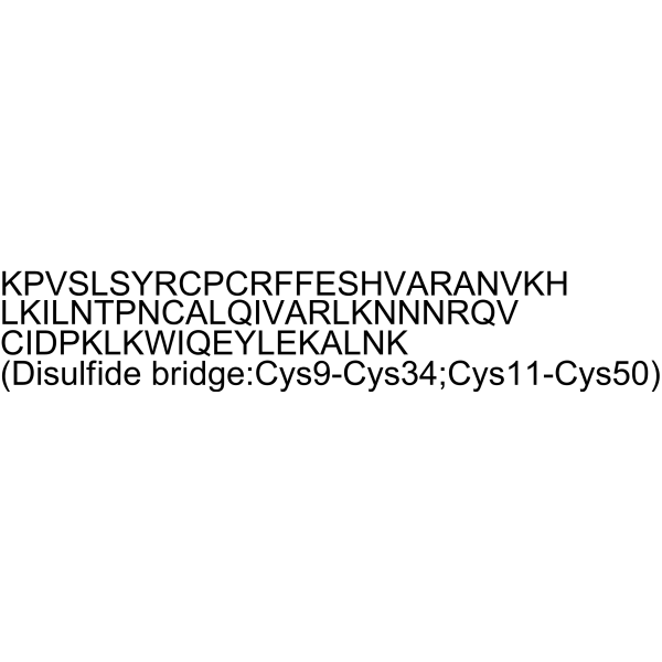
KPVSLSYRCPCRFFESHVARANVKHLKILNTPNCALQIVARLKNNNRQVCIDPKLKWIQEYLEKALNK (Disulfide bridge:Cys9-Cys34;Cys11-Cys50)
-
Shipping
Room temperature in continental US; may vary elsewhere.
-
Storage
Please store the product under the recommended conditions in the Certificate of Analysis.


Publications (2)
-
Journal Impact Factor
-
Most Recent
-
-
ACS Appl Mater Interfaces
Advanced Strategies in Bone Tissue Engineering: "Membrane-Jelly" Hydrogel System to Improve Bone Marrow Stem Cell Osteogenic Differentiation and Bone Regeneration. [Abstract]2025 Jun 18;17(24):34982-34996. PMID: 40492576


Purity & Documentation
References
[1]. Gaonkar RH, et al. Development of a New Class of CXCR4-Targeting Radioligands Based on the Endogenous Antagonist EPI-X4 for Oncological Applications. Journal of medicinal chemistry. 2023 Jul 13;66(13):8484-8497. [Content Brief]
[2]. Katzman BM, et al. Tetrahydroisoquinoline CXCR4 Antagonists Adopt a Hybrid Binding Mode within the Peptide Subpocket of the CXCR4 Receptor. ACS medicinal chemistry letters. 2019 Jan 10;10(1):67-73. [Content Brief]

Calculators
Concentration (start) × Volume (start) = Concentration (final) × Volume (final)


SDF-1α (human) Related Classifications
HY-P4911 Related Classifications
 Powered by Bioz