
SH491
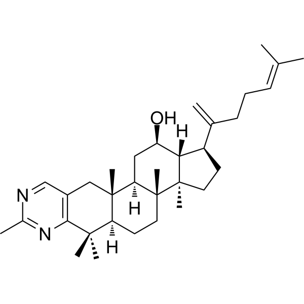
SH491 (Compound 33) is an antiosteoporosis agent. SH491 inhibits RANKL-induced osteoclast differentiation on bone-marrow-derived monocytes (BMMs) (IC50: 11.8 nM). SH491 inhibits the expression of osteoclastogenesis-related marker genes (TRAP, CTSK, MMP-9, and ATPase v0d2) and proteins (TRAP, CTSK, MMP-9).
For research use only. We do not sell to patients.
- CAS No.: 2975285-28-8
- Formula: C33H50N2O
- Molecular Weight:490.76
-
Storage:
Please store the product under the recommended conditions in the Certificate of Analysis.
All Cathepsin Isoforms
More
Biological Activity
Description
Cellular Effect
|
Cell Line
|
Type | Value | Description | References |
|---|---|---|---|---|
| BMMC | IC50 |
10.3 nM
Compound: 33; SH491
|
Anti-osteoclastogenic activity in C57/BL6 mouse BMMC cells assessed as inhibition of RANKL-induced osteoclast differentiation incubated for 5 to 7 days in presence of M-CSF by TRAP staining based microscopic analysis relative to control
|
[PMID: 37597216] |
Chemical Information
-
CAS No. 2975285-28-8
-
Molecular Weight 490.76
-
Formula C33H50N2O
-
SMILES
C[C@]12[C@]([H])(C(C)(C)C3=C(C2)C=NC(C)=N3)CC[C@@]4([C@@]1(C[C@H]([C@]5([C@]4(CC[C@@H]5C(CC/C=C(C)\C)=C)C)[H])O)[H])C
-
Shipping
Room temperature in continental US; may vary elsewhere.
-
Storage
Please store the product under the recommended conditions in the Certificate of Analysis.
Protocols
-
RNA extraction experimental
By lysing cells, releasing RNA, and removing impurities such as proteins and DNA, high-purity RNA products are finally obtained. The commonly used traditional method is the guanidine isothiocyanate/phenol/chloroform method (Trizol), which is suitable for a variety of animal materials including animal tissues, microorganisms, cultured cells, etc., and most plant materials.
-
Mitochondrial membrane-potential fluorescent assay
Mitochondrial membrane potential fluorescent assays estimate ΔΨm in living cells using lipophilic cationic dyes such as TMRM, TMRE, rhodamine 123, and JC-1, which accumulate in mitochondria according to membrane polarization; loss of signal after FCCP or CCCP treatment is interpreted as mitochondrial depolarization. TMRM/TMRE and rhodamine 123 are commonly used for semi-quantitative live-cell microscopy or flow cytometry, while JC-1 can report a shift from red aggregate fluorescence to green monomer fluorescence during depolarization; interpretation requires controls because dye concentration, quenching mode, cell type, dye efflux, and mitochondrial mass can affect fluorescence independently of ΔΨm.
-
Cell Cytotoxicity Assay
Cytotoxicity assays are usually based on the assessment of cell membrane damage, which can also be indirectly detected by measuring cell viability. Detection methods include MTT assay, CKK-8 assay, LDH assay and ATP assay, etc.
-
Mitochondrial membrane-potential and mitochondrial mass staining
Mitochondrial membrane potential staining measures the electrochemical polarization across the mitochondrial inner membrane in live cells using lipophilic cationic fluorescent probes; early rhodamine-based work showed that selective mitochondrial dye accumulation is lost when the mitochondrial transmembrane potential is dissipated. JC-1 reports mitochondrial polarization by shifting from green monomer fluorescence to red J-aggregate fluorescence as dye concentration increases within energized mitochondria; therefore, the red/green fluorescence ratio is used as a relative readout of mitochondrial membrane potential. TMRE or TMRM staining provides a single-channel relative readout because these cationic rhodamine esters accumulate in polarized mitochondria, and lower fluorescence indicates reduced mitochondrial polarization when acquisition and dye-loading conditions are controlled. Mitochondrial mass staining is commonly performed with MitoTracker Green FM or related MitoTracker dyes as
-
Osteoclast differentiation from monocyte/macrophage precursors
Osteoclast differentiation is an in vitro induction assay in which monocyte/macrophage-lineage precursors are exposed to macrophage colony-stimulating factor (M-CSF) and receptor activator of NF-κB ligand (RANKL), generating multinucleated osteoclasts that are commonly identified by tartrate-resistant acid phosphatase (TRAP) staining and functionally confirmed by resorption pits on dentin, bone, or mineralized substrates. M-CSF supports survival and expansion of osteoclast precursors, while RANKL binding to RANK drives osteoclast commitment, fusion, maturation, and resorptive function; osteoprotegerin inhibits this pathway by binding RANKL and preventing RANK activation. The main readouts are the number of TRAP-positive multinucleated cells, formation of F-actin rings, and resorbed surface area; TRAP-positive multinucleated cells indicate osteoclast differentiation, whereas pit formation on dentin, bone, or mineralized coating indicates functional bone-resorbing activity.
Purity & Documentation
References
Calculators
Concentration (start) × Volume (start) = Concentration (final) × Volume (final)

