



Taurolithocholic acid
Based on 6 publication(s) in Google Scholar
Taurolithocholic acid is an orally active bile acid and antiviral agent. Taurolithocholic acid upregulates FADS2 by activating the TGR5-PI3K/AKT-SREBP2 signaling axis, inhibits SFTSV-induced ferroptosis (Ferroptosis), viral replication and viral entry of HBV/HDV, while reducing the release of IL-1β, lipid ROS and LDH. While exerting antiviral protective effects, Taurolithocholic acid also stimulates the recycling of hepatocellular membrane transporters, impairs canalicular bile acid secretion function, and induces hepatocyte cholestasis, apoptosis and acute hepatocellular injury. Taurolithocholic acid serves as an experimental model compound for hepatocellular cholestasis. At concentrations ≤200 μM, Taurolithocholic acid shows no cytotoxicity and does not activate the interferon pathway. Taurolithocholic acid not only protects mice from lethal SFTSV infection but also is suitable for studies related to severe fever with thrombocytopenia syndrome and cholestasis.
For research use only. We do not sell to patients.
- Purity : 98.38%
- CAS No.: 516-90-5
- Formula: C26H45NO5S
- Molecular Weight:483.70
-
Storage:Powder -20°C, 3 years ; In solvent -80°C, 6 months , -20°C, 1 month


To place orders, for customer services and technical support, please contact: MedChemExpress USA
Tel: 609-228-6898 E-mail: [email protected] [email protected]


-
 Biological Activity
-
Chemical Information
-
Solvent & Solubility
- Protocols
- Purity & Documentation
- References
-
 Help & FAQs


-
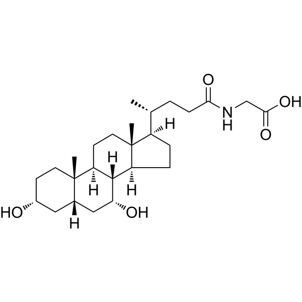
HY-N2334
Glycochenodeoxycholic acidEndogenous Metabolite -
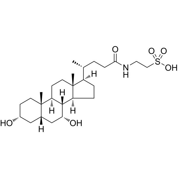
HY-N2027
Taurochenodeoxycholic acidEndogenous Metabolite -
HY-B0172
Lithocholic acidAutophagy Inducer -
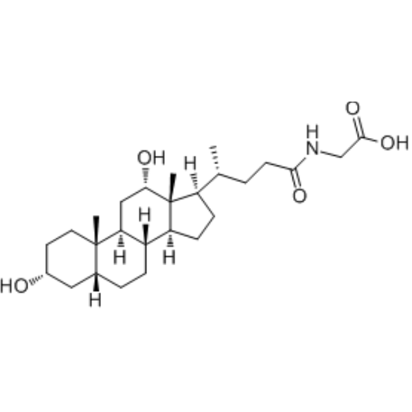
HY-125731
Glycodeoxycholic acidEndogenous Metabolite -
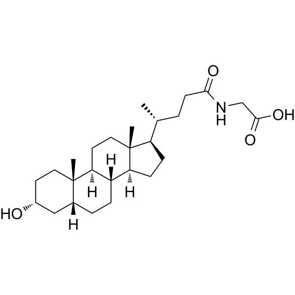
HY-116374
Glycolithocholic acidSecondary Bile Acid -
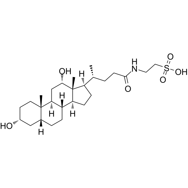
HY-B1899
Taurodeoxycholic acidAnti-inflammatory Agent -
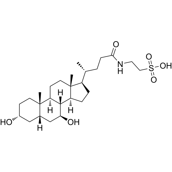
HY-19696
TauroursodeoxycholateEndoplasmic Reticulum Stress Inhibitor -
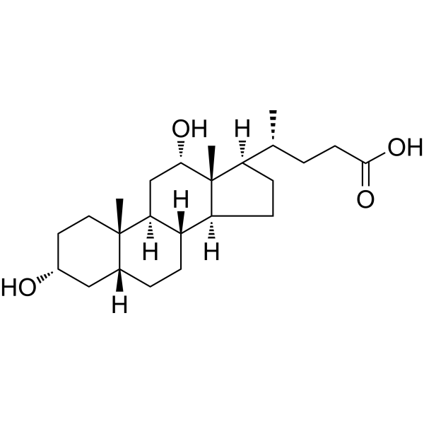
HY-N0593
Deoxycholic acidBile Acid Receptor Activator -
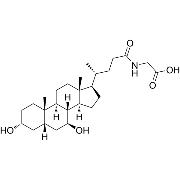
HY-N1424
Glycoursodeoxycholic acidEndogenous Metabolite -
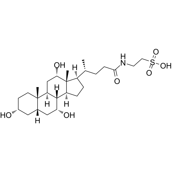
HY-B1788
Taurocholic acidImmunoregulation Agent -
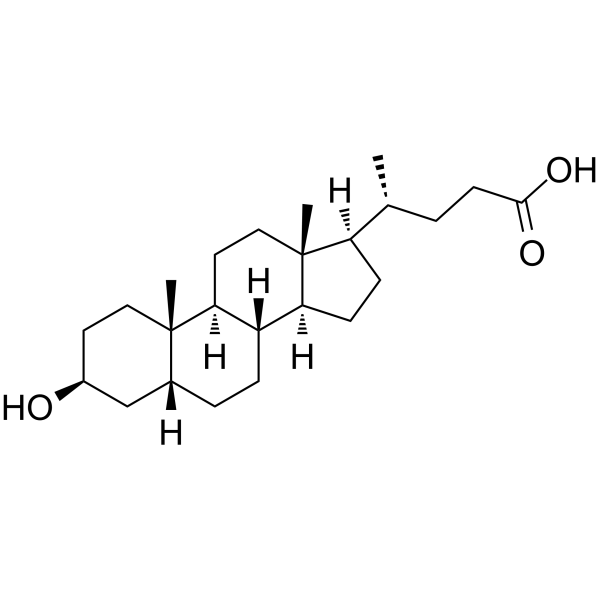
HY-B0172B
Isolithocholic acidBile Acid -
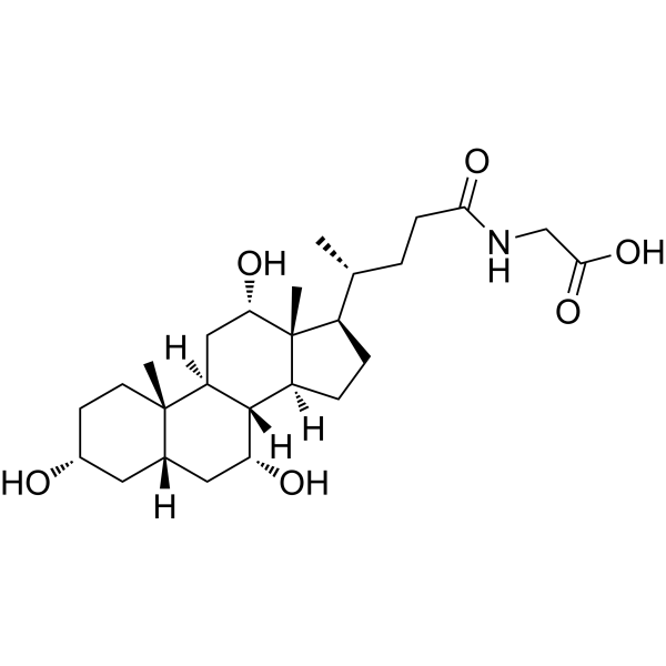
HY-N1423
Glycocholic acidBile Acid Derivative
Publications Citing Use of MedChemExpress (MCE) Taurolithocholic acid
More Customer Validation & Images

-
RT-PCR
All Calcium Channel Isoforms
More

Biological Activity
Description
IC50 & Target
|
Human Endogenous Metabolite |
Cellular Effect
|
Cell Line

|
Type | Value | Description | References |
|---|---|---|---|---|
| CHO | EC50 |
0.29 μM
Compound: 6a, tauro
|
Agonist activity at human TGR5 expressed in CHO cells by luciferase assay
Agonist activity at human TGR5 expressed in CHO cells by luciferase assay
|
[PMID: 18307294] |
| CHO | EC50 |
0.847 μM
Compound: TLCA
|
Agonist activity at recombinant human TGR5 expressed in CHO cells assessed as increase in cAMP accumulation after 30 mins by TR-FRET assay
Agonist activity at recombinant human TGR5 expressed in CHO cells assessed as increase in cAMP accumulation after 30 mins by TR-FRET assay
|
[PMID: 31268316] |
| CHO | EC50 |
330 nM
Compound: TLCA
|
Agonist activity at recombinant human TGR5 expressed in CHO cells assessed as increase in cAMP accumulation in presence of 3-isobutyl-1-methylxanthine after 20 mins
Agonist activity at recombinant human TGR5 expressed in CHO cells assessed as increase in cAMP accumulation in presence of 3-isobutyl-1-methylxanthine after 20 mins
|
[PMID: 31268316] |
| HEK293 | IC50 |
20 μM
Compound: Taurolithocholate
|
TP_TRANSPORTER: inhibition of E217betaG uptake in membrane vesicles from MRP4-expressing HEK-293 cells
TP_TRANSPORTER: inhibition of E217betaG uptake in membrane vesicles from MRP4-expressing HEK-293 cells
|
[PMID: 12523936] |
| HEK-293T | EC50 |
>150 μM
Compound: TLCA
|
Agonist activity at VP16 tagged-VDR-LBD (unknown origin) expressed in HEK293T cells assessed as SRC1 coactivator peptide recruitment after 16 hrs by luciferase reporter gene based two hybrid assay
Agonist activity at VP16 tagged-VDR-LBD (unknown origin) expressed in HEK293T cells assessed as SRC1 coactivator peptide recruitment after 16 hrs by luciferase reporter gene based two hybrid assay
|
[PMID: 26774929] |
| HEK-293T | IC50 |
>50 μM
Compound: TLCA
|
Antagonist activity against VP16 tagged-VDR-LBD (unknown origin) expressed in HEK293T cells assessed as inhibition of 1,25-dihydroxyvitamin D3-induced SRC1 coactivator peptide recruitment after 16 hrs by luciferase reporter gene based two hybrid assay
Antagonist activity against VP16 tagged-VDR-LBD (unknown origin) expressed in HEK293T cells assessed as inhibition of 1,25-dihydroxyvitamin D3-induced SRC1 coactivator peptide recruitment after 16 hrs by luciferase reporter gene based two hybrid assay
|
[PMID: 26774929] |
In Vitro
Taurolithocholic acid (50-200 μM; 24 h) dose-dependently inhibits SFTSV replication in human THP-1 monocytic cells without inducing cytotoxicity[1].
Taurolithocholic acid (50-200 μM; 24 h) dose-dependently inhibits HRTV replication in human THP-1 monocytic cells[1].
Taurolithocholic acid (16 h) inhibits the post-entry replication stage of SFTSV infection in human THP-1 monocytic cells, reducing intracellular viral RNA levels and infection rate by approximately 50% when added post-infection or throughout the 16 h infection period[1].
Taurolithocholic acid (50-200 μM) dose-dependently inhibits SFTSV mini-genome activity in BSR-T7 cells[1].
Taurolithocholic acid (50-200 μM; 24 h) dose-dependently suppresses SFTSV-induced IL-1β production and NF-κB activation in human THP-1 monocytic cells[1].
Taurolithocholic acid (200 μM; 24 h) upregulates FADS2 expression in SFTSV-infected human THP-1 monocytic cells, reversing SFTSV-induced FADS2 downregulation[1].
Taurolithocholic acid (0-24 h) time-dependently activates the TGR5-PI3K/AKT-SREBP2 pathway and upregulates FADS2 in human THP-1 monocytic cells over 0 to 24 h[1].
Taurolithocholic acid (100 μM; 24 h) alleviates SFTSV-induced ferroptosis in murine BMDMs, as measured by reduced lipid ROS and LDH release[1].
Taurolithocholic acid (100 μM; pre-treatment, followed by 24 h incubation with ferroptosis agonists) inhibits Fe2+- and RSL-3-induced ferroptosis in murine BMDMs via upregulation of FADS2, as shown by reduced lipid ROS and LDH release which is reversed by FADS2 inhibition[1].
Taurolithocholic acid (5 μM; 4 h) induces robust apoptosis in Ntcp-transfected HepG2 cells, increasing the percentage of apoptotic cells to 65.5% of total cells[2].
Taurolithocholic acid (2.5-5 μmol/liter; 20 min total incubation) impairs canalicular bile acid secretion by 54% and 60%, respectively, without affecting substrate uptake in isolated rat hepatocyte couplets[3].
Taurolithocholic acid (5 μmol/liter; 5-60 min) induces sustained activation of PKB/Akt, increasing activity to 194% of control levels after 60 min in isolated rat hepatocytes[3].
MedChemExpress (MCE) has not independently confirmed the accuracy of these methods. They are for reference only. Further protocols information, click here.
-
Cell Line:Ntcp-transfected human hepatoma (HepG2) cells
-
Concentration:5 μM
-
Incubation Time:4 h
-
Result:Increased the rate of apoptotic cell death to 65.5 ± 34.1% of total cells, compared to 1.5 ± 1.0% in control cells.
In Vivo
Taurolithocholic acid (final concentration of 1 μM in cerebrospinal fluid; chronic intracerebroventricular infusion; continuous constant-rate administration; treatment duration of 23 days) significantly enhances the uptake of triglyceride-derived fatty acids by brown adipose tissue, upregulates the mRNA expression of thermogenesis-related genes such as Lpl, Tgr5, Dio2 and Ucp1 in brown adipose tissue, and exerts no effect on energy expenditure, body weight or gonadal white adipose tissue weight in male C57Bl/6J mice fed a high-fat diet[4].
MedChemExpress (MCE) has not independently confirmed the accuracy of these methods. They are for reference only.
-
Animal Model:Male C57Bl/6J mice (male, 15 weeks old), fed with high fat diet (HFD, energetic content: 45% fat, 35% carbohydrates, 20% protein)[4]
-
Dosage:Final concentration of 1 μM in cerebrospinal fluid,
-
Administration:chronic intracerebroventricular (i.c.v.) infusion
-
Result:Significantly enhanced the uptake of triglyceride-derived fatty acids from [3H]TO-labeled emulsion particles by subscapular brown adipose tissue (sBAT), and markedly upregulated the mRNA expression of thermogenic genes including Lpl, Tgr5, Dio2 and Ucp1 in interscapular brown adipose tissue (iBAT). It showed a trend of increased Ucp1 mRNA expression in subcutaneous white adipose tissue (sWAT), and had no effect on energy expenditure, body weight, gonadal white adipose tissue (gWAT) weight, and the uptake of triglyceride-derived fatty acids by white adipose tissue depots.


Chemical Information
-
CAS No. 516-90-5
-
Appearance Solid
-
Molecular Weight 483.70
-
Formula C26H45NO5S
-
Color White to off-white
-
SMILES
C[C@@]12[C@](CC[C@]2([H])[C@H](C)CCC(NCCS(=O)(O)=O)=O)([H])[C@@]3([H])[C@@](CC1)([H])[C@@]4([C@](C[C@@H](CC4)O)([H])CC3)C
-
Shipping
Room temperature in continental US; may vary elsewhere.
-
Storage
Powder -20°C 3 years In solvent -80°C 6 months -20°C 1 month


Publications (6)
-
Journal Impact Factor
-
Most Recent
-
Nature
2026 Mar;651(8104):260-267. PMID: 41606328 -
Nature
2025 Jul;643(8070):192-200. PMID: 39695227 -
Nat Microbiol
Taurolithocholic acid protects against viral haemorrhagic fever via inhibition of ferroptosis. [Abstract]2024 Oct;9(10):2583-2599. PMID: 39294459 -
Adv Sci (Weinh)
Targeting FDFT1 Reduces Cholesterol and Bile Acid Production and Delays Hepatocellular Carcinoma Progression Through the HNF4A/ALDOB/AKT1 Axis. [Abstract]2025 Mar;12(12):e2411719. PMID: 39899681
Taurolithocholic acid purchased from MedChemExpress. Usage Cited in: Adv Sci (Weinh). 2025 Mar;12(12):e2411719. [Abstract]
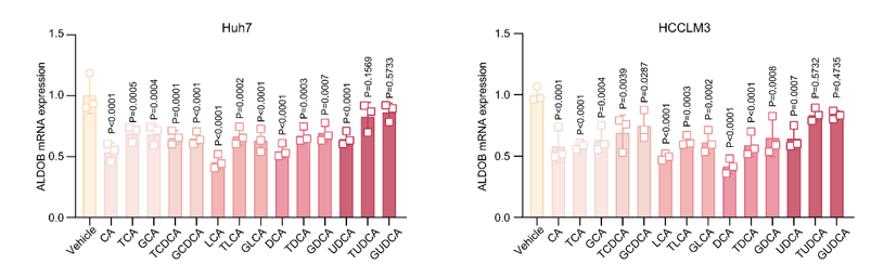
The mRNA levels of ALDOB in Huh7 or HCCLM3 cells treated with various bile acids (100 μM, 24 h). All data are presented as mean ± SD. Data were analyzed by one-way ANOVA with Bonferroni multiple-comparison correction. CA, cholic acid; TCA, taurocholic acid; GCA, glycocholic acid; TCDCA, taurochenodeoxycholic acid; GCDCA, glycochenodeoxycholic acid; LCA, lithocholic acid; TLCA, taurolithocholic acid; GLCA, glycolithocholic acid; DCA, deoxycholic acid; TDCA, taurodeoxycholic acid; GDCA, glycodeoxycholic acid; UDCA, ursodeoxycholic acid; TUDCA, tauroursodeoxycholic acid; GUDCA, glycoursodeoxycholic acid.
-
J Transl Med
Conjugated bile acids alleviate acute pancreatitis through inhibition of TGR5 and NLRP3 mediated inflammation. [Abstract]2024 Dec 20;22(1):1124. PMID: 39707318 -
Biomedicines
2025 Apr 4;13(4):874. PMID: 40299495


Solvent & Solubility
In Vitro:
DMSO : 16.67 mg/mL (34.46 mM; Need ultrasonic; Hygroscopic DMSO has a significant impact on the solubility of product, please use newly opened DMSO)
Please refer to the solubility information to select the appropriate solvent. Once prepared, please aliquot and store the solution to prevent product inactivation from repeated freeze-thaw cycles.
Storage method and period of stock solution: -80°C, 6 months; -20°C, 1 month. When stored at -80°C, please use it within 6 months. When stored at -20°C, please use it within 1 month.
Please refer to the solubility information to select the appropriate solvent. Once prepared, please aliquot and store the solution to prevent product inactivation from repeated freeze-thaw cycles.
Storage method and period of stock solution: -80°C, 6 months; -20°C, 1 month. When stored at -80°C, please use it within 6 months. When stored at -20°C, please use it within 1 month.
Concentration (start) × Volume (start) = Concentration (final) × Volume (final)
In Vivo:
Select the appropriate dissolution method based on your experimental animal and administration route.
- For the following dissolution methods, please ensure to first prepare a clear stock solution using an In Vitro approach and then sequentially add co-solvents:
- To ensure reliable experimental results, the clarified stock solution can be appropriately stored based on storage conditions. As for the working solution for In Vivo experiments, it is recommended to prepare freshly and use it on the same day.
- The percentages shown for the solvents indicate their volumetric ratio in the final prepared solution. If precipitation or phase separation occurs during preparation, heat and/or sonication can be used to aid dissolution.
Add each solvent one by one: 10% DMSO 40% PEG300 5% Tween-80 45% Saline
Solubility: ≥ 1.67 mg/mL (3.45 mM); Clear solution
This protocol yields a clear solution of ≥ 1.67 mg/mL (saturation unknown).
Taking 1 mL working solution as an example, add 100 μL DMSO stock solution (16.7 mg/mL) to 400 μL PEG300, and mix evenly; then add 50 μL Tween-80 and mix evenly; then add 450 μL Saline to adjust the volume to 1 mL.
Preparation of Saline: Dissolve 0.9 g sodium chloride in ddH₂O and dilute to 100 mL to obtain a clear Saline solution.
Add each solvent one by one: 10% DMSO 90% (20% SBE-β-CD in Saline)
Solubility: ≥ 1.67 mg/mL (3.45 mM); Clear solution
This protocol yields a clear solution of ≥ 1.67 mg/mL (saturation unknown).
Taking 1 mL working solution as an example, add 100 μL DMSO stock solution (16.7 mg/mL) to 900 μL 20% SBE-β-CD in Saline, and mix evenly.
Preparation of 20% SBE-β-CD in Saline (4°C, storage for one week): 2 g SBE-β-CD powder is dissolved in 10 mL Saline, completely dissolve until clear.
In Vivo Dissolution Calculator
Please enter the basic information of animal experiments:
-
-
-
-
Recommended: Prepare an additional quantity of animals to account for potential losses during experiments.
Please enter your animal formula composition:
-
%DMSO +
Recommended: Keep the proportion of DMSO in working solution below 2% if your animal is weak.
-
%+
-
+%Tween-80 + +
-
%Saline +
The co-solvents required include: DMSO, . All of co-solvents are available by MedChemExpress (MCE). , Tween 80. All of co-solvents are available by MedChemExpress (MCE).
Working solution concentration: 0.22 mg/mL
Method for preparing stock solution: mg drug dissolved in μL DMSO. Stock solution concentration: mg/mL.
1. Take μL DMSO stock solution;
2. Add μL .
μL , mix evenly;
3. Then add μL Tween 80, mix evenly;
4. Then add μL
Please ensure that the stock solution in the first step is dissolved to a clear state, and add co-solvents in sequence. You can use ultrasonic heating (ultrasonic cleaner, recommended frequency 20-40 kHz), vortexing, etc. to assist dissolution.

Protocols
-
Research Protocol for Infectious Diseases
Infectious-disease experiments test how pathogens interact with host barriers, innate immune receptors, inflammatory signaling, pathogen replication, and tissue injury; pattern-recognition receptors such as TLRs, RIG-I-like receptors, NOD-like receptors, and inflammasomes detect microbial molecules and activate NF-κB, interferon, and cytokine responses. The central hypothesis is that infection severity reflects the balance between pathogen burden and host response: protective inflammation restricts pathogen growth, whereas excessive or mislocalized inflammation contributes to tissue damage and disease phenotype. Unresolved questions include which host pathways are protective versus pathogenic, why some infection models fail to translate to human disease, and which combined readouts best predict clinically relevant infection outcomes.
-
Apoptosis
Apoptosis, also called programmed cell death, is generally characterized by distinct morphological characteristics.
-
TUNEL staining for apoptotic DNA fragmentation
TUNEL staining detects DNA strand breaks by using terminal deoxynucleotidyl transferase to add labeled nucleotides to exposed 3′-OH DNA termini, generating either microscopic staining in fixed cells or tissue sections, or fluorescence/cytometric signal in cell suspensions. TUNEL positivity reflects DNA fragmentation but should not be interpreted alone as definitive apoptosis, because TUNEL can also label necrotic, autolytic, mechanically damaged, or DNA-repair-associated DNA breaks.
-
Cell Cytotoxicity Assay
Cytotoxicity assays are usually based on the assessment of cell membrane damage, which can also be indirectly detected by measuring cell viability. Detection methods include MTT assay, CKK-8 assay, LDH assay and ATP assay, etc.
-
Mammalian live/dead viability and cytotoxicity staining
Live/dead viability and cytotoxicity staining assays are based on the simultaneous detection of intracellular esterase activity in metabolically active (viable) cells and membrane integrity loss in non-viable cells. In commonly used dual-staining approaches, membrane-permeant fluorogenic substrates are converted by intracellular esterases into fluorescent products in live cells, while impermeant DNA-binding dyes selectively enter cells with compromised plasma membranes and label nucleic acids in dead or dying cells, enabling discrimination between viable and non-viable populations by fluorescence microscopy or flow cytometry.
-
Annexin V plus membrane-impermeant dye apoptosis staining
Annexin V-based apoptosis assays rely on the detection of phosphatidylserine (PS) externalization from the inner leaflet of the plasma membrane to the outer leaflet, an early biochemical hallmark of apoptosis. Fluorescently labeled Annexin V binds PS in a calcium-dependent manner, enabling identification of early apoptotic cells by flow cytometry or fluorescence microscopy. When combined with a membrane-impermeant DNA-binding dye (e. g. , propidium iodide), this approach allows discrimination between viable (Annexin V−/dye−), early apoptotic (Annexin V+/dye−), and late apoptotic or necrotic (Annexin V+/dye+) cell populations by assessing membrane integrity and PS exposure.
-
ROS/oxidative-stress fluorescent staining
ROS/oxidative-stress fluorescent staining uses cell-permeant fluorogenic probes that become fluorescent after oxidation inside cells or tissues; commonly used examples include DCFH-DA/DCFDA for broad cellular oxidant detection, DHE for superoxide-related signal detection, MitoSOX for mitochondrial superoxide-related signal detection, and CellROX probes for oxidative-stress-associated fluorescence readouts. The assay detects probe oxidation rather than a single ROS species unless the probe and analysis method have been chemically validated for that species. DCFH-DA enters cells, is deacetylated by intracellular esterases to DCFH, and produces fluorescent DCF after oxidation, so the readout is used as an operational measure of total cellular oxidative stress rather than a species-specific ROS measurement. DHE and MitoSOX can report superoxide-related oxidation, but red fluorescence alone can include non-specific ethidium-like oxidation products; HPLC or optimized spectral approaches are
-
Apoptosis Solutions
Apoptosis is a regulated, generally non-lytic cell-death pathway that removes unwanted, damaged, infected, or abnormal cells through coordinated morphological changes, caspase activation, DNA fragmentation, and membrane remodeling. The intrinsic apoptosis pathway is controlled mainly by mitochondrial outer membrane permeabilization, BCL-2 family proteins, cytochrome c release, apoptosome formation, caspase-9 activation, and downstream executioner caspase-3/7 activation. The extrinsic apoptosis pathway is initiated by death receptors such as Fas, TNFR, and TRAIL receptors, which recruit adaptor proteins and activate caspase-8 before engaging executioner caspases or mitochondrial amplification through BID cleavage. Apoptosis is linked to many phenotypes, including cancer cell killing, tissue homeostasis, immune regulation, neurodegeneration, infection response, and treatment-induced cytotoxicity; unresolved questions include how apoptosis interacts with necroptosis, pyroptosis, ferroptos
-
Ferroptosis Solutions
Ferroptosis is an iron-dependent, non-apoptotic form of regulated cell death characterized by lethal lipid peroxidation and sensitivity to suppression by iron chelators or lipophilic radical-trapping antioxidants. The core pathway links cystine uptake through system Xc−, glutathione availability, GPX4-dependent detoxification of phospholipid hydroperoxides, iron-dependent oxidative reactions, and polyunsaturated-phospholipid metabolism into a cell-death program that is biochemically and morphologically distinct from apoptosis, necrosis, and autophagy. The ferroptosis pathway is experimentally linked to phenotype through chemical and genetic perturbation. Erastin induces ferroptosis by inhibiting cystine uptake through system Xc− and weakening antioxidant defenses, while GPX4 inhibition or depletion causes lipid peroxide accumulation and ferroptotic cancer-cell death. ACSL4 and oxidizable arachidonoyl- or adrenoyl-containing phosphatidylethanolamines shape ferroptosis sensitivity by con
-
Pyroptosis Solutions
Pyroptosis is a lytic inflammatory cell-death pathway executed by gasdermin pores, most classically through inflammasome-mediated activation of caspase-1, cleavage of gasdermin D, membrane pore formation, LDH release, and secretion of IL-1β and IL-18. The canonical pathway is commonly modeled by priming cells with an inflammatory signal such as LPS to induce pro-IL-1β and inflammasome components, followed by an activation signal such as ATP or nigericin to activate NLRP3, ASC speck formation, caspase-1 cleavage, GSDMD cleavage, cytokine release, and pyroptotic membrane rupture. The non-canonical pathway is triggered when cytosolic LPS activates mouse caspase-11 or human caspase-4/5, leading to GSDMD cleavage and pyroptosis, and this can secondarily activate NLRP3-dependent IL-1β release. Pyroptosis is linked to inflammatory injury, infection, cancer, liver disease, ocular disease, placental inflammation, and other disease phenotypes, but unresolved questions include which gasdermin fam
-
Cell Viability Determination by MTT Colorimetric Assay
The following protocol uses the MTT colorimetric assay as a classic literature-established method for assessing cell viability/metabolic activity in cultured mammalian cells. MTT[3-(4,5-dimethylthiazol-2-yl)-2,5-diphenyltetrazolium bromide] is reduced by metabolically active cells to a colored formazan product; the amount of formazan is quantified spectrophotometrically and provides an indirect measure of metabolically active viable cells. Importantly, MTT reduction reflects cellular oxidoreductase/metabolic activity rather than an absolute direct count of living cells, so changes in cellular metabolism can alter the signal independently of cell number.


Purity & Documentation
-
Data Sheet (281 KB)
-
SDS (419 KB)
- English - EN (419 KB)
- Français - FR (419 KB)
- Deutsch - DE (419 KB)
- Norwegian - NO (419 KB)
- Español - ES (419 KB)
- Swedish - SV (419 KB)
- Italian - IT (419 KB)
- Korean - KR (419 KB)
- Portuguese - PT (419 KB)
-
Handling Instructions (2659 KB)
References
[1]. Zheng X, et al. Taurolithocholic acid protects against viral haemorrhagic fever via inhibition of ferroptosis. Nat Microbiol. 2024;9(10):2583-2599. [Content Brief]
[2]. Denk GU, et al. Conjugation is essential for the anticholestatic effect of NorUrsodeoxycholic acid in taurolithocholic acid-induced cholestasis in rat liver. Hepatology. 2010;52(5):1758-1768. [Content Brief]
[3]. Beuers U, et al. Taurolithocholic acid exerts cholestatic effects via phosphatidylinositol 3-kinase-dependent mechanisms in perfused rat livers and rat hepatocyte couplets. J Biol Chem. 2003;278(20):17810-17818. [Content Brief]


Complete Stock Solution Preparation Table
Please refer to the solubility information to select the appropriate solvent. Once prepared, please aliquot and store the solution to prevent product inactivation from repeated freeze-thaw cycles.
Storage method and period of stock solution: -80°C, 6 months; -20°C, 1 month. When stored at -80°C, please use it within 6 months. When stored at -20°C, please use it within 1 month.
| Optional Solvent | Concentration Solvent Mass | 1 mg | 5 mg | 10 mg | 25 mg |
|---|---|---|---|---|---|
| DMSO | 1 mM | 2.0674 mL | 10.3370 mL | 20.6740 mL | 51.6849 mL |
| 5 mM | 0.4135 mL | 2.0674 mL | 4.1348 mL | 10.3370 mL | |
| 10 mM | 0.2067 mL | 1.0337 mL | 2.0674 mL | 5.1685 mL | |
| 15 mM | 0.1378 mL | 0.6891 mL | 1.3783 mL | 3.4457 mL | |
| 20 mM | 0.1034 mL | 0.5168 mL | 1.0337 mL | 2.5842 mL | |
| 25 mM | 0.0827 mL | 0.4135 mL | 0.8270 mL | 2.0674 mL | |
| 30 mM | 0.0689 mL | 0.3446 mL | 0.6891 mL | 1.7228 mL |


Taurolithocholic acid Related Classifications
HY-113308 Related Classifications
 Powered by Bioz