



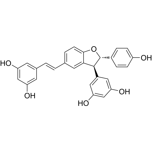
δ-Viniferin
δ-Viniferin is a COX-1/COX-2 inhibitor (with an IC50 of 5 µM for both enzymes). δ-Viniferin induces the expression of SIRT1, catalase and HO-1, promotes the phosphorylation of eNOS, and stimulates nitric oxide (NO) production. δ-Viniferin is applicable to research related to atherosclerosis, downy mildew, Gram-positive bacterial infections, inflammation, infections, cardiovascular diseases and diabetes.
For research use only. We do not sell to patients.
- CAS No.: 681293-15-2
- Formula: C28H22O6
- Molecular Weight:454.47
-
Storage:
Please store the product under the recommended conditions in the Certificate of Analysis.

To place orders, for customer services and technical support, please contact: MedChemExpress USA
Tel: 609-228-6898 E-mail: [email protected] [email protected]


-
 Biological Activity
-
Chemical Information
- Protocols
- Purity & Documentation
- References
-
 Help & FAQs




Biological Activity
Description
|
COX-1 5 μM (IC50) |
COX-2 5 μM (IC50) |
SIRT1 |
HO-1 |
eNOS |
Cellular Effect
|
Cell Line

|
Type | Value | Description | References |
|---|---|---|---|---|
| CCRF-CEM | IC50 |
49 μM
Compound: 4
|
Cytotoxicity against human CEM cells
Cytotoxicity against human CEM cells
|
[PMID: 10650073] |
| HepG2 | IC50 |
48 μM
Compound: 6
|
Cytotoxicity against human HepG2 cells after 3 days by microplate reader analysis
Cytotoxicity against human HepG2 cells after 3 days by microplate reader analysis
|
[PMID: 32705864] |
| MCF-10A | IC50 |
48 μM
Compound: 6
|
Cytotoxicity against human MCF-10A cells after 3 days by microplate reader analysis
Cytotoxicity against human MCF-10A cells after 3 days by microplate reader analysis
|
[PMID: 32705864] |
| RAW264.7 | IC50 |
80.2 μM
Compound: 2a
|
Antiinflammatory activity in mouse RAW264.7 cells assessed as inhibition of LPS/IFNgamma-induced NO production by measuring NO level preincubated for 2 hrs and followed by LPS/IFNgamma addition and measured after 24 hrs by Griess assay
Antiinflammatory activity in mouse RAW264.7 cells assessed as inhibition of LPS/IFNgamma-induced NO production by measuring NO level preincubated for 2 hrs and followed by LPS/IFNgamma addition and measured after 24 hrs by Griess assay
|
[PMID: 31350127] |
In Vitro
δ-Viniferin (5 μM; 24 h) promotes wound repair in porcine aortic vascular endothelial cells and increases nitric oxide production in these cells at a concentration of 5 μM[1].
δ-Viniferin (5 μM; 24 h) induces phosphorylation of endothelial nitric oxide synthase in a time-dependent manner and upregulates the expression of SIRT1, HO-1, and catalase in porcine aortic endothelial cells at a concentration of 5 μM[1].
δ-Viniferin (5-10 μM; 24 h) protects porcine aortic vascular endothelial cells against H2O2-induced reduction in cell viability at a concentration of 5 μM[1].
δ-Viniferin (5 μM) inhibits the activities of cyclooxygenase-1 and cyclooxygenase-2, with an IC50 of 5 μM[3].
MedChemExpress (MCE) has not independently confirmed the accuracy of these methods. They are for reference only. Further protocols information, click here.
-
Cell Line:porcine aortic vascular endothelial cells (VECs)
-
Concentration:5, 10, 20 μM
-
Incubation Time:24 h
-
Result:Stimulated VEC wound repair significantly at 5 μM.
Did not produce a significant stimulatory effect.
-
Cell Line:porcine aortic vascular endothelial cells (VECs)
-
Concentration:5 μM
-
Incubation Time:6, 12, 24 h (phos-eNOS, SIRT1, HO-1 analysis); 1, 3, 6, 12, 24 h (catalase analysis)
-
Result:Induced phosphorylation of eNOS and increased expression of SIRT1 and HO-1 over 6, 12, and 24 hours.
Increased catalase protein expression in a time-dependent manner, with a significant 3-fold increase observed after 24 hours.
-
Cell Line:porcine aortic vascular endothelial cells (VECs)
-
Concentration:5, 10 μM
-
Incubation Time:24 h
-
Result:Reversed the reduction in cell viability caused by H2O2 exposure significantly at 5 μM.
Parmacokinetics
| Species | Dose | Route | AUC0-t | AUC0-∞ | MRT0-t | MRT0-∞ | T1/2β | Tmax | Vz/F | CLz/F | Cmax | Bioavailability |
|---|---|---|---|---|---|---|---|---|---|---|---|---|
| Rat[5] | 70 mg/kg | p.o. | 1162.0 μg/L·h | 1181.9 μg/L·h | 5.6 h | 6.0 h | 4.2 h | 1.7 h | 379.7 L/kg | 60.6 L/h/kg | 399.0 μg/L | 2.3 % |
| Rat[5] | 1.37 mg/kg | i.v. | 1004.7 μg/L·h | 1017.6 μg/L·h | 1.5 h | 2.0 h | 5.8 h | 0.03 h | 12.2 L/kg | 1.4 L/h/kg | 7611.7 μg/L | / |


Chemical Information
-
CAS No. 681293-15-2
-
Molecular Weight 454.47
-
Formula C28H22O6
-
SMILES
OC1=CC(O)=CC(/C=C/C2=CC=C3O[C@H](C4=CC=C(C=C4)O)[C@@H](C5=CC(O)=CC(O)=C5)C3=C2)=C1
-
Structure Classification
-
Initial Source
-
Shipping
Room temperature in continental US; may vary elsewhere.
-
Storage
Please store the product under the recommended conditions in the Certificate of Analysis.

Protocols
-
Kinase activity and phosphorylation assays
Kinase activity assays measure the ability of kinases to transfer phosphate groups from ATP to specific substrates, while phosphorylation assays detect the presence and levels of phosphorylated proteins. Common methods include radiolabeled ATP incorporation (e. g. ,), ADP release detection via bioluminescence (e. g. ,[3]), enzyme-linked immunosorbent assays (ELISA) for phospho-specific epitopes (e. g. ,[6]), and microtiter-based formats for high-throughput screening (e. g. ,[8]). The ADP-Glo assay quantifies kinase activity by measuring ADP produced during phosphorylation using a luciferase-based system. Radiometric assays involve autoradiography or scintillation counting after incorporation of 32P-labeled ATP into substrate proteins. ELISA-based approaches rely on phospho-specific antibodies to detect activated kinases in cell lysates or purified samples.
-
RNA extraction experimental
By lysing cells, releasing RNA, and removing impurities such as proteins and DNA, high-purity RNA products are finally obtained. The commonly used traditional method is the guanidine isothiocyanate/phenol/chloroform method (Trizol), which is suitable for a variety of animal materials including animal tissues, microorganisms, cultured cells, etc., and most plant materials.
-
Western Blot
Western blotting (WB) is a commonly used experimental method in molecular biology, biochemistry, and immunogenetics for identifying and quantifying target proteins. It combines gel electrophoresis with immunoassay, enabling researchers to analyze protein expression, post-translational modifications, and molecular weight.
-
Cell Cytotoxicity Assay
Cytotoxicity assays are usually based on the assessment of cell membrane damage, which can also be indirectly detected by measuring cell viability. Detection methods include MTT assay, CKK-8 assay, LDH assay and ATP assay, etc.
-
Research Protocol for Cardiovascular Diseases
Cardiovascular disease can be modeled as maladaptive cardiac remodeling, where ischemic injury or pressure overload activates inflammatory signaling, fibroblast activation, extracellular-matrix deposition, cardiomyocyte hypertrophy, vascular remodeling, and progressive ventricular dysfunction. The TGF-β/SMAD axis is a central profibrotic pathway after myocardial injury and pressure overload, while innate immune and cytokine pathways regulate leukocyte recruitment, scar formation, and adverse remodeling. Key unresolved questions include which inflammatory signals are reparative versus harmful, when fibrosis is protective versus maladaptive, and whether pathway inhibition improves function without weakening necessary infarct healing or compensatory remodeling.
-
Protocol for Kinase activity and phosphorylation assays
Kinase activity assays measure transfer of phosphate from ATP to a protein or peptide substrate, generating phosphorylated substrate, ADP, or incorporated radiolabeled phosphate as the readout; phosphorylation assays measure site-specific phosphorylation in cells or tissues as a proxy for kinase-pathway activation, inhibition, or substrate regulation. Phosphorylation can be detected by phospho-specific Western blot, immunoprecipitation kinase assay, phospho-immunofluorescence, phospho-flow cytometry, luminescent ADP detection, radiolabeled ATP incorporation, or reporter-based pathway assays, and these readouts can be applied to cancer cells, primary neurons, mouse tumors, organoids, inflammatory macrophages, ferroptosis studies, and mitophagy studies when the kinase target is biologically relevant.
-
Gram Staining of Tissue Sections
Gram staining of tissue sections is a histochemical technique used to differentiate Gram-positive and Gram-negative bacteria within histological specimens based on differences in bacterial cell wall structure and dye retention, adapted from classical bacteriological Gram staining into tissue-compatible “histological Gram stain” variants. In tissue applications, modifications of the Brown-Hopps and Brown-Brenn methods are commonly used to improve differentiation of microorganisms embedded within host connective tissue and to reduce overstaining or loss of Gram-negative signal, which are known limitations of earlier approaches. The principle relies on crystal violet-iodine complex retention in Gram-positive organisms and subsequent decolorization and counterstaining steps that allow contrast visualization of Gram-negative organisms against tissue background.
-
Research Protocol for Inflammation-related Diseases
The NLRP3 inflammasome is a cytosolic innate immune signaling platform that integrates priming signals and danger-signal activation to promote caspase-1 activation, maturation of IL-1β and IL-18, and gasdermin D-mediated pyroptotic cell death. The core experimental logic is to determine whether inflammatory disease phenotypes are driven by increased NLRP3 expression, ASC-containing inflammasome assembly, caspase-1 cleavage, GSDMD cleavage, and extracellular release of IL-1β/IL-18 rather than by nonspecific cell injury alone. The pathway is strongly linked to inflammation-related disease phenotypes because monosodium urate crystals activate NALP3/NLRP3 inflammasome signaling in gout-like crystal inflammation, cholesterol crystals activate NLRP3 inflammasomes in atherogenesis models, and DSS-induced intestinal inflammation has been reported to involve NLRP3 inflammasome activity. However, experimental colitis studies also show context-dependent protective effects of NLRP3 inflammasome co
-
Research Protocol for Metabolic Diseases
AMP-activated protein kinase, AMPK, is a conserved cellular energy sensor that responds to reduced cellular energy status and coordinates metabolism by increasing ATP-generating catabolic pathways while suppressing ATP-consuming anabolic processes. In metabolic disease research, the AMPK pathway is experimentally relevant because it regulates hepatic lipid synthesis, fatty acid oxidation, glucose production, skeletal-muscle glucose disposal, mTORC1-linked biosynthesis, autophagy, mitochondrial homeostasis, and whole-body energy balance. The central pathway logic is that energy stress, metformin, exercise-like stimulation, or direct AMPK activators increase AMPKα Thr172 phosphorylation and downstream substrate phosphorylation, including ACC and RAPTOR. Phosphorylation of ACC suppresses lipogenesis and supports fatty acid oxidation, whereas phosphorylation of RAPTOR suppresses mTORC1 signaling and links cellular energy status to growth and protein synthesis control. The pathway is linked


Purity & Documentation
References

Calculators
Concentration (start) × Volume (start) = Concentration (final) × Volume (final)


δ-Viniferin Related Classifications
HY-N3841A Related Classifications
 Powered by Bioz