



BT-PROTAC
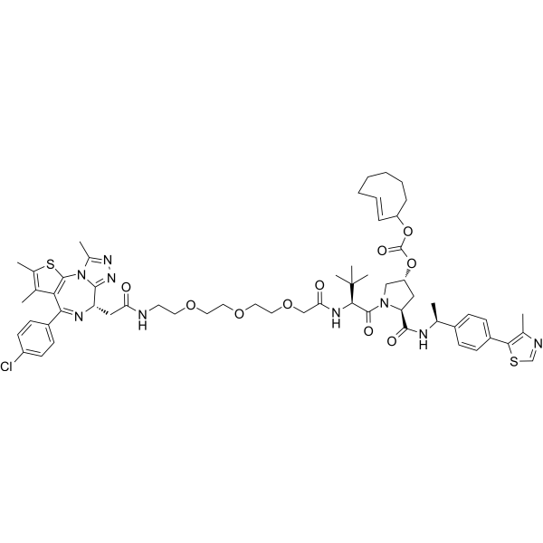
BT-PROTAC is a bioorthogonally activatable BRD4/BRD3 PROTAC degrader prodrug. BT-PROTAC is inactive when present alone, but upon activation by tetrazine compounds, it promotes the degradation of BRD4 and BRD3 via the ubiquitin-proteasome system, inhibits c-Myc expression, activates caspase-3, and induces apoptosis in breast cancer cells. BT-PROTAC can be used for breast cancer research.
(Pink: BRD4 and BRD3 ligand (HY-13030); Blue: VHL ligand (HY-125845); Black: linker (HY-140189)).
Nos produits utilisent uniquement pour la recherche. Nous ne vendons pas aux patients.
- CAS No.: 3032848-64-6
- Formule: C59H74ClN9O10S2
- Masse moléculaire:1168.86
-
Stockage:
Please store the product under the recommended conditions in the Certificate of Analysis.

To place orders, for customer services and technical support, please contact: MedChemExpress USA
Tel: 609-228-6898 E-mail: [email protected] [email protected]


-
 Activité biologique
-
Chemical Information
- Protocole
- Pureté et documentation
- Références
-
 Help & FAQs


Voir tous les produits spécifiques à Isoform PROTACs
MoreVoir tous les produits spécifiques à Isoform Caspase
More

Activité biologique
Description
IC50 & Target
[1]|
BRD4 |
BRD3 |
VHL |
Caspase 3 |
In Vitro
BT-PROTAC (48 h) exhibits low cytotoxicity when acting alone on MDA-MB-231 breast cancer cells, with an IC50 of 652.2 nM; however, its cytotoxicity is restored after pre-incubation with BODIPY-TZ, and the IC50 decreases to 326.5 nM[1].
BT-PROTAC (48 h) exhibits low cytotoxicity when applied alone to MCF-7 breast cancer cells, with an IC50 of 1319.0 nM; however, its cytotoxicity is restored after pre-incubation with BODIPY-TZ, and the IC50 decreases to 263.6 nM[1].
BT-PROTAC (0-2.0 μM; 48 h) alone does not degrade BRD4 or BRD3 in T47D, MB-231, and MCF-7 breast cancer cells even at concentrations as high as 2.0 μM, but achieves degradation of BRD4 and BRD3 via the ubiquitin-proteasome system after pre-incubation with BODIPY-TZ[1].
MedChemExpress (MCE) has not independently confirmed the accuracy of these methods. They are for reference only.
-
Cell Line:T47D, MDA-MB-231 and MCF-7 breast cancer cells
-
Concentration:0, 0.2, 0.4, 0.8, 1 and 2 μM (BT-PROTAC alone); 0, 25, 100, 200, 400 and 1000 nM (BT-PROTAC after 20.0 μM BODIPY-TZ preincubation)
-
Incubation Time:48 h (BT-PROTAC incubation); 12 h (BODIPY-TZ preincubation)
-
Result:Did not degrade BRD4 or BRD3 at concentrations up to 2.0 μM when used alone.
Significantly reduced BRD4 and BRD3 protein levels when combined with BODIPY-TZ.
Had its degradation effect blocked by 10.0 μM MG132 (HY-13259) or 3.0 μM MLN4924 (HY-70062), confirming dependence on the ubiquitin-proteasome system.
In Vivo
MedChemExpress (MCE) has not independently confirmed the accuracy of these methods. They are for reference only.
-
Animal Model:BALB/c nude mice (female, 6-8 weeks old)[1]
-
Dosage:20 mg/kg (preceded by 50 mg/kg IR808-TZ 2 hours earlier)
-
Administration:i.v.; single administration
-
Result:Reduced tumor volume increase to 1.74-fold at 23 days post-injection, compared to 5.98-fold in the BT-PROTAC-only group and 6.32-fold in the PBS group.
Significantly reduced tumor weight compared to PBS and BT-PROTAC-only groups.
Significantly reduced BRD4 protein levels in tumor tissue, while BT-PROTAC alone had negligible effect on BRD4 levels.
Induced obvious apoptosis via cleaved caspase-3 staining, with no apoptosis signal detected in the BT-PROTAC-only group.
Significantly inhibited tumor cell proliferation via Ki67 staining compared to BT-PROTAC-only and PBS groups.


Chemical Information
-
CAS No. 3032848-64-6
-
Masse moléculaire 1168.86
-
Formule C59H74ClN9O10S2
-
SMILES
O=C([C@@H](NC(COCCOCCOCCNC(C[C@H]1C2=NN=C(N2C3=C(C(C4=CC=C(C=C4)Cl)=N1)C(C)=C(C)S3)C)=O)=O)C(C)(C)C)N5[C@@H](C[C@H](C5)OC(OC6CCCCC/C=C/6)=O)C(N[C@H](C7=CC=C(C=C7)C8=C(N=CS8)C)C)=O
-
Livraison
Room temperature in continental US; may vary elsewhere.
-
Stockage
Please store the product under the recommended conditions in the Certificate of Analysis.


Pureté et documentation
Références

Calculators
Concentration (start) × Volume (start) = Concentration (final) × Volume (final)


BT-PROTAC Related Classifications
HY-155371 Related Classifications
 Powered by Bioz