



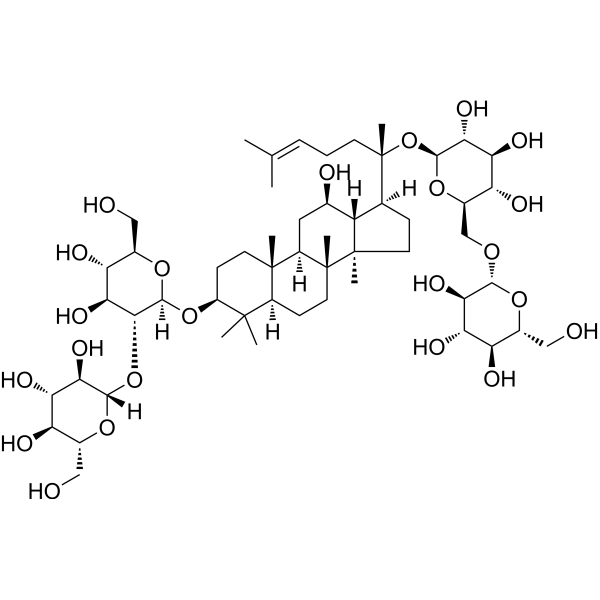
Ginsenoside Rb1
Based on 18 publication(s) in Google Scholar
Ginsenoside Rb1, a main constituent of the root of Panax ginseng, inhibits Na+, K+-ATPase activity with an IC50 of 6.3±1.0 μM. Ginsenoside also inhibits IRAK-1 activation and phosphorylation of NF-κB p65 .
Nos produits utilisent uniquement pour la recherche. Nous ne vendons pas aux patients.
- Pureté : 99.75%
- CAS No.: 41753-43-9
- Formule: C54H92O23
- Masse moléculaire:1109.29
-
Stockage:Powder -20°C, 3 years , 4°C, 2 years ; In solvent -80°C, 6 months , -20°C, 1 month

To place orders, for customer services and technical support, please contact: MedChemExpress USA
Tel: 609-228-6898 E-mail: [email protected] [email protected]


-
 Activité biologique
-
Chemical Information
-
Solvant et solubilité
- Protocole
- Pureté et documentation
- Références
-
 Help & FAQs


-
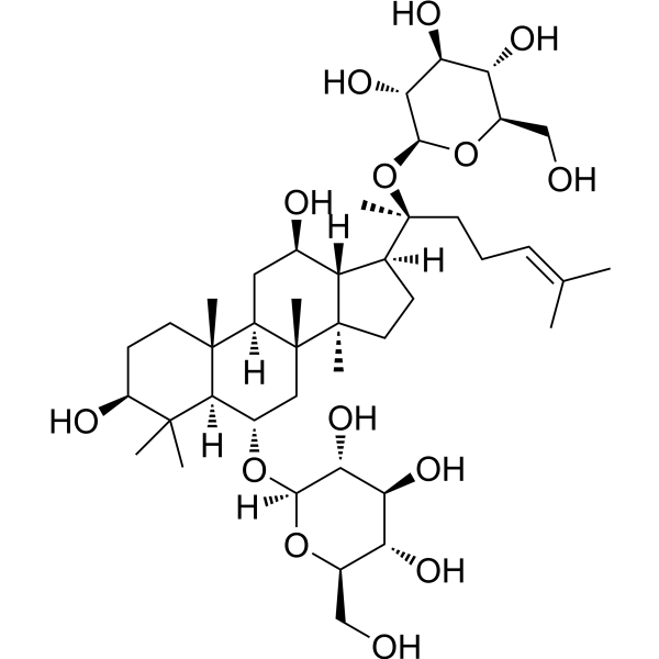
HY-N0045
Ginsenoside Rg1Apoptosis Inducer -
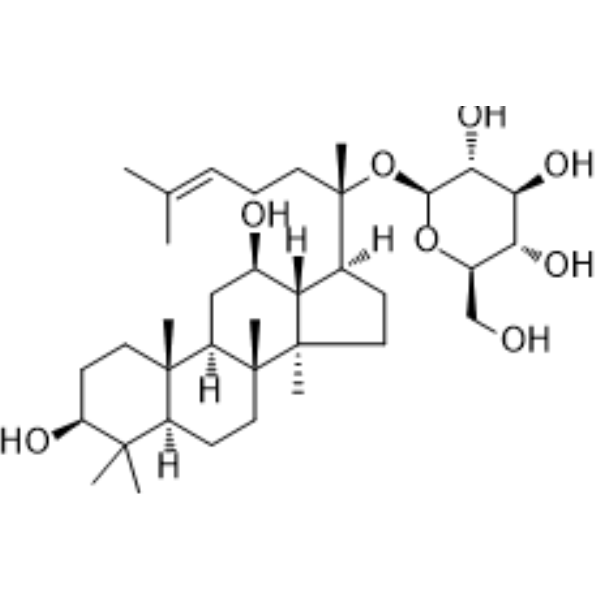
HY-N0904
Ginsenoside C-KNO Synthase/COX Inhibitor -
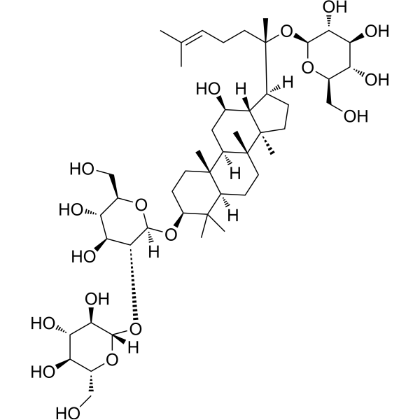
HY-N0043
Ginsenoside RdCytochrome P450 Inhibitor -
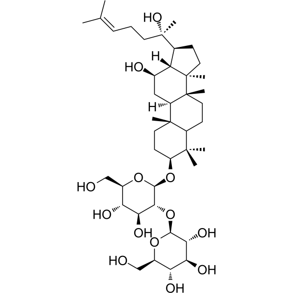
HY-N0603
20(S)-Ginsenoside Rg3Sodium Channel Inhibitor -
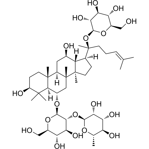
HY-N0044
Ginsenoside ReEndogenous Metabolite -
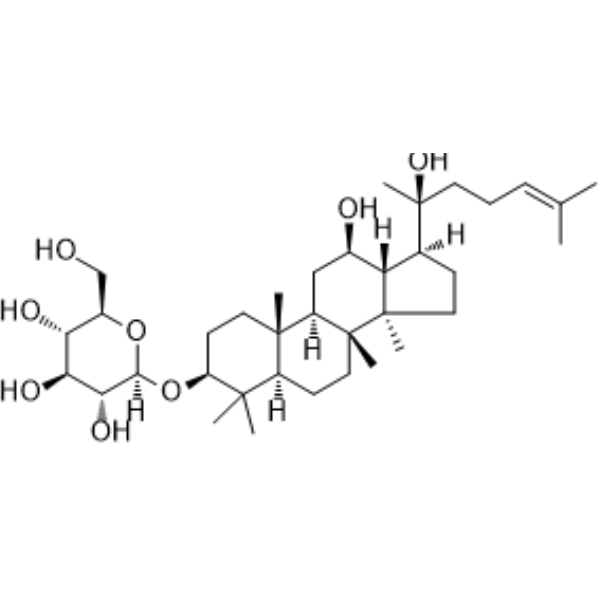
HY-N0605
Ginsenoside Rh2Caspase Activator -
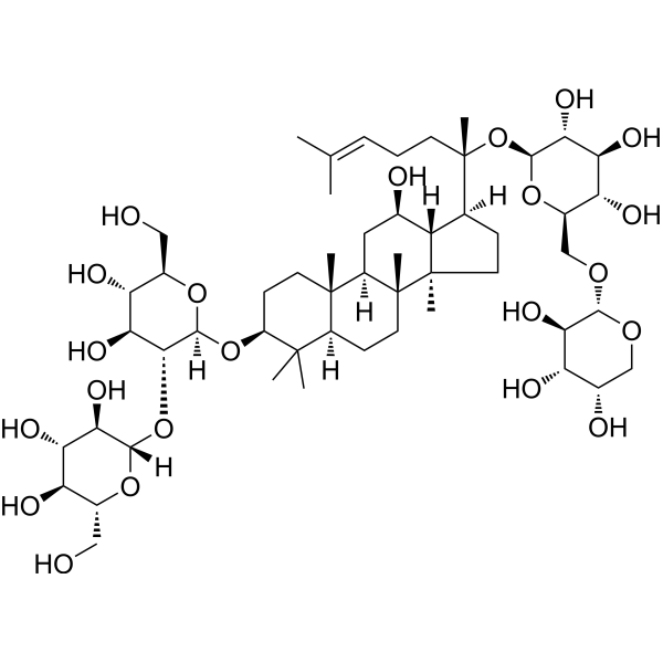
HY-N0040
Ginsenoside Rb2Influenza Virus Inhibitor -
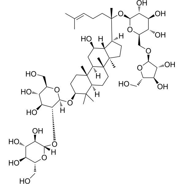
HY-N0042
Ginsenoside RcTNF Receptor Inhibitor -
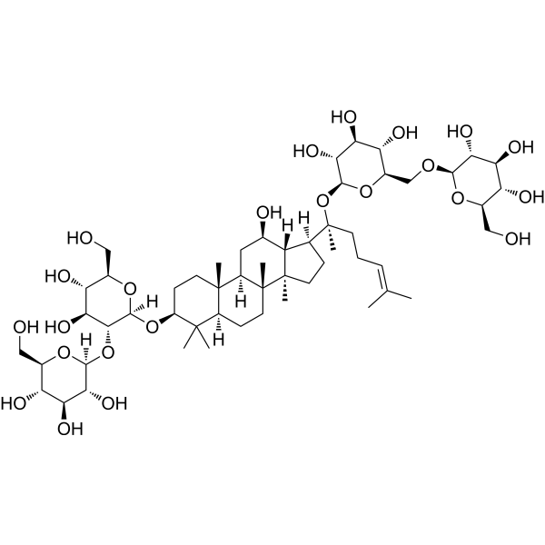
HY-N0039R
Ginsenoside Rb1 (Standard) -
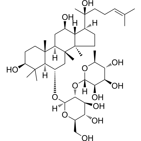
HY-N0602
Ginsenoside Rg2NF-κB Inhibitor -
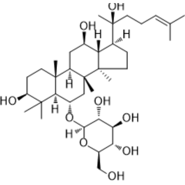
HY-N0604
Ginsenoside Rh1Endogenous Metabolite -
HY-N0431
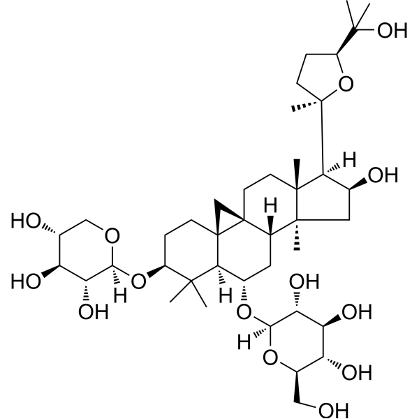
Astragaloside IVMMP Inhibitor
Publications Citing Use of MedChemExpress (MCE) Ginsenoside Rb1
More- J Adv Res. 2026 Jul 3:S2090-1232(26)00527-8.
- J Nanobiotechnology. 2025 Jun 13;23(1):443. [Abstract]
- Cell Death Dis. 2022 Sep 26;13(9):824. [Abstract]
- Phytomedicine. 2026 Apr 26. [Abstract]
- Phytomedicine. 2023 Dec:121:155083. [Abstract]
- Mol Med. 2024 Dec 2;30(1):240. [Abstract]
- Mater Des. 2024 Sep.
- Br J Pharmacol. 2024 Dec;181(23):4822-4844. [Abstract]
- Chin Med. 2026 Jan 8;21(1):19. [Abstract]
- Drug Des Devel Ther. 2026 Apr 23:20:602453. [Abstract]
- Int J Mol Sci. 2025 Jan 26;26(3):1075. [Abstract]
- Front Pharmacol. 2021 Apr 27:12:643188. [Abstract]
- Sci Rep. 2026 Feb 1;16(1):6880. [Abstract]
- Biol Proced Online. 2025 Jun 16;27(1):21. [Abstract]
- Vet Microbiol. 2026 May:316:110992. [Abstract]
- Pharmacol Res Perspect. 2026 Oct;14(5):e70324.
- Research Square Print. September 1st, 2022.
- Research Square Preprint. 2021 Feb.
 Customer Validation & Images

-
In Vivo Efficacy Study
-
Histological Imaging/Staining

-
IHC

-
Cell Proliferation/Viability Assay
-
Bio/Physico-chemical Assay


Activité biologique
Description
|
Na+, K+-ATPase 6.3 μM (IC50) |
IRAK-1 |
p65 |
Autophagy |
Mitophagy |
HSV-1 |
Cellular Effect
|
Cell Line

|
Type | Value | Description | References |
|---|---|---|---|---|
| Erythrocyte | IC50 |
3.18 μM
Compound: 70
|
Inhibition of AAPH-induced hemolysis of human erythrocytes
Inhibition of AAPH-induced hemolysis of human erythrocytes
|
[PMID: 32006794] |
In Vitro
Rat brain microsomal Na+, K+-ATPase activity is inhibited significantly and rapidly by Ginsenoside Rb1. The IC50 of Ginsenoside Rb1 for Na+,K+-ATPase is 6.3±1.0 μM. The inhibition is enhanced with increasing the concentration of Ginsenoside Rb1 or decreasing that of Na+ and K+. Kinetic analysis reveals that Ginsenoside is an uncompetitive inhibitor with respect to ATP[1]. Ginsenoside Rb1 significantly inhibits the activation of interleukin-1 receptor-associated kinase-1 (IRAK-1), IKK-β, NF-κB, and MAP kinases (ERK, JNK, and p-38); however, interaction between LPS and Toll-like receptor-4, IRAK-4 activation and IRAK-2 activation are unaffected[2]. Ginsenoside Rb1 is an ingredient of a Chinese medicine Panax ginseng. Ginsenoside Rb1 is a major bioactive compound in the regulating pregnane X receptor (PXR)/NF-κB signaling. Ginsenoside Rb1 is the compound with potent anti-inflammatory activity in ginseng saponin extract (GSE). The concentration for Ginsenoside Rb1 (10 μM) is optimized from a preliminary study to ensure sufficient anti-inflammatory activity and without apparent cytotoxicity. Ginsenoside Rb1 significantly reduces TNF-α-induced upregulation of IL-1β and iNOS mRNA levels, and restores the mRNA levels of PXR and CYP3A4 in LS174T cells. TNF-α causes a significant reduction in PXR protein levels and increase in the ratio of phosphorylated to total NF-κB p65, both of which are significantly abrogated by Ginsenoside Rb1[3].
MedChemExpress (MCE) has not independently confirmed the accuracy of these methods. They are for reference only.
In Vivo
MedChemExpress (MCE) has not independently confirmed the accuracy of these methods. They are for reference only.


Chemical Information
-
CAS No. 41753-43-9
-
Appearance Solid
-
Masse moléculaire 1109.29
-
Formule C54H92O23
-
Color White to off-white
-
SMILES
C[C@@]([C@@]12C)(CC[C@@]3([H])C4(C)C)[C@@](C[C@@H](O)[C@]1([H])[C@]([C@@](CC/C=C(C)/C)(C)O[C@@H]([C@@H]([C@@H](O)[C@@H]5O)O)O[C@@H]5CO[C@@H]([C@@H]([C@@H](O)[C@@H]6O)O)O[C@@H]6CO)([H])CC2)([H])[C@]3(CC[C@@H]4O[C@@](O[C@H](CO)[C@@H](O)[C@@H]7O)([H])[C@@H]7O[C@]([C@@H]([C@@H](O)[C@@H]8O)O)([H])O[C@@H]8CO)C
-
Synonyms
Gypenoside III
-
Structure Classification
-
Initial Source
-
Livraison
Room temperature in continental US; may vary elsewhere.
-
Stockage
Powder -20°C 3 years 4°C 2 years In solvent -80°C 6 months -20°C 1 month


Publications (18)
-
Journal Impact Factor
-
Most Recent
-
-
J Nanobiotechnology
Overcoming acquired immunotherapy resistance in non-small cell lung cancer using ginsenoside Rb1-loaded, peptide-enhanced exosome delivery systems. [Abstract]2025 Jun 13;23(1):443. PMID: 40514658
Ginsenoside Rb1 purchased from MedChemExpress. Usage Cited in: J Nanobiotechnology. 2025 Jun 13;23(1):443. [Abstract]
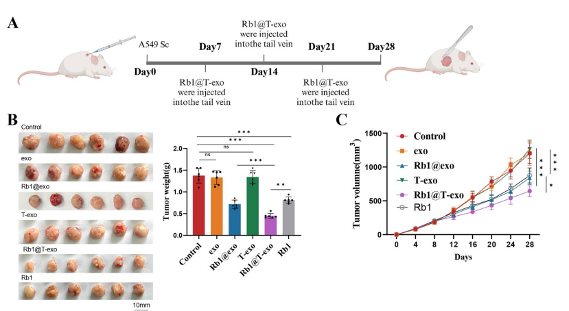
Ginsenoside Rb1 (5 mg/kg; via tail vein injection on days 7, 14, and 21 post-tumor implantation) in BALB/c nude mice (PI3K-mutated A549 cells) exhibited significantly decreased tumor weight and reduced tumor growth rate compared to the Control group. A Schematic timeline of the animal experiment; B Images and weights of dissected tumors from subcutaneous xenograft model mice in each group. C Tumor growth curves for mice in each group;
Ginsenoside Rb1 purchased from MedChemExpress. Usage Cited in: J Nanobiotechnology. 2025 Jun 13;23(1):443. [Abstract]
Ginsenoside Rb1 (5 mg/kg; via tail vein injection on days 7, 14, and 21 post-tumor implantation) in BALB/c nude mice (PI3K-mutated A549 cells) reduced infiltration of inflammatory cells, tighter cell arrangement, more normal cell morphology, uniform cell nuclei, and clear nucleoli. H&E staining images of tumors from each group.
Ginsenoside Rb1 purchased from MedChemExpress. Usage Cited in: J Nanobiotechnology. 2025 Jun 13;23(1):443. [Abstract]
Ginsenoside Rb1 (5 mg/kg; via tail vein injection on days 7, 14, and 21 post-tumor implantation) in BALB/c nude mice (PI3K-mutated A549 cells) compared to the Control group, with a more substantial decrease in Ki67 expression in the Rb1@T-exo group.
-
Cell Death Dis
A dual Keap1 and p47phox inhibitor Ginsenoside Rb1 ameliorates high glucose/ox-LDL-induced endothelial cell injury and atherosclerosis. [Abstract]2022 Sep 26;13(9):824. PMID: 36163178
Ginsenoside Rb1 purchased from MedChemExpress. Usage Cited in: Cell Death Dis. 2022 Sep 26;13(9):824. [Abstract]
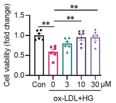
EA.hy926 ECs were pretreated with Ginsenoside Rb1 (Rb1; 3, 10, and 30 μM) for 30 min before ox-LDL/HG for 24 h, Ginsenoside Rb1 dose-dependently restored the declined cell viability in ox-LDL/HG-challenged ECs.
Ginsenoside Rb1 purchased from MedChemExpress. Usage Cited in: Cell Death Dis. 2022 Sep 26;13(9):824. [Abstract]
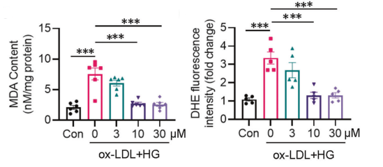
EA.hy926 ECs were pretreated with Ginsenoside Rb1 (Rb1; 3, 10, and 30 μM) for 30 min before ox-LDL/HG for 24 h. Exposure to ox-LDL/HG promoted the generation of MDA, a marker of oxidative stress, while this was concentrationdependently reversed by Rb1 pretreatment.
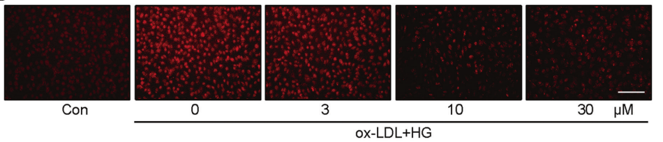
Ginsenoside Rb1 purchased from MedChemExpress. Usage Cited in: Cell Death Dis. 2022 Sep 26;13(9):824. [Abstract]
EA.hy926 ECs were pretreated with Ginsenoside Rb1 (Rb1; 3, 10, and 30 μM) for 30 min before ox-LDL/HG for 24 h. Representative images of DHE staining.
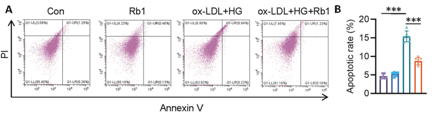
Ginsenoside Rb1 purchased from MedChemExpress. Usage Cited in: Cell Death Dis. 2022 Sep 26;13(9):824. [Abstract]
EA.hy926 ECs were pretreated with Ginsenoside Rb1 (Rb1; 10 μM) for 30 min before ox-LDL/HG for 24 h. A Cell apoptosis measured by flow cytometer. B Apoptosis ratio.
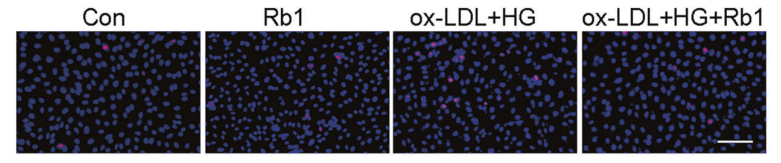
Ginsenoside Rb1 purchased from MedChemExpress. Usage Cited in: Cell Death Dis. 2022 Sep 26;13(9):824. [Abstract]
EA.hy926 ECs were pretreated with Ginsenoside Rb1 (Rb1; 10 μM) for 30 min before ox-LDL/HG for 24 h. Cell apoptosis measured by TUNEL assay.
-
Phytomedicine
Rare ginsenoside Rk1 protects against cisplatin-induced auditory damage by regulating the MST1/LONP1 pathway. [Abstract]2026 Apr 26. PMID: 42097003 -
Phytomedicine
Ginsenoside Rb1 protects hippocampal neurons in depressed rats based on mitophagy-regulated astrocytic pyroptosis. [Abstract]2023 Dec:121:155083. PMID: 37722244 -
Mol Med
Ginsenoside Rb1 affects mitochondrial Ca2+ transport and inhibits fat deposition and fibrosis by regulating the wnt signaling pathway to treat rotator cuff tears via docking with SFRP1. [Abstract]2024 Dec 2;30(1):240. PMID: 39617900 -
-
Br J Pharmacol
The plant extract PNS mitigates atherosclerosis via promoting Nrf2-mediated inhibition of ferroptosis through reducing USP2-mediated Keap1 deubiquitination. [Abstract]2024 Dec;181(23):4822-4844. PMID: 39228119 -
Chin Med
2026 Jan 8;21(1):19. PMID: 41508002 -
Drug Des Devel Ther
Cellular and Molecular Mechanisms of Hyperglycemia-Induced Atherosclerosis and Intervention Strategies of Chinese Herbal Medicine. [Abstract]2026 Apr 23:20:602453. PMID: 42057976 -
Int J Mol Sci
2025 Jan 26;26(3):1075. PMID: 39940841 -
Front Pharmacol
Frutescone O from Baeckea frutescens Blocked TLR4-Mediated Myd88/NF-κB and MAPK Signaling Pathways in LPS Induced RAW264.7 Macrophages. [Abstract]2021 Apr 27:12:643188. PMID: 33986676 -
Sci Rep
Multi-omics analysis reveals that ginsenoside Rb1 improves prognostic outcomes in sepsis by modulating mitochondrial metabolism MTHFD2 targets. [Abstract]2026 Feb 1;16(1):6880. PMID: 41622321 -
Biol Proced Online
Establishment of a mouse lung cancer organoid model and its applications for therapeutic screening. [Abstract]2025 Jun 16;27(1):21. PMID: 40524168 -
Vet Microbiol
The Chinese medicine monomer Schisandrin C inhibits PRRSV infection by regulating the OGT-PI3K/AKT/mTOR signaling pathway. [Abstract]2026 May:316:110992. PMID: 41865607 -
-
-


Solvant et solubilité
In Vitro:
DMSO : 100 mg/mL (90.15 mM; Need ultrasonic; Hygroscopic DMSO has a significant impact on the solubility of product, please use newly opened DMSO)
H2O : ≥ 33.33 mg/mL (30.05 mM)
* "≥" means soluble, but saturation unknown.
Please refer to the solubility information to select the appropriate solvent. Once prepared, please aliquot and store the solution to prevent product inactivation from repeated freeze-thaw cycles.
Storage method and period of stock solution: -80°C, 6 months; -20°C, 1 month. When stored at -80°C, please use it within 6 months. When stored at -20°C, please use it within 1 month.
* Note: If you choose water as the stock solution, please dilute it to the working solution, then filter and sterilize it with a 0.22 μm filter before use.
Please refer to the solubility information to select the appropriate solvent. Once prepared, please aliquot and store the solution to prevent product inactivation from repeated freeze-thaw cycles.
Storage method and period of stock solution: -80°C, 6 months; -20°C, 1 month. When stored at -80°C, please use it within 6 months. When stored at -20°C, please use it within 1 month.
* Note: If you choose water as the stock solution, please dilute it to the working solution, then filter and sterilize it with a 0.22 μm filter before use.
Concentration (start) × Volume (start) = Concentration (final) × Volume (final)
In Vivo:
Select the appropriate dissolution method based on your experimental animal and administration route.
- For the following dissolution methods, please ensure to first prepare a clear stock solution using an In Vitro approach and then sequentially add co-solvents:
- To ensure reliable experimental results, the clarified stock solution can be appropriately stored based on storage conditions. As for the working solution for In Vivo experiments, it is recommended to prepare freshly and use it on the same day.
- The percentages shown for the solvents indicate their volumetric ratio in the final prepared solution. If precipitation or phase separation occurs during preparation, heat and/or sonication can be used to aid dissolution.
Add each solvent one by one: 10% DMSO 40% PEG300 5% Tween-80 45% Saline
Solubility: ≥ 2.5 mg/mL (2.25 mM); Clear solution
This protocol yields a clear solution of ≥ 2.5 mg/mL (saturation unknown).
Taking 1 mL working solution as an example, add 100 μL DMSO stock solution (25.0 mg/mL) to 400 μL PEG300, and mix evenly; then add 50 μL Tween-80 and mix evenly; then add 450 μL Saline to adjust the volume to 1 mL.
Preparation of Saline: Dissolve 0.9 g sodium chloride in ddH₂O and dilute to 100 mL to obtain a clear Saline solution.
Add each solvent one by one: 10% DMSO 90% (20% SBE-β-CD in Saline)
Solubility: ≥ 2.5 mg/mL (2.25 mM); Clear solution
This protocol yields a clear solution of ≥ 2.5 mg/mL (saturation unknown).
Taking 1 mL working solution as an example, add 100 μL DMSO stock solution (25.0 mg/mL) to 900 μL 20% SBE-β-CD in Saline, and mix evenly.
Preparation of 20% SBE-β-CD in Saline (4°C, storage for one week): 2 g SBE-β-CD powder is dissolved in 10 mL Saline, completely dissolve until clear.
In Vivo Dissolution Calculator
Please enter the basic information of animal experiments:
-
-
-
-
Recommended: Prepare an additional quantity of animals to account for potential losses during experiments.
Working solution concentration: 0.22 mg/mL
This product has good water solubility, please refer to the measured solubility data in water/PBS/Saline for details.


Pureté et documentation
-
Fiche technique (290 KB)
-
SDS (396 KB)
- English - EN (396 KB)
- Français - FR (396 KB)
- Deutsch - DE (396 KB)
- Norwegian - NO (396 KB)
- Español - ES (396 KB)
- Swedish - SV (396 KB)
- Italian - IT (396 KB)
- Korean - KR (396 KB)
- Portuguese - PT (396 KB)
-
Instruction de manipulation (2659 KB)
Références
[1]. Cao J, et al. Inhibitory effects of ginsenoside Rg1 and Rb1 on rat brain microsomal Na+,K+-ATPase activity. Zhongguo Yao Li Xue Bao. 1990 Jan;11(1):10-4. [Content Brief]
[2]. Zhang J, et al. Ginsenosides Regulate PXR/NF-κB Signaling and Attenuate Dextran Sulfate Sodium-Induced Colitis. Drug Metab Dispos. 2015 Aug;43(8):1181-9. [Content Brief]
[3]. Joh EH, et al. Ginsenoside Rb1 and its metabolite compound K inhibit IRAK-1 activation--the key step of inflammation. Biochem Pharmacol. 2011 Aug 1;82(3):278-86. [Content Brief]
[4]. Jiang Y, et al. Ginsenoside Rb1 Treatment Attenuates Pulmonary Inflammatory Cytokine Release and Tissue Injury following Intestinal Ischemia Reperfusion Injury in Mice. Oxid Med Cell Longev. 2015;2015:843721. [Content Brief]
[5]. Zhang Y, et al. Ginsenoside Rb1 ameliorates lipopolysaccharide-induced albumin leakage from rat mesenteric venules by intervening in both trans- and paracellular pathway. Am J Physiol Gastrointest Liver Physiol. 2014 Feb 15;306(4):G289-300. [Content Brief]


Complete Stock Solution Preparation Table
Please refer to the solubility information to select the appropriate solvent. Once prepared, please aliquot and store the solution to prevent product inactivation from repeated freeze-thaw cycles.
Storage method and period of stock solution: -80°C, 6 months; -20°C, 1 month. When stored at -80°C, please use it within 6 months. When stored at -20°C, please use it within 1 month.
| Optional Solvent | Concentration Solvent Mass | 1 mg | 5 mg | 10 mg | 25 mg |
|---|---|---|---|---|---|
| H2O / DMSO | 1 mM | 0.9015 mL | 4.5074 mL | 9.0148 mL | 22.5369 mL |
| 5 mM | 0.1803 mL | 0.9015 mL | 1.8030 mL | 4.5074 mL | |
| 10 mM | 0.0901 mL | 0.4507 mL | 0.9015 mL | 2.2537 mL | |
| 15 mM | 0.0601 mL | 0.3005 mL | 0.6010 mL | 1.5025 mL | |
| 20 mM | 0.0451 mL | 0.2254 mL | 0.4507 mL | 1.1268 mL | |
| 25 mM | 0.0361 mL | 0.1803 mL | 0.3606 mL | 0.9015 mL | |
| 30 mM | 0.0300 mL | 0.1502 mL | 0.3005 mL | 0.7512 mL | |
| DMSO | 40 mM | 0.0225 mL | 0.1127 mL | 0.2254 mL | 0.5634 mL |
| 50 mM | 0.0180 mL | 0.0901 mL | 0.1803 mL | 0.4507 mL | |
| 60 mM | 0.0150 mL | 0.0751 mL | 0.1502 mL | 0.3756 mL | |
| 80 mM | 0.0113 mL | 0.0563 mL | 0.1127 mL | 0.2817 mL |
* Note: If you choose water as the stock solution, please dilute it to the working solution, then filter and sterilize it with a 0.22 μm filter before use.


Ginsenoside Rb1 Related Classifications
HY-N0039 Related Classifications
 Powered by Bioz